
Hermatobates lingyangjiaoensis Luo, Chen & Wang, 2019
|
publication ID |
https://doi.org/ 10.11646/zootaxa.4679.3.6 |
|
publication LSID |
lsid:zoobank.org:pub:C42FEC2C-F721-47F8-BF66-4D16AB859862 |
|
persistent identifier |
https://treatment.plazi.org/id/E76E1C21-09A3-48AD-BAAE-CF315A017DB1 |
|
taxon LSID |
lsid:zoobank.org:act:E76E1C21-09A3-48AD-BAAE-CF315A017DB1 |
|
treatment provided by |
Plazi |
|
scientific name |
Hermatobates lingyangjiaoensis Luo, Chen & Wang |
| status |
sp. nov. |
Hermatobates lingyangjiaoensis Luo, Chen & Wang , sp. n.
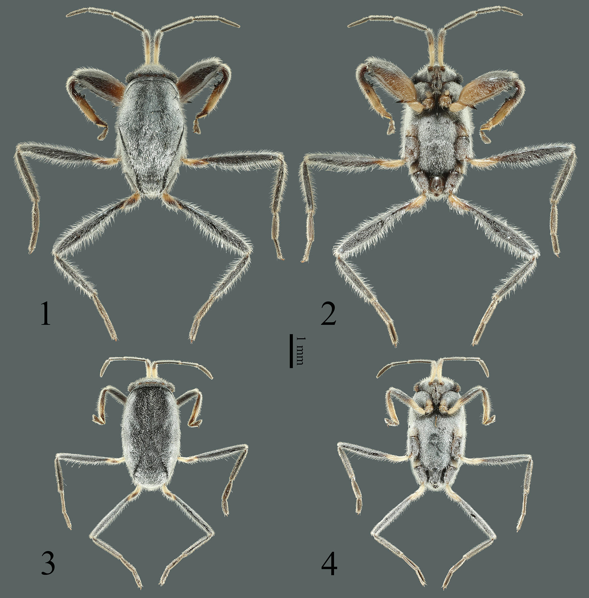
( Figs. 1–22 View FIGURES 1–4 View FIGURES 5–6 View FIGURES 7–12 View FIGURES 13–14 View FIGURES 15–16 View FIGURES 17–22 )
urn:lsid:zoobank.org:act:E76E1C21-09A3-48AD-BAAE-CF315A017DB1
Type material. Holotype (♂) CHINA: Hainan Province, Sansha Prefecture, Xisha, Lingyang Jiao Reef : 16°28’03”N, 111°35’58”E, 0 m at sea level, 2019-V-7, leg. Qiang XIE, mounted on card ( SYSBM) GoogleMaps . Paratypes (5♂♂, 5♀♀): same data as holotype, mounted on cards ( SYSBM) GoogleMaps .
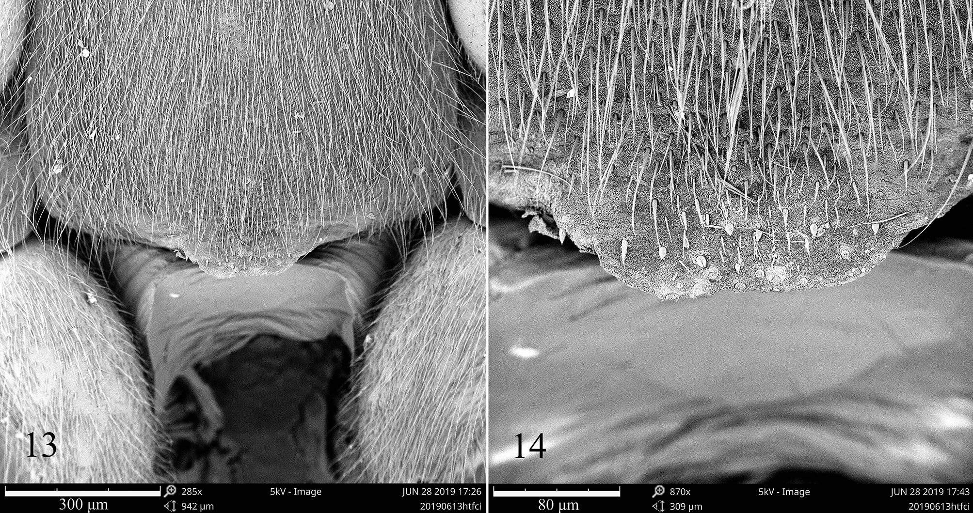
Description. Size: Male, length 3.2–3.9 mm, greatest width (across meso- and metathorax) 1.6–1.9 mm; female, length 3.3–3.4 mm, greatest width (across meso- and metathorax) 1.7–1.8 mm. Color and body surface: Body and most of head above uniformly black, dull, sometimes with bluish tinges ( Figs 1, 3 View FIGURES 1–4 ). Pleural and ventral surfaces of body and head as in dorsal surface ( Figs. 2, 4 View FIGURES 1–4 ). Two pairs of pale areas on vertex along with a transverse suture. Eyes blackish brown to nearly black. Antenna shiny, blackish brown, basal half to two-thirds of first segment light yellowish brown. Anteclypeus, genal lobe and rostrum shiny, blackish brown. Prothorax, mesothorax and metathorax episternal lobes stramineous to yellowish brown ( Figs. 2, 4 View FIGURES 1–4 ). Legs shiny, brown to dark brown, fore coxa and trochanter, base of fore femur, middle of fore tibia yellowish brown, fore tarsus brown; middle and hind coxae brown, basal half of trochanters yellowish brown, tarsi brown. Genital segments shiny, brown to dark brown ( Figs. 5–6 View FIGURES 5–6 ). Pilosity: Body and appendages with fine silver pubescence ( Figs. 1–4 View FIGURES 1–4 ), head and body surface with a dense layer of peglike microtrichia ( Fig. 16 View FIGURES 15–16 ).
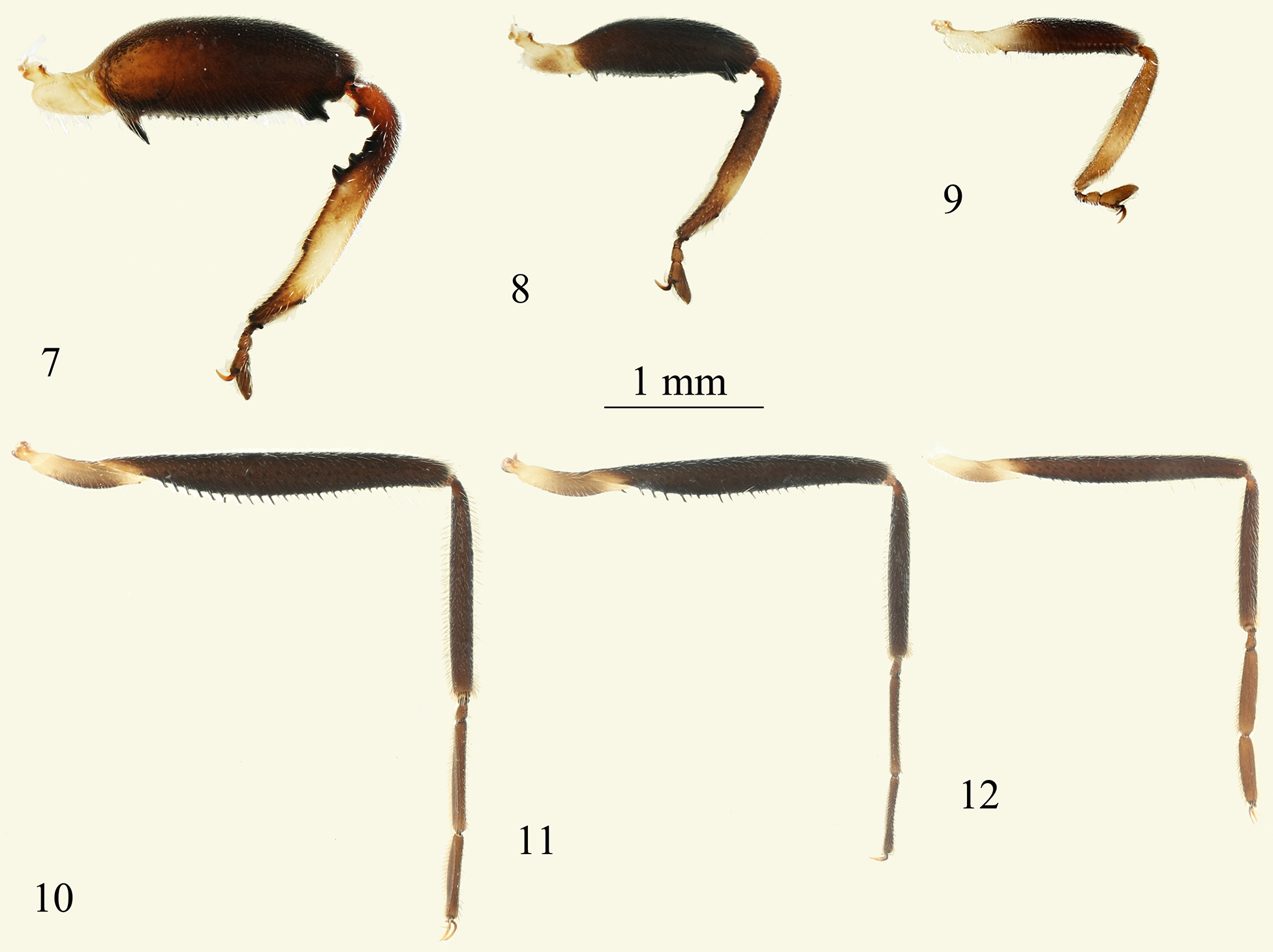
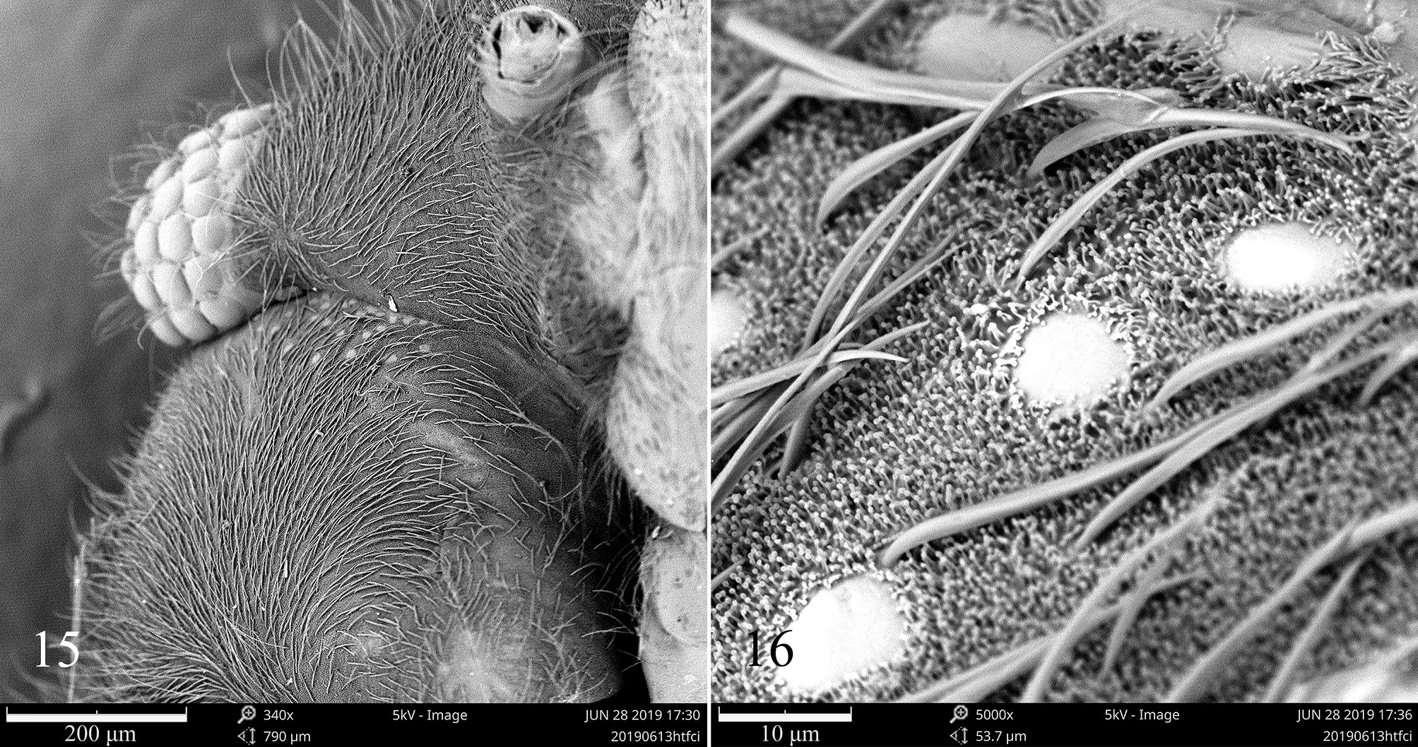
Apterous male. Structures: Body fusiform. Body length 1.9–2.0× greatest width across thorax. Head. Head short, greatest width across eyes 4.4–5.3× median length; width of eyes 0.25–0.28× width of head between eyes; antennal length 0.86–0.96× body length, segments I and II, and segments III and IV subequal in length, first segment slightly thicker than other segments. Apex of rostrum extends posteriorly beyond fore coxa. Thorax. Pronotum very short, median length about 0.5× eye width; anterior and posterior margins almost parallel, curved laterally.About 25 glabrous spots (possibly sense organ) at anterior area of propleuron ( Figs. 15–16 View FIGURES 15–16 ). Meso- and metathorax prolonged and fused, posterior margin of mesothorax visible laterally over insertion of middle coxa and position of metathoracic spiracle. Posterior margin of metasternum with a trapezoidal process medially ( Fig. 5 View FIGURES 5–6 ). Legs. Fore trochanter bearing a small black tooth ventrally at about one-third from base to apex. Fore femur moderately to strongly incrassate, greatest width 0.31–0.42× length, with following armature: inner side of femur with a sharp, black tooth at base obliquely directed apically, a broad, distally bifid, black tooth near apex, between these two large teeth is a row of 10–14 small, black teeth. Base of fore tibia curved, a dark brown tooth within the curve, inner margin with two low tubercles beyond basal curve, followed by a broad, black tooth; a black tubercle at about one-third to apex, a row of small tubercles near apex of tibia ( Figs. 7–8 View FIGURES 7–12 ). Outer margin of tibia with a comb of hairs at about one-fifth to apex, and two small tubercles at both ends of the hairs comb. Middle and hind coxae enlarged. Trochanter of middle leg with a small black tooth near apex of ventral side. Middle femur slightly thickened ventrally, with a row of 24–34 black spines which are longer and hooked from base and shorter and straight toward apex ( Figs. 10–11 View FIGURES 7–12 ). Ventral side of femur bearing several brown, scattered long hairs with length exceeding widest diameter of femur. Middle tibia and tarsus subequal in length. Hind femur slightly thickened ventrally, unarmed. Hind tibia and tarsus subequal in length, a round process at apex of tibia.
External genitalia. Abdominal segment 8 with lateral hook-like projection ( Figs. 17–19 View FIGURES 17–22 ); pygophore globular with triangular basal margin curved upwards ( Figs. 20–22 View FIGURES 17–22 ); parameres stunted, asymmetrical, small and short, with blunt apices ( Figs. 21–22 View FIGURES 17–22 ).
Apterous female. Structures: As in male, except for the following: Body length about 1.9× greatest width across thorax. Head. Head conspicuously shorter than in male, greatest width across eyes 6.9–8.5× median length, eye width 0.24–0.26× width of head between eyes. Antenna relatively shorter than in male, length 0.74–0.76× body length of insect. Thorax. Pronotum anterior and posterior margins almost parallel in the middle, laterally narrow. Meso- and metathorax prolonged and fused, medially with a deep membranous furrow. Posterior margin of metasternum straight, with no process ( Fig. 6 View FIGURES 5–6 ). Legs relatively shorter and slender than in male. Fore trochanter unarmed, fore femur moderately thickened, greatest width 0.20–0.23× length, except for a row of 17–19 small, black teeth on ventral surface of femur, a black tooth separated and larger than other teeth at base of femur ( Fig. 9 View FIGURES 7–12 ). Base of fore tibia curved and unarmed. Middle trochanter unarmed. Middle femur with about 12 very small black teeth on ventral side, and several brown, scattered long hairs with length exceeding widest diameter of femur ( Fig. 12 View FIGURES 7–12 ). Hind femur slightly thickened ventrally, unarmed.
Diagnosis. Based on morphological features, Polhemus & Polhemus (2012) divided Hermatobates into five species groups. We consider that the new species should be placed in H. weddi group, which currently includes H. weddi , H. marchei , H. singaporensis , H. kula , H. armatus and H. schuhi . All the members of this group share the following characteristics: well developed trapezoidal to quadrate posterior metasternal process, male fore tibia with variations on a common basal armature consisting of multiple large teeth, and complex male endosoma with prominent lateral lobes ( Polhemus & Polhemus 2012). The new species can be easily distinguished from H. marchei , H. singaporensis and H. armatus by the trapezoidal shaped process on the posterior margin of the male metasternum; and can be distinguished from H. kula by the metasternum of the male not tumid posteriorly. Hermatobates lingyangjiaoensis sp. n. is closest in morphology to H. weddi and H. schuhi .
Hermatobates lingyangjiaoensis sp. n. is similar to H. weddi in size and habitus, but it can be distinguished from the latter species by a) the posterior margin of the male trapezoidal shaped process on the male metasternum of H. lingyangjiaoensis sp. n. (~0.17) is wider than H. weddi (~0.10, based on an illustration in Polhemus & Polhemus 2012, fig. 2F), (the ratio of width of the posterior margin of the trapezoidal shaped process on the male metasternum to width of the posterior margin of the male metasternum); b) the ventral side of the female middle femur with a row of ~12 black, very small teeth ( Fig. 12 View FIGURES 7–12 ), whereas in H. weddi , the middle femur has only a few tiny black teeth basally on the ventral margin; c) the color of the fore tibia is brown except for the middle part yellowish brown ( Figs. 1–4 View FIGURES 1–4 , 7–9 View FIGURES 7–12 ), whereas the fore tibia of H. weddi is yellowish brown except for the basal part brown; d) the antenna is blackish brown, the basal half to two-thirds of antennal segment I light yellowish brown ( Figs. 1–4 View FIGURES 1–4 ), whereas in H. weddi , the antenna is blackish brown with the basal third of segment I pale; e) the ventral side of the female fore femur bears a tooth which is larger and separated from the other teeth ( Fig. 9 View FIGURES 7–12 ), whereas in the female of H. weddi , the fore femur bears only a row of 15–18 small black teeth along the ventral margin.
Hermatobates lingyangjiaoensis sp. n. is similar to H. schuhi in habitus, but it can be distinguished by a) individuals of the new species are distinctly smaller in both male (length 3.2–3.9 mm, width 1.6–1.9 mm) and female (length 3.3–3.4 mm, width 1.7–1.8 mm), whereas in H. schuhi , the male (length 4.0– 4.2 mm, width 1.7–2.0 mm) and female (length 3.55–3.60 mm, width 2.0– 2.1 mm) are larger; b) the papillae on the posteromedial metasternal process of the male are relatively sparse and somewhat elongate, whereas in H. schuhi , these papillae are very numerous but smaller and more rounded; c) the ventral side of the female middle femur has a row of ~12 black, very small teeth ( Fig. 12 View FIGURES 7–12 ), whereas in H. schuhi , the ventral side of the female middle femur has a row of ~7 black, very small teeth; d) the ventral side of the male middle femur bears a row of 24–34 small, black teeth ( Figs. 10–11 View FIGURES 7–12 ), whereas in H. schuhi , the ventral side of the male middle femur bears a row of 20–24 small teeth; e) the ventral side of the male fore femur bears a row of 10–14 small, black teeth ( Figs. 7–8 View FIGURES 7–12 ), whereas in H. schuhi , the ventral side of the male fore femur bears a row of 14–16 small, black teeth.
Variation. Male individuals of this species have relatively large variations in the size of the fore leg, from moderately incrassate to less incrassate, and the greatest width of the fore femur is positively correlated to the length of body (the specific data of all seven males in mm, body length/greatest width of fore femur = 3.80/0.70, 3.78/0.65, 3.69/0.62, 3.65/0.66, 3.38/0.57, 3.30/0.42, 3.20/0.38). In moderately incrassate individuals, the depression between the bifid tooth and large tooth along the inner margin of the basal part of the fore tibia is subequal to the width of the large tooth ( Fig. 7 View FIGURES 7–12 ); and the same depression is approximately 2× the width of the large tooth in less incrassate individuals ( Fig. 8 View FIGURES 7–12 ). The width of the process on the posterior margin of the metasternum also varies among males.
Etymology. This species is named after the Lingyangjiao Reef in the South China Sea, where the specimens were collected.
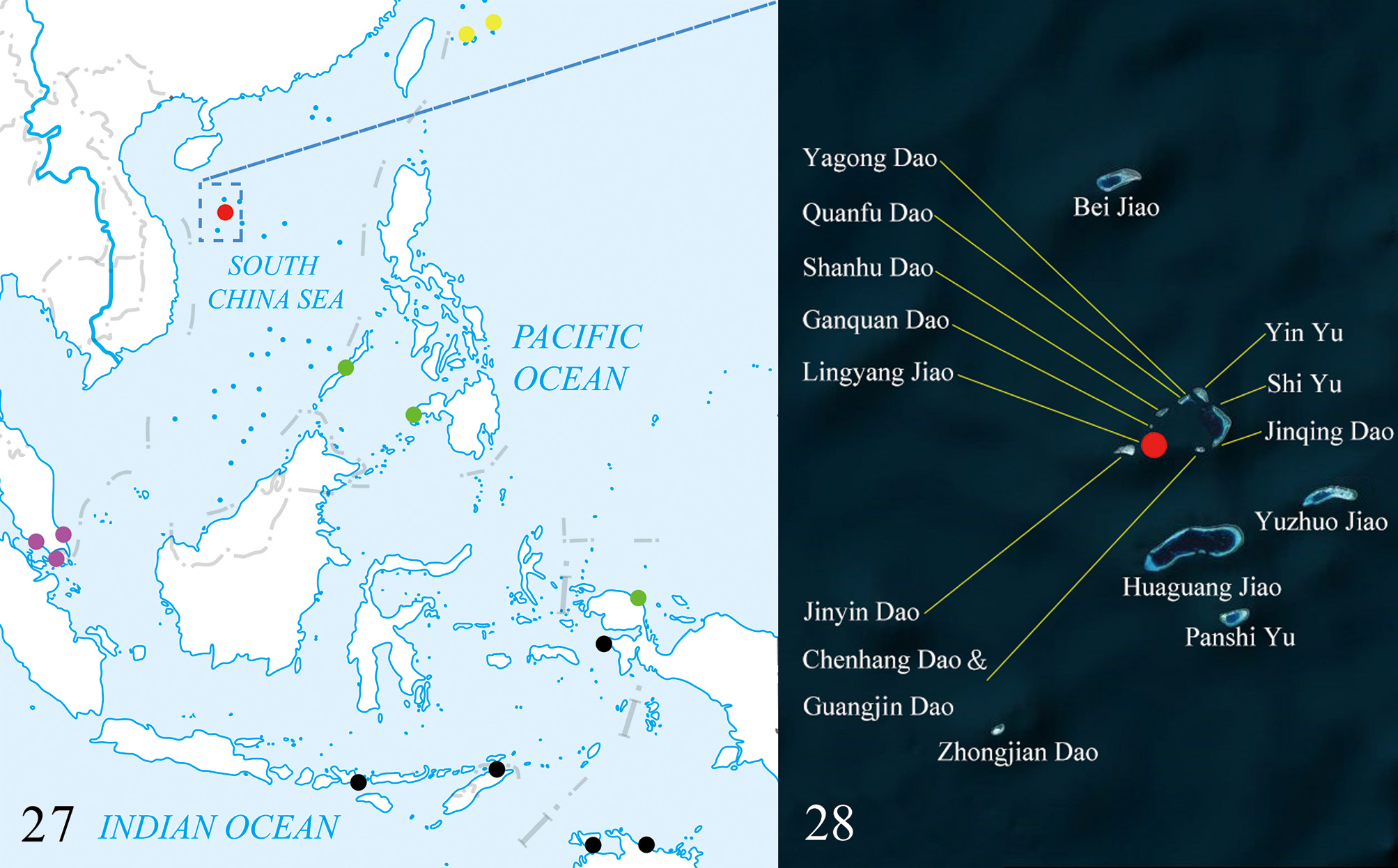
Distribution. This species is so far known from only the type locality at Lingyangjiao Reef (= Antelope Island) in South China Sea. Lingyangjiao Reef is one of the reefs of the Yongle Islands ( Figs. 27–28 View FIGURES 27–28 ), The last author has made an intensive field survey around the Yongle Islands. However, Hermatobates was found only at Lingyangjiao Reef where individuals could regularly be collected.
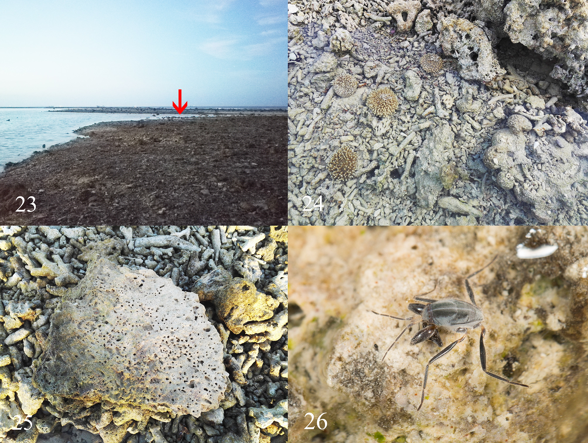
Biology. The biology and ecology of Hermatobatidae are poorly known and are based on the observations of Esaki (1947), Cheng (1977), Foster (1989), and Polhemus (1990). Esaki found Hermatobates living together with many Halovelia septentrionalis Esaki , collembolans, staphylinid beetles, and marine chironomid midges; Cheng found Hermatobates living together with Halovelia , collembolans, staphylinid beetles, and mites; Foster observed them feeding on recently dead adult chironomid midges; Polhemus found they were in company with various species of Halobates . Our specimens of Hermatobates were collected at an intertidal reef at low tide, without plants ( Figs. 23–24 View FIGURES 23–26 ). When the reef was exposed, Hermatobates appeared on the surface of shallow water, and could also be found under coral rocks ( Fig. 25 View FIGURES 23–26 ). The tidal range between high tide and low tide at the collecting locality was generally over one meter, and the reefs remain out of the water for no more than 8 hours. In the same habitat where Hermatobates were collected, many Halovelia , mites, and a few Halobates were also found. Hermatobates lingyangjiaoensis sp. n. was found preying on Halovelia sp. ( Fig. 26 View FIGURES 23–26 ), which is the first direct evidence of their feeding habit.


No known copyright restrictions apply. See Agosti, D., Egloff, W., 2009. Taxonomic information exchange and copyright: the Plazi approach. BMC Research Notes 2009, 2:53 for further explanation.
|
Kingdom |
|
|
Phylum |
|
|
Class |
|
|
Order |
|
|
Family |
|
|
Genus |