
Parastenocaris enckelli, Reddy, Yenumula Ranga, Totakura, Venkateswara Rao & Shaik, Shabuddin, 2016
|
publication ID |
https://doi.org/ 10.11646/zootaxa.4066.2.2 |
|
publication LSID |
lsid:zoobank.org:pub:814E71CA-6313-49F3-B989-3D903DEEFA4A |
|
DOI |
https://doi.org/10.5281/zenodo.6087670 |
|
persistent identifier |
https://treatment.plazi.org/id/4177F156-FF9F-FFBB-4BF0-F730FF05775F |
|
treatment provided by |
Plazi |
|
scientific name |
Parastenocaris enckelli |
| status |
sp. nov. |
Parastenocaris enckelli n. sp.
( Figs. 2–7 View FIGURE 2 View FIGURE 3 View FIGURE 4 View FIGURE 5 View FIGURE 6 View FIGURE 7 )
Type locality. River Krishna (water temperature 31°C, pH 7.0) near Kanaka Durga Varadhi at Vijayawada city (16°29′13.0″N, 80°37′ 38.6″E; elevation ca 10 m) in Krishna District, Andhra Pradesh state, southeastern India ( Fig. 1 View FIGURE 1 ).
Type material examined. Holotype male (MNHN-IU-2013-11923), dissected on 3 slides; allotype female (MNHN-IU-2013-11924), dissected on 4 slides; 1 male paratype (MNHN-IU-2013-11925) and 1 female paratype (MNHN-IU-2013-11926) whole-mounted on 1 slide each; 2 juveniles (1 male and 1 female, both of copepodid V stage) whole-mounted on 1 slide each in TVR’s personal collections. 31 August 1998, Coll. Y. Ranga Reddy.
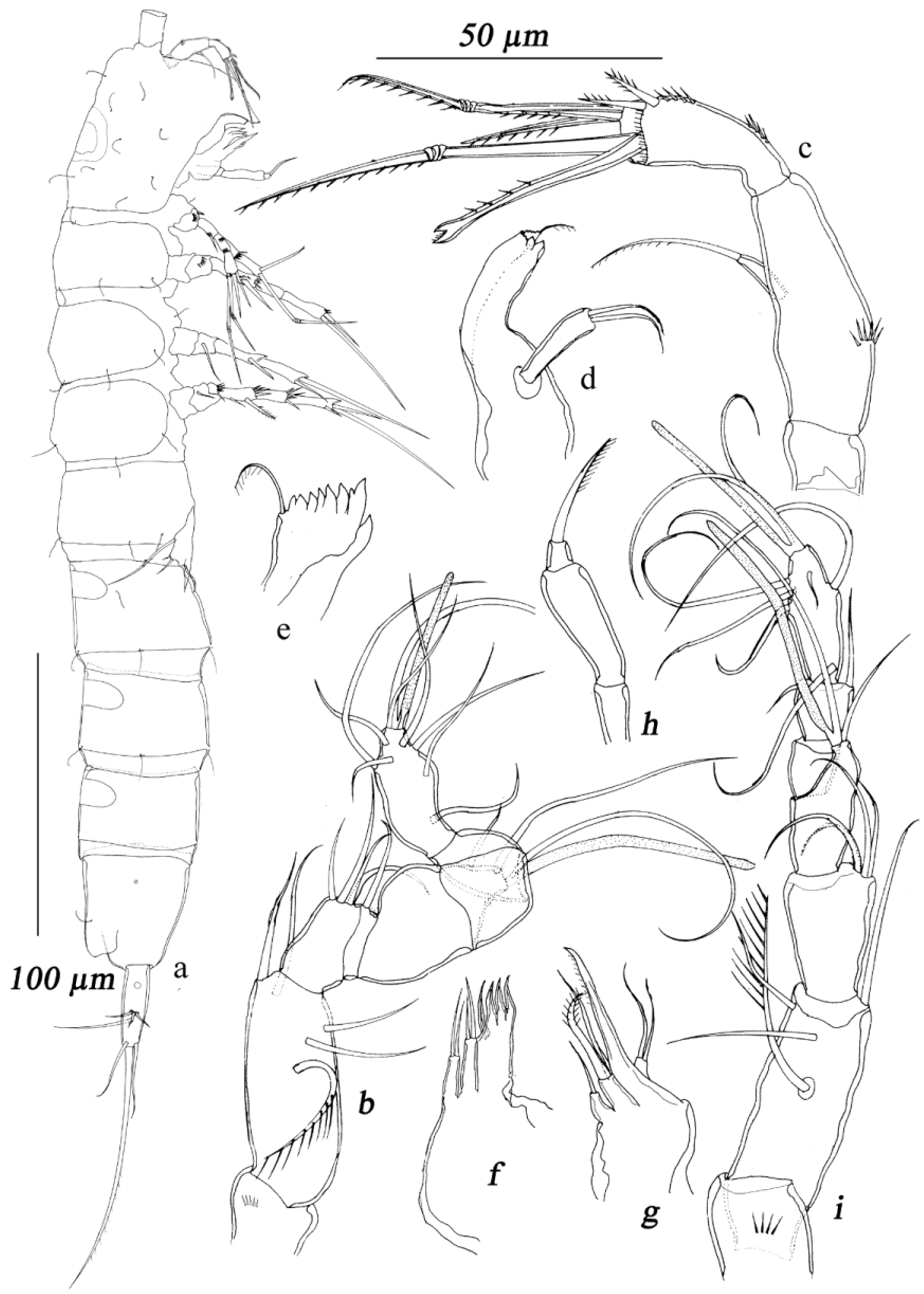
Description of adult male. Total body length, measured from tip of rostrum to posterior margin of caudal rami (excluding caudal setae) 362–378 Μm. Preserved specimens colourless. Nauplius eye absent. Habitus ( Fig. 2 View FIGURE 2 a, b) cylindrical and very slender, with inconspicuous podoplean boundary between prosome and urosome; prosome/ urosome ratio about 0.6 in dorsal view; greatest width in dorsal view at fifth pedigerous somite. Free pedigerous prosomites narrower than distal half of cephalothorax. Body length/width ratio about 8.3; cephalothorax 1.4 times as wide as genital somite in lateral view. Free pedigerous somites without any lateral or dorsal expansions, all connected by well developed arthrodial membranes. Hyaline fringes of all somites smooth, very narrow and hard to distinguish from arthrodial membranes. Integument smooth, ornamented only with sensilla and pores. Cephalothorax with obovate, dorsal, cuticular double-window at about midlength. Pleural areas of cephalothorax and free pedigerous somites moderately developed; cephalic appendages and coxa of leg 1 clearly exposed in lateral view ( Fig. 2 View FIGURE 2 b). Rostrum ( Fig. 2 View FIGURE 2 a) small, subtriangular, membranous, not demarcated at base, barely reaching midlength of first antennular segment and ornamented with 2 small, dorsal sensilla. Cephalothorax ( Fig. 2 View FIGURE 2 a, b) somewhat dilated behind, about 1.4 times as long as wide in dorsal view, representing 16.4% of total body length. Surface of cephalic shield ornamented with 8 pairs of large sensilla besides dorsal double-window. Second pedigerous somite 0.9 times as wide as posterior half of cephalothorax in dorsal view, with 2 pairs of large sensilla (1 dorsal, 1 lateral). Third pedigerous somite slightly wider and longer than second pediger, with 3 pairs of large sensilla (2 dorsal, 1 lateral). Fourth pedigerous somite slightly wider and longer than third prosomite and with 3 pairs of large posterior sensilla.
Urosome: first urosomite widest of all urosomites but slightly shorter than fourth prosomite, and with 2 pairs of large posterior sensilla. Second urosomite about as wide as first urosomite but slightly shorter, with 2 pairs of posterior sensilla and with small elliptical dorsal cuticular window in anterior half. Third urosomite about as long as first urosomite but slightly narrower, with wider dorsal cuticular window, and with 2 pairs of large posterior sensilla. Fourth urosomite slightly narrower than third one, with 2 pairs of large posterior sensilla, and with dorsal cuticular window. Preanal somite slightly narrower and longer than fourth urosomite, and without any surface ornamentation except for dorsal cuticular window. Anal somite ( Fig. 3 View FIGURE 3 a, b) about as long as, but slightly narrower than, preanal somite, 1.2 times as long as wide and ornamented with 2 large dorsal sensilla, 1 lateral cuticular pore ( Fig. 2 View FIGURE 2 b) in anterior half. A single large, longitudinally placed spermatophore ( Figs. 2 View FIGURE 2 a, 3b) visible through fifth pediger and genital somite, about 2.7 times as long as wide, bean-shaped, with narrow and curved neck. Anal operculum moderately developed, ornamented with 1 transverse row of ventro-distal spinules, with slightly concave distal margin, not reaching posterior end of anal somite and representing 68% of somite's width. Anal sinus wide open.
Caudal rami ( Figs. 2 View FIGURE 2 a, b, 3a, b): slightly divergent, distal third narrow, about 3 times as long as greatest width at subproximal level in dorsal view, 2.6 times as long in lateral view, and about 0.7 times as long as anal somite; full complement of setae (3 lateral, 1 dorsal, 2 apical, and 1 outer) and ornamented with large lateral cuticular pore anteriorly ( Fig. 2 View FIGURE 2 b). Dorsal seta (VII) slender and smooth, inserted close to inner margin at about ¾ of ramus length at the level of setae I–III, about 0.8 times as long as caudal ramus, biarticulate basally. Inner apical seta (VI) smooth, inserted close to ventral margin, about 0.7 times as long as ramus. Middle apical seta (V) strongest, without breaking plane, unipinnate, about 4 times as long as ramus, directed distally. Outer apical seta (IV) also without breaking plane and unipinnate, about 1.2 times as long as ramus, inserted subapically close to dorsal surface and directed laterally.
Antennule ( Fig. 4 View FIGURE 4 b): slightly longer than cephalothorax, 8-segmented, prehensile, ‘pocket-knife type’ sensu Schminke 2010, and digeniculate; geniculation between segments 3 and 4, and 6 and 7; antepenultimate segment sharply bent inwards, forming sickle-shaped structure with its preceding segment; segment 8 directed medially. First segment short, ornamented with 1 row of spinules. Segments 5 and 6 moderately dilated, segment 5 without any proximal spinous process on anterior surface, with slender, elongate aesthetasc having blunt tip, overreaching ultimate segment, and fused basally to simple seta; shorter, slender, staff-like apical aesthetasc on segment 8, fused basally to 2 setae (acrotheck). Setal formula: 0.5.4.1.3+aes.0.0.9+aes. All setae slender, smooth except proximalmost seta on second segment unipinnate with long setules along outer margin. Length ratio of antennular segments from proximal to distal end and along caudal margin 1.0:2.7:1.4:0.2:1.3:0.9:0.8:1.5.
Antenna ( Fig. 4 View FIGURE 4 c): relatively stout and composed of coxa, allobasis, 1-segmented endopod, and 1-segmented exopod. Coxa very short, unornamented. Allobasis about 2.6 times as long as maximum width, unarmed but ornamented with 1 crescentic row of spinules on anterior surface. Exopod small, cylindrical, about 3.2 times as long as wide, unornamented and armed with 1 apical unipinnate seta, which is 2.7 times as long as segment. Endopod 0.6 times as long as allobasis and 2.3 times as long as wide, with surface frill subdistally, ornamented with 2 spinular rows on inner margin, and armed with 2 short bipinnate similar spines laterally and 5 strong elements (2 spines, 2 geniculate and 1 unipinnate transformed setae) subapically.
Mandible ( Fig. 4 View FIGURE 4 d, e): cutting edge narrow on elongate coxa and with 2 complex teeth ventrally, 1 unipinnate seta dorsally and several smaller teeth. Palp 1-segmented, subcylindrical, about 3.7 times as long as wide, unornamented and armed with 2 smooth, apical setae.
Maxillule ( Fig. 4 View FIGURE 4 f): praecoxal arthrite trapezoidal, about 1.4 times as long as wide in lateral view; armed with 1 strong, smooth lateral seta and 3 strong, apical spinous processes. Coxal endite armed with 2 smooth apical setae. Basis slightly longer than coxal endite and armed with 2 smooth apical setae.
Maxilla ( Fig. 4 View FIGURE 4 g): composed of syncoxa, basis, and 1-segmented endopod. Syncoxa unornamented and with 2 endites; proximal endite short, armed with 1 smooth apical seta; distal endite armed with 1 smooth seta and 1 strong pinnate seta apically. Allobasis prolonged into strong claw with serrulate inner margin and without seta at base. Endopod represented by small segment, armed with 2 smooth, subequal apical setae.
Maxilliped ( Fig. 4 View FIGURE 4 h): with short and slender syncoxa, unarmed and unornamented. Basis slender, 2.6 times as long as wide and 2.1 times as long as syncoxa, unarmed and unornamented; endopod small, with unipinnate claw, slightly shorter than basis.
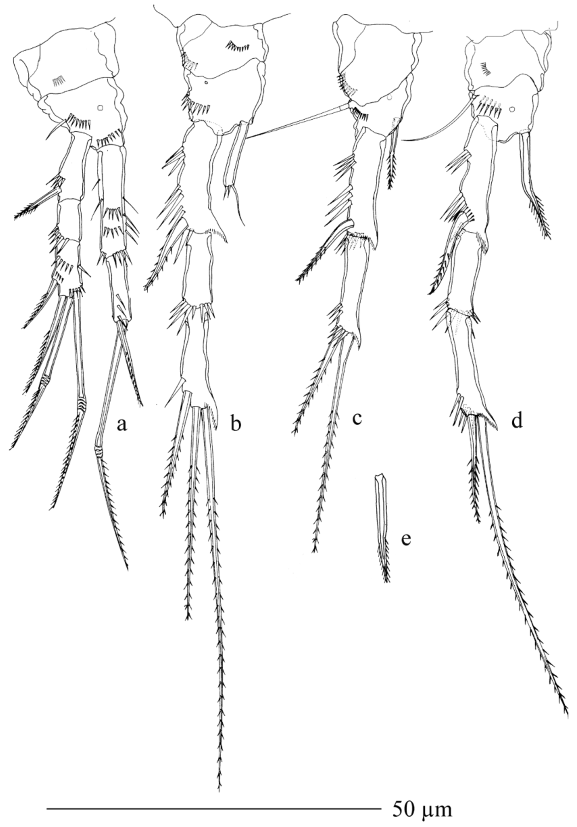
Leg 1 ( Fig. 5 View FIGURE 5 a): coxa ornamented with 1 row of spinules near outer margin on dorsal surface. Basis shorter than coxa, trapezoidal; armed with 1 slender seta on outer margin and 1 spiniform seta on inner margin; ornamented with 1 row of large spinules at base of exopod, 1 ventral row of spinules at base of endopod, 1 row near inner margin and 1 cuticular pore on anterior surface. Exopod 3-segmented; first segment 0.8 times as long as next 2 segments combined, armed with 1 outer bipinnate spine on first segment; second segment unarmed and third segment with 4 elements (1 outer spine, 1 apical seta and 2 apical geniculate setae); ornamented with spinular rows along outer margin of all exopodal segments, as illustrated. Endopod 2-segmented, about as long as exopod; first segment reaching distal margin of second exopodal segment, 3 times as long as wide, unarmed and ornamented with 1 row of spinules on outer margin and 2 rows of spinules on inner margin; second segment ornamented with 1 transverse row of spinules on outer margin and armed apically with 1 long geniculate seta and 1 short spine; endopodal geniculate seta 1.5 times as long as entire endopod, 1.2 times as long as larger geniculate exopodal seta and almost 2.4 times as long as outer spine on endopod. All exopodal and endopodal armature elements unipinnate along outer margin except bipinnate spine on first exopodal segment.
Leg 2 ( Fig. 5 View FIGURE 5 b): coxa large, unarmed and ornamented with 1 row of small spinules on outer margin. Basis much smaller than coxa, unarmed, ornamented with 1 row of spinules at base of exopod and 1 pore on anterior surface. Exopod 3-segmented; first segment 0.7 times as long as next 2 segments combined and slightly curved inwards; all segments ornamented with rows of spinules along outer margin, as illustrated, and segments 1 and 3 with hyaline frill each at inner distal corner; inner corner of second segment with 1 row of spinules; segment 1 armed with outer bipinnate spine; segment 2 unarmed. Segment 3 longer than segment 2, armed with 3 long elements: 1 subapical unipinnate spine and 2 apical bipinnate setae. Endopod 1-segmented, subcylindrical and distally dilated, almost 2.6 times as long as wide, about half as long as first exopodal segment; apical margin armed with 1 smooth seta, which is 0.8 times as long as segment and pointing inwards, and ornamented with 2 spinules.
Leg 3 ( Fig. 5 View FIGURE 5 c): coxa trapezoidal, ornamented with arched row of spinules near mid-distal margin ventrally. Basis robust, ornamented with 1 ventral row of minute spinules near outer margin and 1 pore on anterior surface; armed with moderately long, slender seta on outer margin. Endopod represented by smooth, slender seta, inserted at 3/4 of inner margin of basis. Both exopodal segments fused; ancestral proximal segment moderately stout, 3.5 times as long as wide, swollen at subproximal outer margin, only slightly curved inwards; small prominence on inner distal margin; 1 spinular row on posterior surface of outer distal corner; ancestral distal segment (apophysis) ladle-shaped with hyaline membrane, somewhat bent inwards, longer than thumb, unornamented and unarmed; thumb slender, spiniform, inwardly curved and reaching 3/4 of apophysis.
Leg 4 ( Fig. 5 View FIGURE 5 d): coxa rectangular, ornamented with short oblique spinular row near outer distal corner. Basis shorter, trapezoidal in anterior view and armed with moderately long outer seta; ornamented with 1 pore on anterior surface. Exopod 3-segmented, ornamented with spinules along outer margins of all segments; segments 1 and 3 with hyaline frill each at inner distal corner; segment 2 with 1 row of spinules at inner distal corner; segment 1 stout, about as long as next 2 segments combined and armed with strong bipinnate outer spine; segment 2 unarmed; segment 3 armed with 1 outer spine and 1 inner bipinnate seta; inner apical seta 1.8 times as long as outer seta, 2.4 times as long as third exopodal segment, 0.8 times as long as entire exopod. Inner distal margin of basis with large chitinized plate, below which lie 2 hyaline, outwardly directed, blunt structures at outer corner (arrowed in Fig. 5 View FIGURE 5 d); also, with 1 smooth, moderately strong, somewhat claw-like spine at distal inner corner. Endopod proper as acutely pointed conical hyaline membrane, ornamented with 3 spinules at mid-outer margin.
Leg 5 ( Figs. 2 View FIGURE 2 b, 3a): simple, elongated, rhomboidal plate; both legs fused at base, pointing caudally and slightly overreaching end of the somite, inner distal corner produced into long, acute spiniform process, ornamented with cuticular pore on anterior surface and armed with basal seta and 2 inner smooth setae on oblique distal margin; basal seta longer than entire leg; outer seta about 0.3 times as long as leg 5, and 1.2 times as long as inner seta.
Leg 6 ( Fig. 3 View FIGURE 3 a): smooth, unarmed, forming simple operculum covering gonopore and elliptical in ventral view.
Description of adult female. Body length, measured as in male, 385–400 Μm. Habitus ( Fig. 4 View FIGURE 4 a): slightly less slender than in male, prosomites, colour and nauplius eye similar to male, except genital and first abdominal somites fused into double-somite.
Genital double-somite ( Figs. 4 View FIGURE 4 a, 6a, b): shorter than maximum width (ventral view), without any trace of subdivision, with oval dorsal cuticular window in anterior half. Genital complex occupying anteroventral half of genital double-somite; genital apertures paired, each covered by vestigial sixth legs; copulatory pore medial; seminal receptacles small, hard to distinguish from internal tissue and gut content; copulatory duct very short and weakly sclerotized ( Fig. 6 View FIGURE 6 c). Sensilla similar to those on male third urosomite, while 2 sensilla of male second urosomite missing. Third urosomite, preanal somite, and anal somite very similar to male.
Caudal rami ( Figs. 4 View FIGURE 4 a, 6a, b): 0.7 times as long as anal somite, about 2.8 times as long as wide in ventral view, with armature and ornamentation as in male.
Antennule ( Fig. 4 View FIGURE 4 i): 7-segmented, segment 1 ornamented with 4 minute spinules disto-ventrally, aesthetasc on segment 4 slender, overreaching tip of appendage, and that on segment 7 more slender, staff-like and fused basally to 2 apical setae; setal formula: 0.4.4.2+aes.0.2.8+aes. All setae except proximalmost one on segment 2 smooth. Length ratios of antennular segments from proximal to distal end and along caudal margin 1.0: 2.5: 2.2: 2.2: 0.5: 0.5: 1.3.
Antenna, labrum, mandible, maxillule, maxilla, maxilliped, and leg 5 similar to male.
Leg 1 ( Fig. 7 View FIGURE 7 a): coxa rectangular, ornamented with 1 row of spinules near distal margin; basis trapezoidal, ornamented with 1 row of spinules near base of exopod, 1 row at base of endopod, 1 pore on proximal surface; armed with 1 small seta each on outer margin; exopod and endopod almost as in male.
Leg 2 ( Fig. 7 View FIGURE 7 b): coxa trapezoidal, ornamented with 1 arched row of minute spinules near inner margin and another row at outer distal corner ventrally. Basis larger than coxa, unarmed, and ornamented with arched spinular row near outer distal corner and 1 pore proximally; exopodal segments as in male. Endopod cylindrical, 4.5 times as long as wide; apical margin armed with 1 seta and ornamented with 2 spinules.
Leg 3 ( Fig. 7 View FIGURE 7 c): coxa with arched row of ventral spinules on outer distal margin. Basis ornamented with 1 arched spinular row near outer margin and armed with long and smooth outer seta, which is about 0.6 times as long as entire exopod. Exopod 2-segmented, ornamented with large spinules along outer margin, each segment with hyaline frill distally on inner distal corner; segment 1 armed with single outer spine; segment 2 with outer spine and strong apical seta; seta 1.8 times as long as spine; all elements bipinnate. Endopod 1-segmented, slender, 0.6 times as long as first exopodal segment, tapering to pointed tip and with spinulose disto-lateral margins.
Leg 4 ( Fig. 7 View FIGURE 7 d): exopod similar to male. Endopod lanceolate, slightly shorter than first exopodal segment; distal third bent inwards, gradually tapering to acuminate point and with serrulate lateral margins.
Leg 6 ( Fig. 6 View FIGURE 6 c) vestigial, fused into simple cuticular flap, covering gonopores; unarmed and unornamented.
Etymology. The new species is named in honour of Dr. P. H. Enckell, who was the first to describe parastenocaridids from the Indian subcontinent. The specific epithet, enckelli , is a noun in the genitive singular.
Distribution and ecology. This species is so far known from the type locality, co-occurring with the following taxa: a species each of Harpacticoida , Cyclopoida and Cladocera, some rotifers, oligochaetes and chironomid larvae.


No known copyright restrictions apply. See Agosti, D., Egloff, W., 2009. Taxonomic information exchange and copyright: the Plazi approach. BMC Research Notes 2009, 2:53 for further explanation.