
Rhodochlanis suaedicola Cho, Burckhardt & Lee
|
publication ID |
https://doi.org/ 10.11646/zootaxa.4028.3.4 |
|
publication LSID |
lsid:zoobank.org:pub:919062AB-8560-4677-A486-9C59A05E1E5E |
|
DOI |
https://doi.org/10.5281/zenodo.6102674 |
|
persistent identifier |
https://treatment.plazi.org/id/431CE626-FFCF-FFF4-46B6-FDE44C7AF8F0 |
|
treatment provided by |
Plazi |
|
scientific name |
Rhodochlanis suaedicola Cho, Burckhardt & Lee |
| status |
sp. nov. |
Rhodochlanis suaedicola Cho, Burckhardt & Lee View in CoL , sp. nov.
( Figs 1−21 View FIGURES 1 − 6 View FIGURES 7 − 9 View FIGURES 10 − 12 View FIGURES 13 − 18 View FIGURES 19 − 21 )
Type material. Holotype ♂, South Korea: Incheon-si, Namdong-gu, Nonhyun-dong, Sorae Wetlands Ecological Park, 5.ix.2014, Suaeda japonica (G.H. Cho) , ( SNU, dry mounted).
Paratypes. South Korea: 38 ♂, 35 ♀, 1 immature, same data as holotype ( SNU, NHMB, dry and slide mounted, 95% ethanol); 19 ♂, 18 ♀, Jeollanam-do, Suncheon-si, Daedae-dong, Suncheon Bay Ecological Park, 13.ix.2014, Suaeda japonica (G.H. Cho) ; ( SNU, NHMB, dry and slide mounted, 95% ethanol).
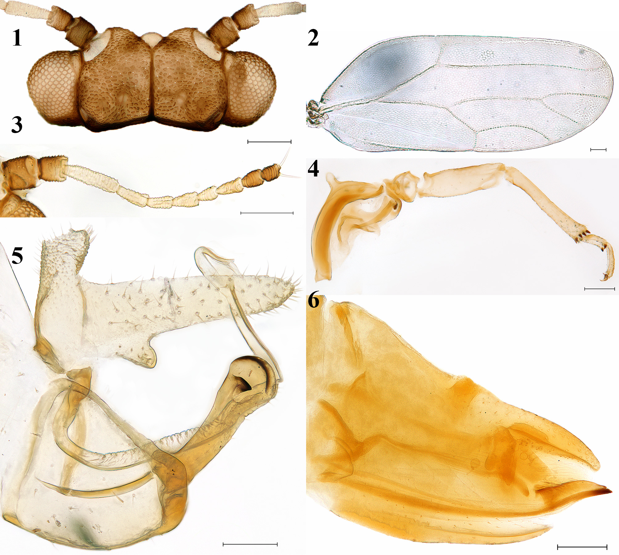
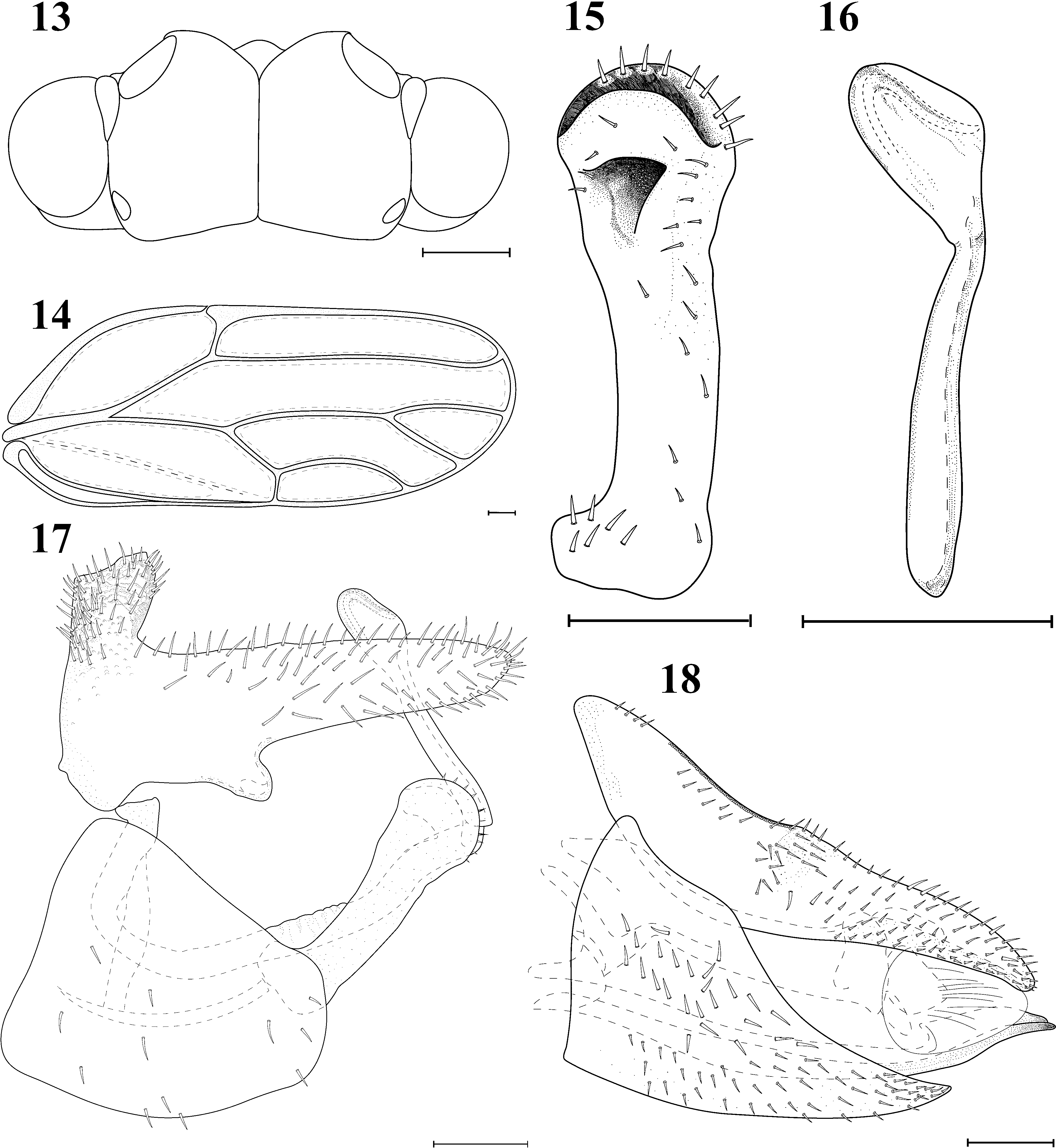
Diagnosis. Forewing oblong-oval ( Figs. 2 View FIGURES 1 − 6 , 14 View FIGURES 13 − 18 ) lacking colour pattern; surface spinules arranged in cellular pattern forming small hexagons; vein Rs almost straight, curved towards fore margin apically; vein Cu1b short, almost straight, cell cu1 narrow and long with vein Cu1a weakly curved. Paramere ( Fig. 15 View FIGURES 13 − 18 ) clavate, apically rounded, bearing large, sclerotised tooth on the inner surface in apical third. Female terminalia and circumanal ring relatively long ( Figs. 6 View FIGURES 1 − 6 , 18 View FIGURES 13 − 18 ), apex of subgenital plate ending in a single point.
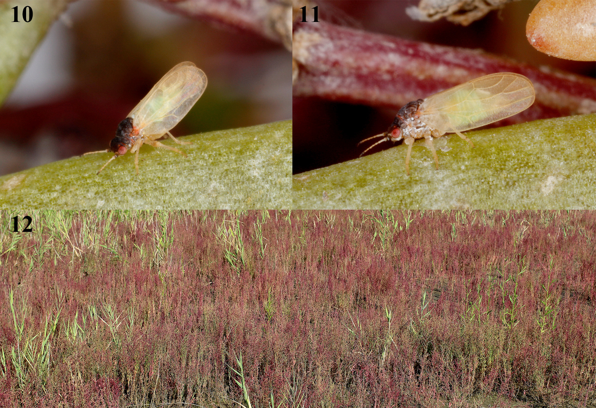
Description. Adult. Coloration ( Figs. 10−11 View FIGURES 10 − 12 ). General body colour light yellowish brown, except for head and thorax. Eyes reddish brown, ocelli light brown. Antenna off-white; antennal segment 1 dark brown; segment 2 brown, white apically; segments 9−10 brown to dark brown; terminal setae white. Head, pronotum, mesopraescutum, mesonotum and metanotum dark brown to almost black dorsally. Thorax light reddish brown laterally. Forewing with pale yellow veins; membrane semitransparent, indistinctly yellow. Hindwing transparent, whitish. Legs light yellowish brown; metatibia yellowish brown with dark brown to almost black apical tibial spurs, tarsal spurs and claws. Abdomen light green to greenish yellow. Terminalia yellowish brown or ochreous. Female and young specimens generally lighter in colour.
Structure. Measurements and ratios as in Table 1. Head ( Figs. 1 View FIGURES 1 − 6 , 13 View FIGURES 13 − 18 ) as broad as thorax; preocular sclerite small, posteriorly not reaching to the middle of vertex; genae not produced into processes; vertex short, 0.44−0.48 times as long as wide, anterior margin, in dorsal view, concave. Antenna ( Fig. 3 View FIGURES 1 − 6 ) 10-segmented, short, with subapical rhinarium on each of segments 4−9; segment 3 the longest; two terminal setae of segment 10 about as long as the segment. Thorax curved moderately. Forewing ( Figs. 2 View FIGURES 1 − 6 , 14 View FIGURES 13 − 18 ) oblong-oval; pterostigma short; costal break present; vein C+Sc strongly curved in the middle; cell c+sc large, broadest near the middle; vein Rs almost straight, curved towards fore margin apically; vein M evenly curved with relatively long diverging branches; vein M1+2 weakly curved, meeting middle of wing margin; vein M3+4 shorter than vein M1+2, almost straight; cell cu1 narrow and long; vein Cu1a long, moderately curved; vein Cu1b short, almost straight; surface spinules present in all cells, forming regular cellular pattern; forewing margin and veins sparsely covered with short setae. Hindwing simple and membraneous. Hind legs ( Fig. 4 View FIGURES 1 − 6 ) saltatorial, longer than fore and mid legs; metacoxa with small, straight, and swollen meracanthus; metafemur shorter than metacoxa; metatibia longer than metafemur, with 6 equidistant strongly sclerotised short apical spurs forming an open crown; apex of metatibia dilated; genual spine absent; metabasitarsus with two small sclerotised tarsal spurs; metatarsus with apical segment longer than basal segment, with two falcate claws. Male terminalia as in Figs. 5 View FIGURES 1 − 6 , 17 View FIGURES 13 − 18 . Proctiger with a pair of elongated posterior lobes bearing an inwards directed hook in the basal third of the lower margin; apical portion of proctiger, in profile, tubular thick, with conspicuous stout setae entirely. Subgenital plate large, pentagonal in profile, somewhat concave at dorsal margin, sparsely hairy. Paramere ( Fig. 15 View FIGURES 13 − 18 ) clavate, rounded apically, 0.42−0.45 times as long as head width; outer surface sparsely covered with short setae; inner surface with a strongly sclerotised tooth in apical third. Distal segment of aedeagus ( Fig. 16 View FIGURES 13 − 18 ) flat, apical half weakly inflated, apex narrowly rounded, 0.36−0.43 times as long as head width. Female terminalia ( Figs. 6 View FIGURES 1 − 6 , 18 View FIGURES 13 − 18 ) relatively long, sparsely hairy. Proctiger 0.99−1.13 times as long as head width; dorsal margin, distal to circumanal ring, evenly concave, curved apically. Circumanal ring long, with 2 complete rows of unequel pores, 0.32−0.42 times as long as proctiger. Subgenital plate slim and long, cuneate, 0.77−0.85 times as long as proctiger, ending in a single point. Lateral valvulae triangular, rounded apically; dorsal valvulae cuneate, weakly curved, ventral valvulae spear-like shape, lacking teeth.
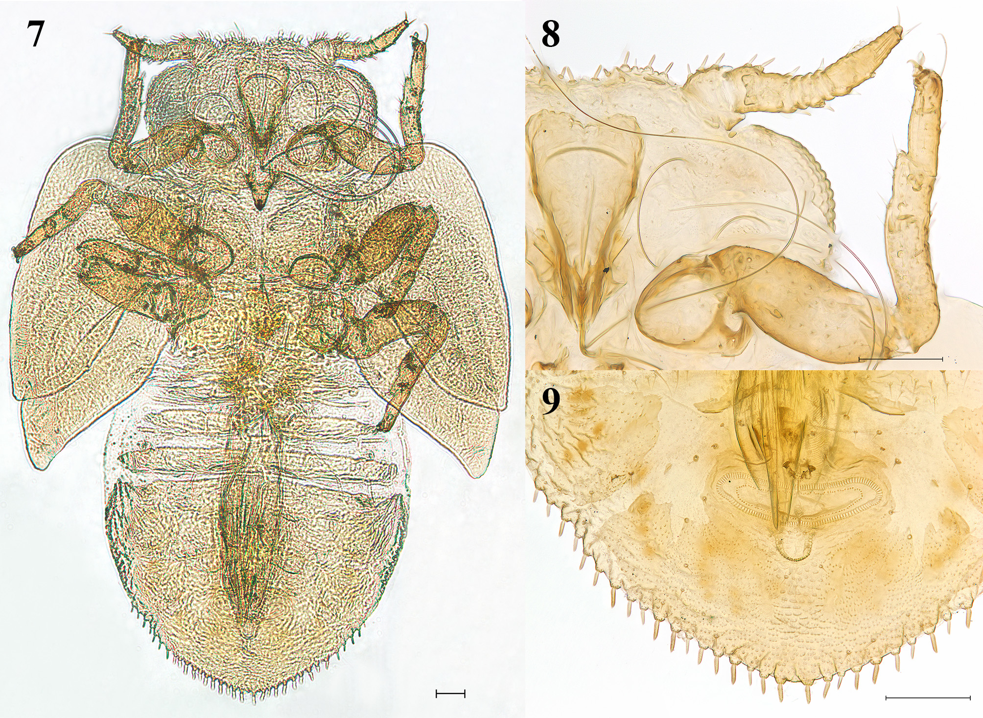
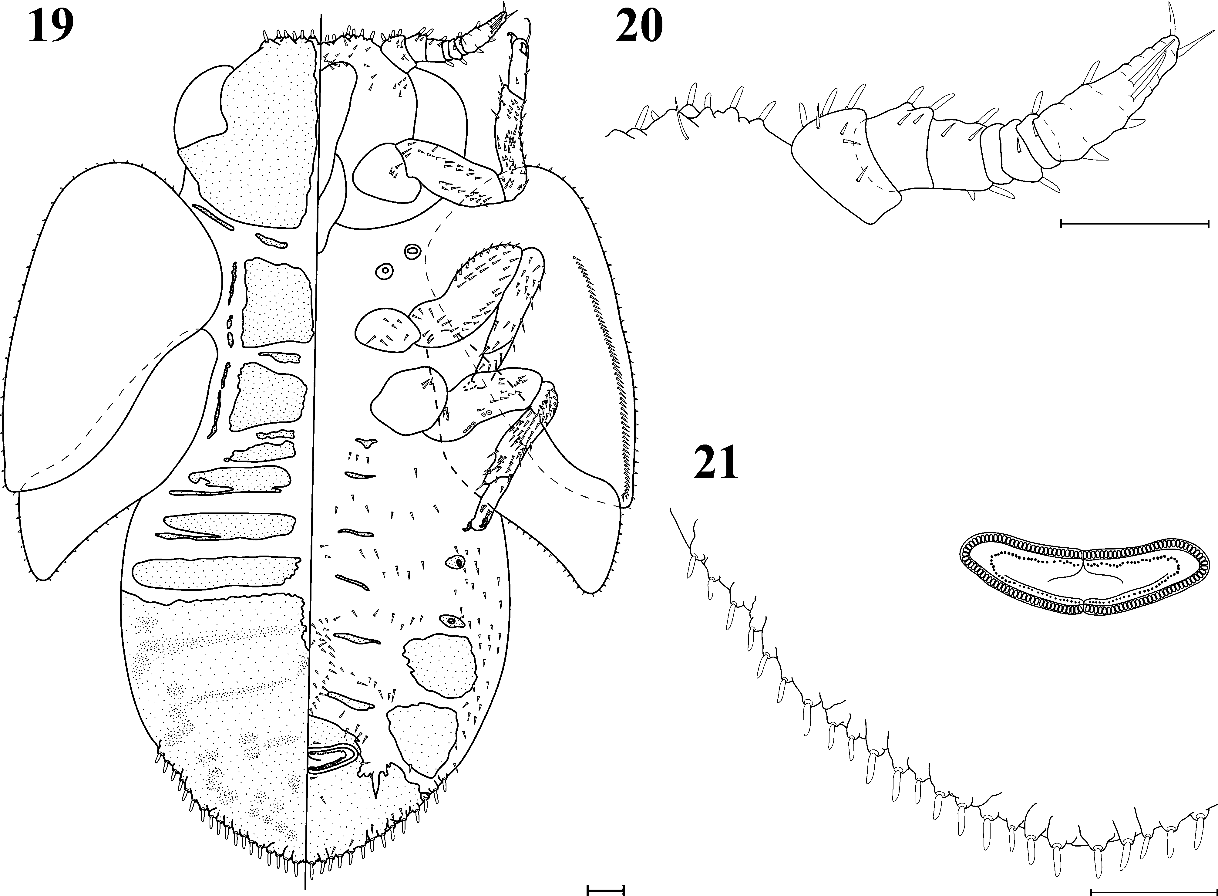
Fifth instar immature ( Figs. 7−9 View FIGURES 7 − 9 , 19−21 View FIGURES 19 − 21 ). Measurements and ratios as in Table 2. General body colour pale yellow; claws and sclerotised spurs dark brown. Body fairly slender, with relatively short antenna and legs. Body margin ( Figs. 7 View FIGURES 7 − 9 , 19 View FIGURES 19 − 21 ) with 12 small slender lanceolate setae on head ( Figs. 8 View FIGURES 7 − 9 , 20 View FIGURES 19 − 21 ) between insertion of antennae, and 32 slender lanceolate equidistant setae on caudal plate margin ( Figs. 9 View FIGURES 7 − 9 , 21 View FIGURES 19 − 21 ), starting approximately in the middle of caudal plate margin. Small setae present on forewing pad surface and margin, and on hindwing pad margin. Eye large. Antenna ( Figs. 8 View FIGURES 7 − 9 , 20 View FIGURES 19 − 21 ) short, stout and curved, 7−segmented; 3 small lanceolate setae on each of segments 1 and 2, 2 small lanceolate setae on each of segments 3, 4 and 7, 1 small lanceolate seta on each of segments 5 and 6, 2 long simple setae on segment 8, 2 apical simple setae on segment 9. Hind leg short and stout; metatibia longer than metafemur. Abdomen dorsally with 4 free pre-caudal tergites and fused caudal plate. Caudal plate large and rounded. Anus located on venter, away from the apex of the abdomen; circumanal ring ( Fig. 9 View FIGURES 7 − 9 , 21 View FIGURES 19 − 21 ) small, transverse, narrowly oval; outer ring composed of a single row of pores.
TABLE 1. Measurements and ratios for adults of Rhodochlanis suaedicola sp. nov. *MSD = mean ± standard deviation, R = range. Abbreviations: HW = head width; VW = vertex width; VL = vertex length; F1 = first antennal flagellomere length; AL = antenna length; WL = forewing length; WW = forewing width; PT = pterostigma length; Rs = vein Rs length; RC = length of line connecting apices of vein Rs and vein Cu1a; a = length of line connecting base and apex of vein M1+2; b = length of line connecting apices of veins M1+2 and M3+4; c = length of line connecting apices of vein Cu1a and Cu1b; d = length of line connecting base and apex of vein Cu1b; TL = metatibia length; MP = male proctiger length; PL = paramere length; DL = length of distal segment of aedeagus; FP = female proctiger length; CL = circumanal ring length; SL = female subgenital plate length.
Characters Fifth instar immature (n = 1) BL 1.70
BW 1.32
AL 0.28
FL 0.74
TL 0.34
CL 0.56
CW 0.78
RW 0.20
BL/BW 1.29
AL/FL 0.38
CW/CL 1.40
CW/RW 3.92
Etymology. The name of the host plant genus and the Latin suffix -cola = inhabitant.
Distribution. South Korea.
Host plant. Suaeda japonica Makino (Amaranthaceae) .
Remarks. Rhodochlanis suaedicola belongs to the R. bicolor group as defined by Burckhardt (1989), containing R. ancistrocalis Li, 1996 , R. bicolor ( Scott, 1880) , R. orientalis Loginova, 1964 and R. qixianana Li, 2011 . R. suaedicola differs from these species in the paramere bearing a characteristic tooth in apical third on the inner face. This species resembles R. ancistrocalis in the male proctiger shape, tooth on the inner face of paramere and female terminalia shape. It is easily diagnosed by its forewing shape and venation, setation of male proctiger. From R. orientalis , it differs also in the forewing which has a narrow cell cu1 and a short and straight vein Cu1b (in R. orientalis cell cu1 broad and vein Cu1b relatively long and curved). In R. suaedicola the female circumanal ring is longer than in R. orientalis .
Conservation. Rhodochlanis suaedicola develops on Suaeda japonica , which is mostly distributed at the inner side of tidal mudflats, especially of the supralittoral zone and closed salterns. For recent decades, closed salterns and salt marshes of South Korea have been under constant pressure from developments of the waterfront, and the areas of salt marshes in Korea has steadily decreased. The new Rhodochlanis species is known from two conservation area, Suncheon Bay Ecological Park and Sorae Wetlands Ecological Park, and, for the sustainability of the species, conservation of salt marshes should be considered.
| NHMB |
Naturhistorisches Museum, Basel |
No known copyright restrictions apply. See Agosti, D., Egloff, W., 2009. Taxonomic information exchange and copyright: the Plazi approach. BMC Research Notes 2009, 2:53 for further explanation.