
Ophlitaspongia Bowerbank, 1866
|
publication ID |
https://doi.org/ 10.11646/zootaxa.5297.1.2 |
|
publication LSID |
lsid:zoobank.org:pub:1F89880E-AFBB-4392-87F6-913C99EBCABD |
|
DOI |
https://doi.org/10.5281/zenodo.7990959 |
|
persistent identifier |
https://treatment.plazi.org/id/446ADD2C-FFFF-DA03-F9B4-FEB332A0FE44 |
|
treatment provided by |
Plazi |
|
scientific name |
Ophlitaspongia Bowerbank, 1866 |
| status |
|
Parent Ophlitaspongia Bowerbank, 1866 View in CoL View at ENA
Orig. name Ophlitaspongia (?) horrida Row, 1911:349-351 , pl. 40 fig. 26, text fig. 23
Accepted name Clathria (Clathria) horrida ( Row, 1911)
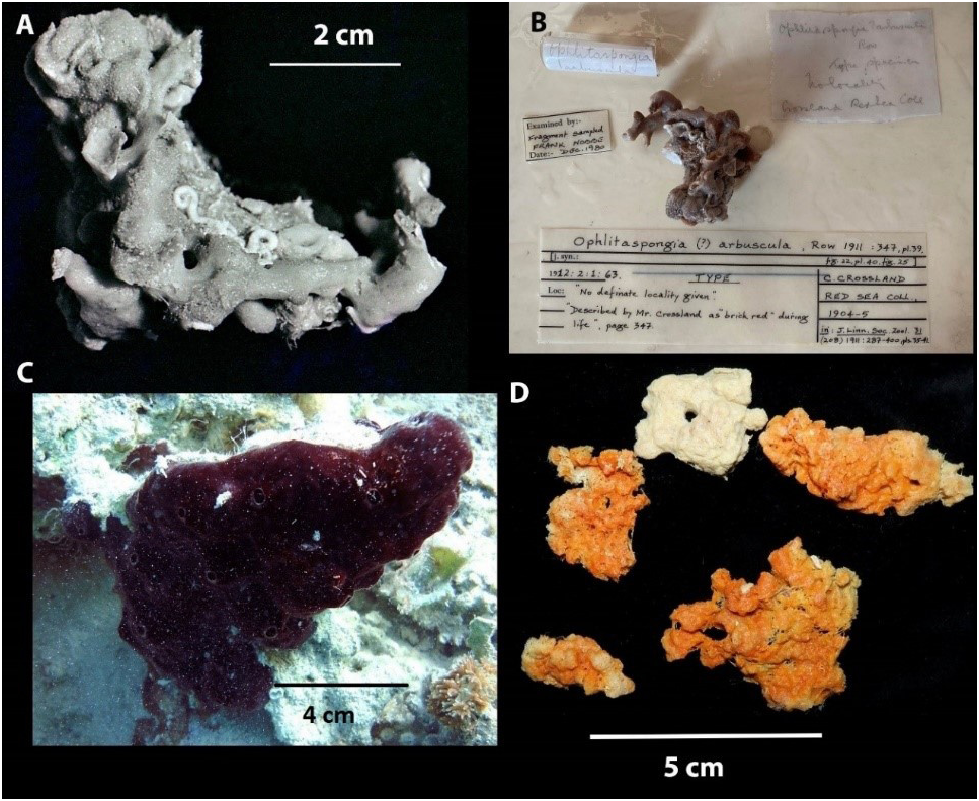
Material examined: Holotype BMNH 1912.2 .1.63, We Shubuk , Sudanese Red Sea, Dredge , 16.5m Coll. C. Crossland, 1904-1905. Slide of the holotype BMNH 1912.2 .1.65, housed at the Queensland Museum. QM G339443 , Hurghada, Egypt, Red Sea, 27 o 17.80’ N, 33 o 46.26’ E, 5-7 m. SCUBA, Coll. M. A. L. H. Ezz El-Arab, October 2018, NIOF FD100 . QM G339444 , same collection details as QM G339443 . GoogleMaps
Distribution: Known only from the Red Sea.
Description: Thin spreading sheets up to 4 mm thick encrusting on corals and calcareous shells. The sponge mass may deviate and protrude from the encrusting level to be a small protrusion without a basal structure as is often the case in the rest of the sponge ( Fig 4 View FIGURE 4 ). The small protrusion may reach to 2 cm length. The actual surface of the sponge is quite smooth, being covered by a very delicate dermal membrane.
The sponge has irregularly dispersed oscula 2–3 mm in diameter and 2.5–3 cm apart. The color is dark-red during life and reddish-brown color in alcohol.
The texture is compressible and resilient when separate off the substrate, can be cut and torn easily.
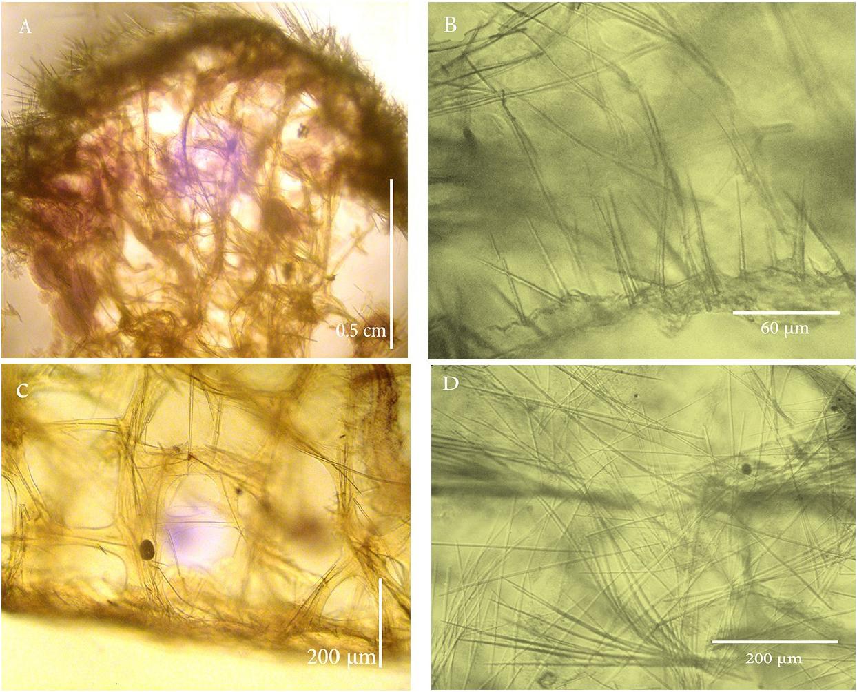
Skeleton: A flattened basal layer of spongin measures 0.3 mm lying on the substratum, and a hymedesmioid of auxiliary styles or/and echinating (acantho)styles or tylotes oriented perpendicular to the spongin ( Fig. 6B View FIGURE 6 ). Tylotes almost confined to the head regions, project from this basal layer, while the heads of the styles are buried in the spongin. In NIOF FD100, there is a flattened basal layer of spongin with no hymedesmioid of auxiliary styles, or the auxiliary styles in the basal layer may be scarce and hard to find ( Fig. 6C View FIGURE 6 ).
The choanosome of all collected specimens consists of (subtylo)styles and styles cored fibres (0.08 mm in diameter), perpendicular to the substratum which ascend through the sponge from the basal layer to the dermal layer. The fibres are strongly coated with spongin and the reticulation through the choanosome is fairly recognisable, where a distinction into primary and secondary fibres can be made. The primary ascending fibres are abundantly cored by (subtylo)styles and styles, echinated by auxiliary styles or echinating (acantho)styles. Unispicular or paucispicular smaller secondary spongin fibres are perpendicular to and connect with the primary fibres to form choanosomal elongate or square meshes. The fibres in the sub-ectosome are condensed and irregularly oriented. The ectosomal skeleton consists mainly of densely matted brushes of (subtylo)styles arranged tangentially with the tips of the (subtylo) styles projecting from the surface ( Fig. 6D View FIGURE 6 ). As a result, the ectosomal skeleton forms a dense felting over the surface of the sponge.
Loose interstitial styles and toxas are scattered irregularly through the sponge between the fibres.
Spicules:
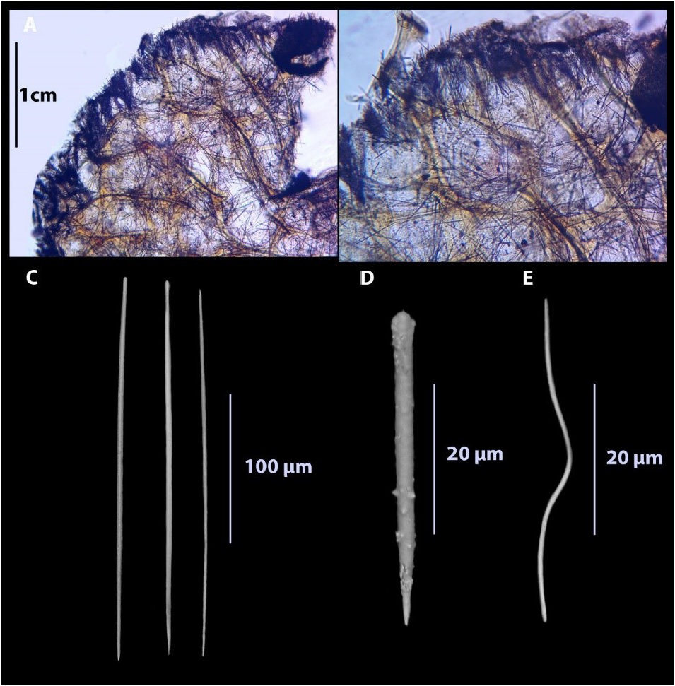
Megascleres: The megascleres are predominately (subtylo)styles, which are frequently curved, sometimes with subtle swellings, but usually well rounded with slight necks ( Fig. 5C View FIGURE 5 ). They range in length from 61 to 400 µm in the fresh material, and 80 to 335 µm in the holotype ( Table 2). The original description ( Row 1911) recorded only 300 µm ( Table 2). They are slender, and range in width between 1 and 7 µm in the fresh material ( Table 2), and from 1 and 6 µm in the holotype ( Table 2). The original description ( Row, 1911), simply recorded 2 µm in width ( Table 2). The (subtylo)styles in almost all the fibres form a slender core. However, they are frequently arranged in a slightly plumose manner within the sponging-fibre, but they never project outside it. A few spicules occur with oxeote or stylote ends.
(Acantho)styles were also found in some of the fresh material and in the holotype ( Table 2) ( Fig. 5D View FIGURE 5 ). (Acantho)styles are relatively strongly spined, and they range in length from 55.6 to 120 µm in the fresh material and 27.5 to 56 µm in the holotype. (Acantho)styles were not recorded in the original description ( Row, 1911).
Microscleres: Toxas were found in all the samples including the holotype and the original description. However, they are very uncommon to rare in the samples. Oxhorn micro-toxas and wing-shaped short toxas with a range of length from 19 to 300 µm, were found in the fresh material and 200 µm was recorded in the original description ( Row, 1911) ( Table 2) ( Fig. 5E View FIGURE 5 ). Rare oxeas (75 to 120 µm in length), sigmas (15 to 30 µm in length) and palmate isochelas (20 µm in length), were also found in some of the material in Egypt (not listed here), but due to their absence in the type material and the material examined in this study (i.e. Table 2) were regarded as non-native.
Remarks: The holotype of Ophlitaspongia horrida (BMNH 1912.2.1.63) described by Row (1911) as a low irregularly branching mass, creeping on coral and calcareous shells. The recently collected specimens are encrusting, from which arise at frequent intervals stout and short processes which frequently branch. These small protrusions do not have basal structures (hymedesmoid architecture) as is often the case in the rest of the sponge.
In these new specimens (QM G339443 and QM G339444) the skeletal structure has a flattened basal layer of spongin lying on the substratum, and a hymedesmioid of small styles or/and (acantho)styles or tylotes) oriented through the spongin at right angles. Row (1911), in the original description of Ophlitaspongia horrida stated only (subtylo)styles as megascleres, and a few toxas as microscleres. It has been found that there is a consensus between Row (1911) and the recent specimens (QM G339443 and QM G339444) in the presence of the (subtylo)styles and convergence in their dimensions. However, the new material recorded (acantho)styles not mentioned in Row (1911).
| QM |
Queensland Museum |
No known copyright restrictions apply. See Agosti, D., Egloff, W., 2009. Taxonomic information exchange and copyright: the Plazi approach. BMC Research Notes 2009, 2:53 for further explanation.
|
Kingdom |
|
|
Phylum |
|
|
Class |
|
|
Order |
|
|
Family |