
Clevelandella parapanesthiae ( Kidder, 1937 )
|
publication ID |
https://doi.org/ 10.5852/ejt.2020.697 |
|
publication LSID |
lsid:zoobank.org:pub:8962B6E6-B278-4EF5-9E62-3E858726E2F2 |
|
DOI |
https://doi.org/10.5281/zenodo.4329008 |
|
persistent identifier |
https://treatment.plazi.org/id/44755610-B00E-FFBB-FD99-FEE7BC0C7212 |
|
treatment provided by |
Valdenar |
|
scientific name |
Clevelandella parapanesthiae ( Kidder, 1937 ) |
| status |
|
Clevelandella parapanesthiae ( Kidder, 1937)
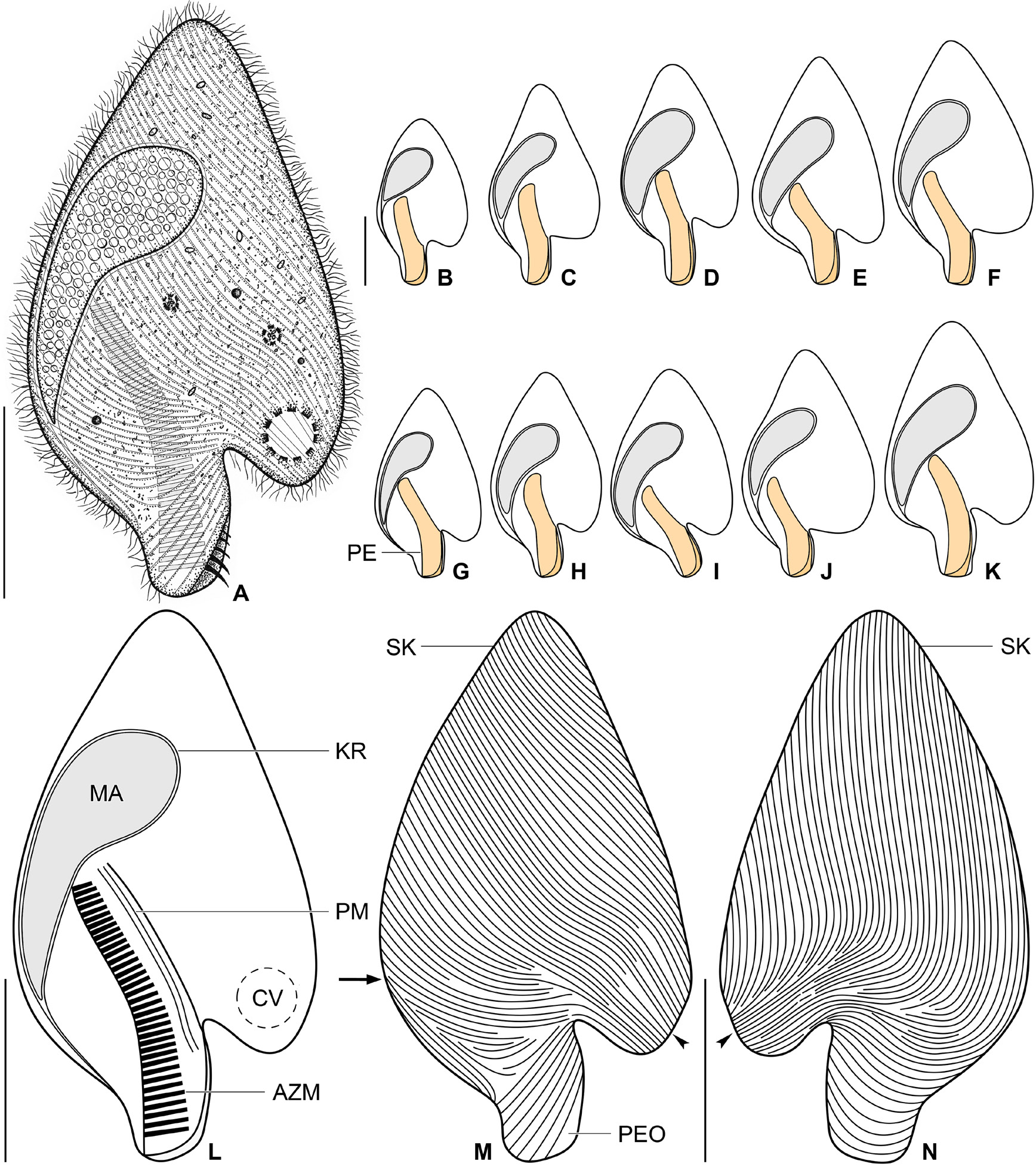
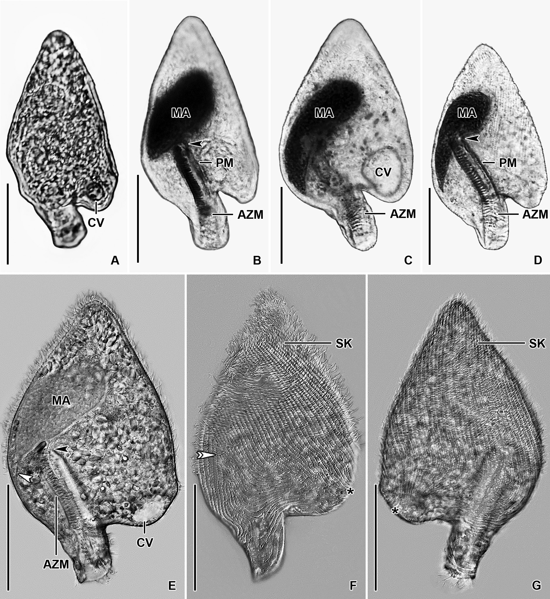
Figs 9–11 View Fig View Fig View Fig
Description of Thai I population
Size in vivo about 70–110 × 40–65 μm, usually 90 × 50 μm, as calculated from some in vivo measurements and morphometric data; length:width ratio very stable, i.e., ranging from 1.7:1 to 1.9: 1 in protargol preparations ( Table 4). Body broadly spade-shaped, widest slightly above posterior third of body, i.e., just above level of contractile vacuole.Anterior end bluntly pointed; posterior body portion differentiated into a conspicuous, moderately long peristomial projection; left margin distinctly notched at base of peristomial projection and recurved posteriorly toward peristomial projection, forming a distinct lobe; dorsoventrally flattened 1.3:1 ( Figs 9 View Fig A–N, 11C–D). Macronucleus located in second and third fourth of body, close to right body margin; tear-shaped with a length:width ratio of 2.1–3.4: 1 in protargol preparations; anterior end broadly rounded and near cell’s midline, posterior end acute and in parallel with right body margin; 27–47 × 12–17 μm in size after protargol impregnation; filled with numerous globular structures (presumably nucleoli) 0.6–1.6 μm in diameter after protargol impregnation, well observable in vivo and after protargol impregnation. Karyophore attached to right body margin near base of peristomial projection ( Table 4; Figs 9 View Fig A–L, 11C–D). Micronucleus not observed. Contractile vacuole in lobe of left body side, i.e., at level of base of peristomial projection ( Figs 9A, L View Fig , 11C View Fig ). Cortex flexible, no cortical granules recognizable. Cytoplasm colorless; finely granulated; numerous refractile bodies scattered throughout cytoplasm; some (symbiotic?) bacteria and/or archaea freely scattered throughout main body portion; few food vacuoles, about 2.5–5.2 μm across and containing prey prokaryotes ( Fig. 9A View Fig ). Swims slowly; dies quickly on microscope slides, possibly due to presence of oxygen; body shape changes in dying and strongly squeezed cells, i.e., lobe of left side becomes less noticeable.
Somatic ciliature holotrichous; cilia about 4.5–6.0 μm long in vivo and very narrowly arranged. About 90 ciliary rows narrowly spaced over entire body surface and about 25 ciliary rows running onto peristomial projection. Peristomial ciliary rows in a form of strongly oblique lines in ventral view, while in a form of shallow arcs in dorsal view ( Fig. 9 View Fig M–N). Almost all body ciliary rows begin from a whorl (posterior suture) on left body side slightly above base of peristomial projection, i.e., near location of contractile vacuole ( Fig. 9 View Fig M–N, arrowheads) to radiate over ventral and dorsal sides toward right body margin; some kineties shortened anteriorly or posteriorly. Right suture extends from base of peristomial projection to anterior body end; formed by obliquely abutting ventral and dorsal ciliary rows ( Fig. 9M View Fig , arrow).
Peristomial projection conspicuous because it occupies on average 24% of body length and measures 15–23 × 10–12 μm in protargol preparations. Peristomial opening situated on left side of peristomial projection, roughly triangular and slender, i.e., about 21% of body length and 13–20 × 7–9 μm in size after protargol impregnation ( Figs 9 View Fig A–M, 11C–D). Peristomial funnel about 37 μm long in protargol preparations. Adoral zone extends slightly obliquely from distal end of peristomial projection across right side of peristomial funnel to terminate about in half of body length; occupies 41% to 57% of body length; composed of on average 38 membranelles; cilia of distalmost membranelles about 8 μm long in vivo and projecting out of peristomial funnel ( Table 4; Figs 9A, L View Fig , 11 View Fig C–D). Paroral membrane diplostichomonad, i.e., composed of two rows of basal bodies; extends in parallel with adoral zone but on opposite side of peristomial funnel; commences about at level of proximal end of peristomial opening and terminates near cytostome at proximal end of peristomial funnel ( Fig. 9L View Fig ). Pharyngeal fibres originate from proximal end of adoral zone and paroral membrane, run transversely leftwards forming a conical funnel about 25–30 μm long in vivo.
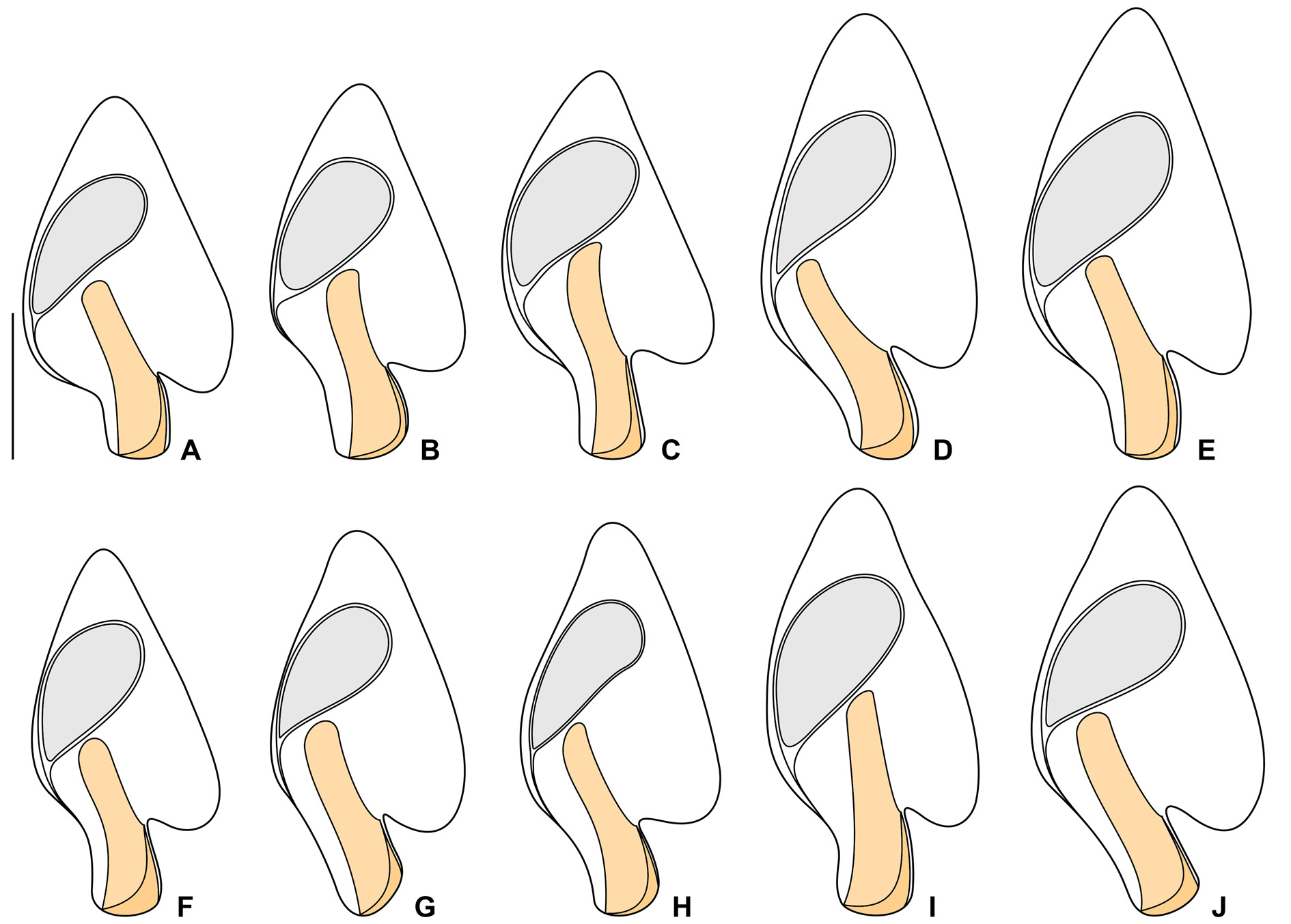
Notes on Vietnamese population
The Vietnamese population exhibits similar morphological features as the Thai population. However, the Vietnamese population shows a slightly narrower range in the body length (70–90 μm vs 70–110 μm) and is slightly more slender (length:width ratio 1.8–2.1:1 vs 1.7–1.9:1) than the Thai population. The macronucleus is shorter in some Vietnamese specimens, but its variability completely falls within the range of the Thai population (length 26–35 μm vs 27–47 μm). The variability of body size and shape of the Vietnamese population is summarized in Table 4 and shown in Figs 10 View Fig A–J, 11A–B, E–G.
No known copyright restrictions apply. See Agosti, D., Egloff, W., 2009. Taxonomic information exchange and copyright: the Plazi approach. BMC Research Notes 2009, 2:53 for further explanation.