
Clevelandella panesthiae ( Kidder, 1937 )
|
publication ID |
https://doi.org/ 10.5852/ejt.2020.697 |
|
publication LSID |
lsid:zoobank.org:pub:8962B6E6-B278-4EF5-9E62-3E858726E2F2 |
|
DOI |
https://doi.org/10.5281/zenodo.4329001 |
|
persistent identifier |
https://treatment.plazi.org/id/44755610-B015-FFB1-FDBC-FC67B8D0733C |
|
treatment provided by |
Valdenar |
|
scientific name |
Clevelandella panesthiae ( Kidder, 1937 ) |
| status |
|
Clevelandella panesthiae ( Kidder, 1937)
Figs 6–8 View Fig View Fig View Fig
Description of Thai I population
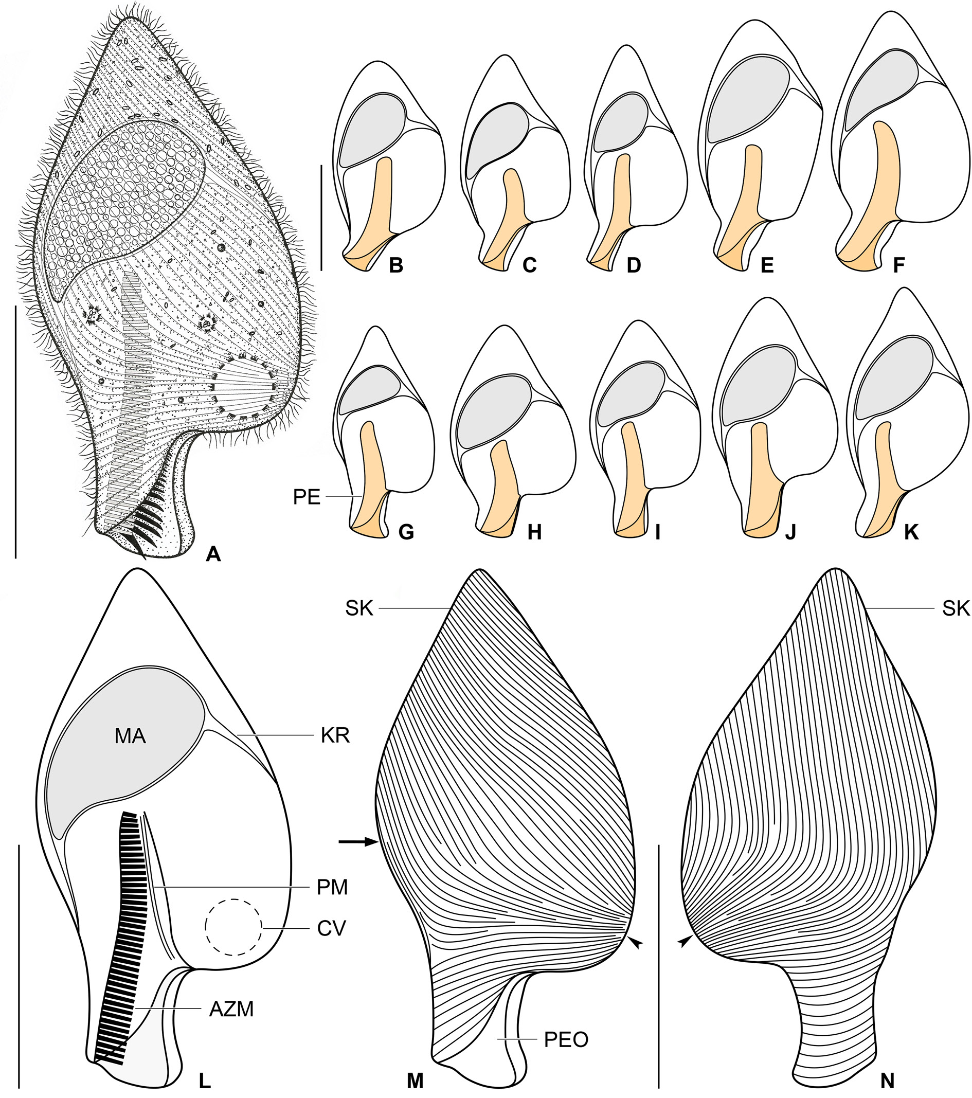
Size in vivo about 95–120 × 45–65 μm, usually 105 × 55 μm, as calculated from some in vivo measurements and morphometric data; length:width ratio rather stable, ranging from 1.7:1 to 2.2:1 after protargol impregnation ( Table 3). Body shape obcordiform in ventral view, widest about at midportion, i.e., usually near level of proximal end of peristomial funnel. Anterior end pointed or bluntly pointed; posterior body portion differentiated into a short but conspicuous peristomial projection; left body margin markedly convex ( Figs 6 View Fig A–N, 8B–D). Macronucleus located in anterior half of body; tearshaped, with a length:width ratio of 1.8–2.3: 1 in protargol preparations; anterior end broadly rounded, posterior end narrowly rounded and oriented toward right body margin; 34–44 × 17–23 μm in size after protargol impregnation; filled with numerous globular structures (very likely nucleoli) 0.9–1.9 μm across after protargol impregnation, well recognizable in vivo and after protargol impregnation. Karyophore attached to right and left cell’s margins ( Table 3; Figs 6 View Fig A–L, 8B–D). Micronucleus not observed. Contractile vacuole at left side of body, slightly above base of peristomial projection ( Figs 6A, L View Fig , 8B, D View Fig ). Cortex flexible, no cortical granules recognizable. Cytoplasm colorless; finely granulated; divided by karyophore into an anterior and a posterior part; compartment anterior to macronucleus contains few oval, refractile bodies (very likely paraglycogen platelets) recognizable in vivo and after protargol impregnation; compartment posterior to macronucleus contains some free (symbiotic?) bacteria and/or archaea scattered throughout cytoplasm and few food vacuoles only about 1.8–2.9 μm in diameter with prey prokaryotes ( Fig. 6A View Fig ). Swims slowly; dies quickly on microscope slides, possibly due to presence of oxygen; body shape changed in dying and strongly squeezed cells, i.e., body becomes thicker and peristomial projection shortens.
Somatic ciliature holotrichous; cilia about 4.0–6.0 μm long in vivo and very narrowly arranged. Approximately 100 ciliary rows narrowly spaced over entire body surface and about 20 ciliary rows encroaching onto peristomial projection. Ciliary rows on peristomial projection appear as oblique lines in ventral view, while as very shallow arcs in dorsal view ( Fig. 6 View Fig M–N). Almost all ciliary rows begin from a whorl (posterior suture) on left body side slightly above base of peristomial projection, i.e., near location of contractile vacuole ( Fig. 6 View Fig M–N, arrowheads), radiate across ventral and dorsal sides toward right body margin; some kineties shortened anteriorly or posteriorly. Right suture spreads from base of peristomial projection to anterior body end; formed by obliquely abutting ventral and dorsal ciliary rows ( Fig. 6M View Fig , arrow).
Peristomial projection short but conspicuous, occupying on average 21% of body length and measuring 16–22 × 12–19 μm in protargol preparations. Peristomial opening situated on the left ventral border of the peristomial projection, roughly triangular and long, i.e., occupying about 21% of body length and measuring 18–22 × 8–12 μm after protargol impregnation ( Figs 6 View Fig A–M, 8B–D). Peristomial funnel about 44 μm long after protargol impregnation. Adoral zone extends slightly obliquely from distal end of peristomial projection across right side of peristomial funnel, terminating near posterior end of macronucleus; occupies from 41% to 55% of body length; composed of on average 45 membranelles; cilia of distalmost membranelles about 8 μm long in vivo and projecting out of peristomial funnel ( Table 3; Fig. 6A, L View Fig ). Paroral membrane diplostichomonad, i.e., composed of two rows of basal bodies; runs in parallel with adoral zone on opposite side of peristomial funnel; commences about at level of proximal end of peristomial opening and terminates near cytostome at proximal end of peristomial funnel ( Fig. 6L View Fig ).
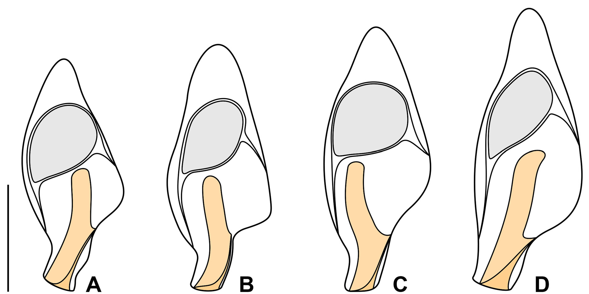
Notes on Vietnamese population
The Vietnamese population displays a similar cell organization as the Thai population. However, the former population is more slender and has a longer peristomial projection. On the other hand, the macronucleus is markedly wider in the Vietnamese population than in the Thai population. The variability of body size and shape of the Vietnamese population is summarized in Table 3 and shown in Figs 7 View Fig A–D, 8A, E–G.

No known copyright restrictions apply. See Agosti, D., Egloff, W., 2009. Taxonomic information exchange and copyright: the Plazi approach. BMC Research Notes 2009, 2:53 for further explanation.