
Clevelandella constricta ( Kidder, 1937 )
|
publication ID |
https://doi.org/ 10.5852/ejt.2020.697 |
|
publication LSID |
lsid:zoobank.org:pub:8962B6E6-B278-4EF5-9E62-3E858726E2F2 |
|
DOI |
https://doi.org/10.5281/zenodo.4329003 |
|
persistent identifier |
https://treatment.plazi.org/id/44755610-B01C-FFA8-FDBA-FB9EBDB8733C |
|
treatment provided by |
Valdenar |
|
scientific name |
Clevelandella constricta ( Kidder, 1937 ) |
| status |
|
Clevelandella constricta ( Kidder, 1937)
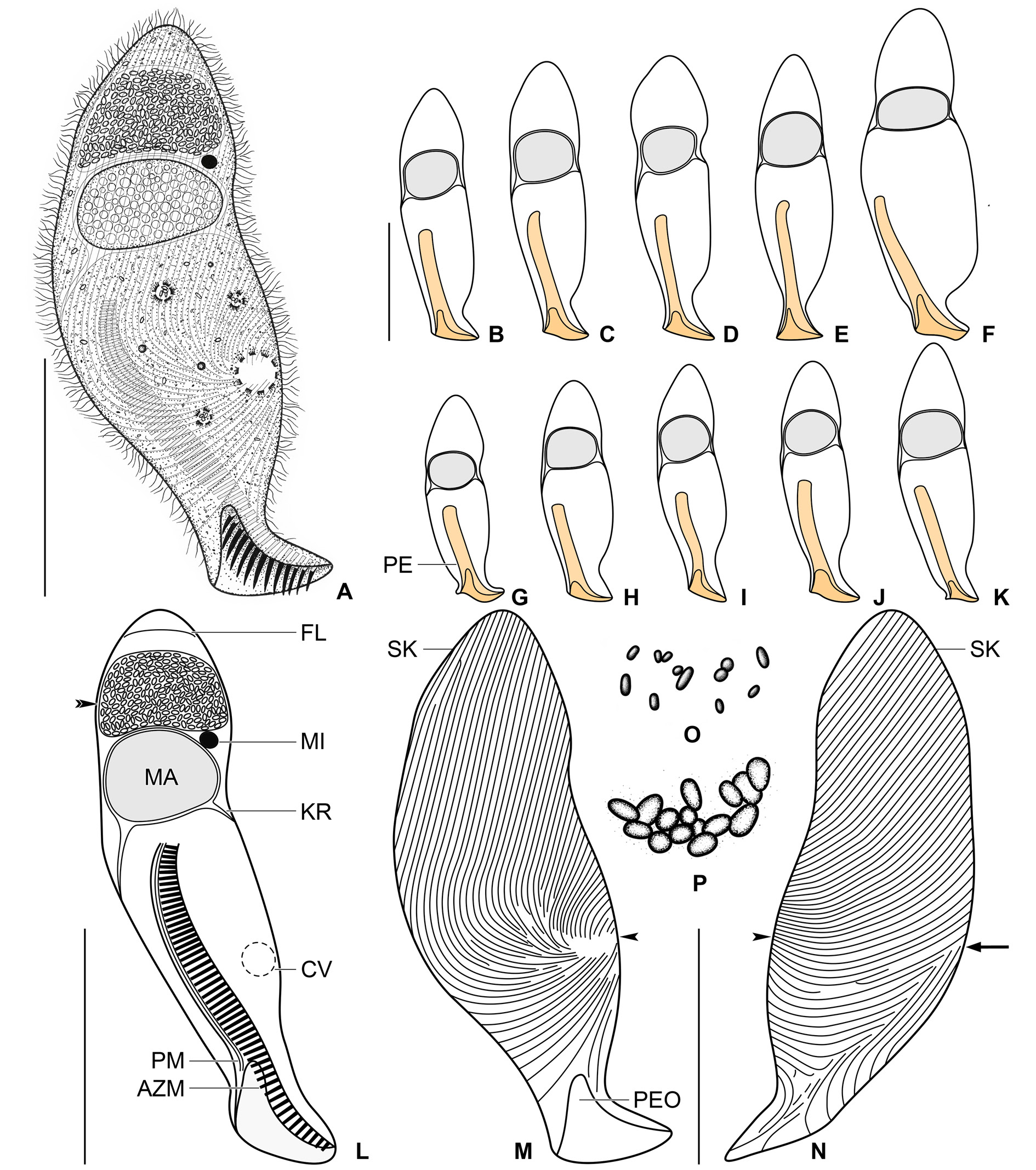
Figs 1–3 View Fig View Fig View Fig
Description of Vietnamese population
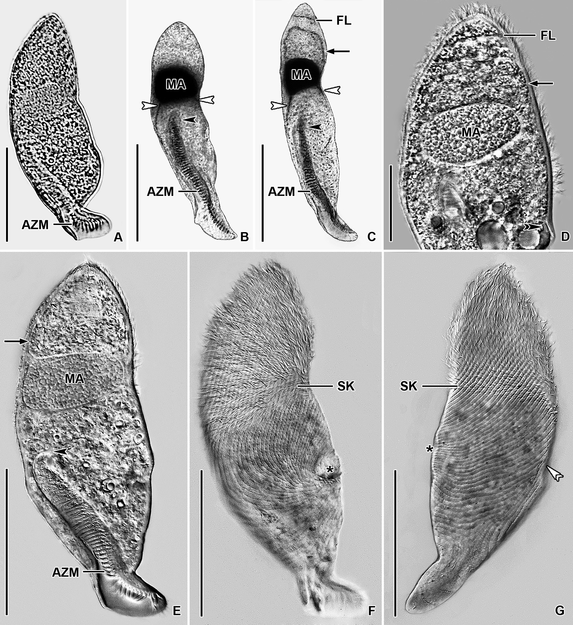
Size in vivo about 100–155 × 25–50 μm, usually 120 × 35 μm, as calculated from some in vivo measurements and morphometric data; length:width ratio ranging from 3:1 to 4.2:1 after protargol impregnation ( Table 1 View Table 1 ). Body spindle-shaped, more or less distinctly constricted in anterior third, usually widest at mid-portion, i.e., about at level of proximal end of adoral zone of membranelles; dorsoventrally flattened 1.4:1. Anterior end bluntly pointed; posterior body portion differentiated into a short, inconspicuous peristomial projection, distinctly constricted at its base; left and right body margins slightly concave at level of macronucleus ( Figs 1 View Fig A–N, 3A, E–G). Macronucleus located in anterior second fifth of body length; ellipsoidal to almost spherical, with a length:width ratio of 1.1– 1.8: 1 in protargol preparations; 20–28 × 15–20 μm in size after protargol impregnation; filled with numerous globular structures (very likely nucleoli) 0.7–1.8 μm across after protargol impregnation, well recognizable in vivo and sometimes in lightly impregnated specimens. Karyophore attached to right and left cell’s margins in middle third of body, usually at level of macronucleus, rarely in midbody. Micronucleus attached to anterior side of macronucleus, i.e., near the place where longitudinal cell axis crosses macronucleus or on its left side; globular and about 4.1–4.3 × 4.5–4.7 μm in size in vivo ( Table 1 View Table 1 ; Figs 1 View Fig A–L, 3E). Contractile vacuole near left body margin in posterior third of cell, i.e., close to canal leading to cytopyge ( Fig. 1A, L View Fig ). Cortex flexible, no cortical granules recognizable. Cytoplasm colorless; finely granulated; divided by karyophore into an anterior and a posterior part; cytoplasm anterior to macronucleus contains a frontal lamina transversely stretching slightly posterior to anterior body end and densely packed, oval, refractile bodies (probably paraglycogen platelets) observable in vivo and after protargol impregnation; compartment posterior to macronucleus contains some (symbiotic?) bacteria and/or archaea freely scattered throughout the main body portion and food a Data based on mounted, protargol-impregnated, and randomly selected specimens. Measurements in µm.
vacuoles about 2.0–5.2 μm across and encompassing prey prokaryotes ( Figs 1A, L View Fig , O–P, 3E). Swims slowly; dies quickly on microscope slides, possibly due to presence of oxygen.
Somatic ciliature holotrichous; cilia about 4.5–6.5 μm long in vivo and narrowly arranged.Approximately 80 ciliary rows narrowly spaced over entire body surface and about 10 ciliary rows encroaching onto peristomial projection. Almost all ciliary rows commence from a whorl (posterior suture) on left side near contractile vacuole to radiate over ventral and dorsal sides toward right body margin; some kineties shortened anteriorly or posteriorly ( Figs 1 View Fig M–N, 3F–G). Right suture extends from base of peristomial
projection to anterior body end; formed by obliquely abutting ventral and dorsal ciliary rows ( Figs 1N View Fig , arrow, 3G, white double arrowhead).
Peristomial projection occupies on average 16% of body length and measures 12–23 × 15–20 μm in protargol preparations. Peristomial opening situated on ventral side of peristomial projection, triangular and short, i.e., about 11% of body length and 10–15 × 13–18 μm in size after protargol impregnation ( Figs 1 View Fig A–M, 3A, E). Peristomial funnel approximately 50 μm long after protargol impregnation. Adoral zone extends obliquely from distal end of peristomial projection along left side of peristomial funnel to terminate about in mid-portion of cell; occupies 41% to 55% of body length; composed of on average 55 membranelles; cilia of distalmost membranelles about 9 μm long in vivo and projecting out of peristomial opening ( Table 1 View Table 1 ; Figs 1A, L View Fig , 3E View Fig ). Paroral membrane diplostichomonad, i.e., composed of two rows of basal bodies; extends in parallel with adoral zone but on opposite side of peristomial funnel; commences at level of proximal end of peristomial opening and terminates near cytostome at proximal end of peristomial funnel ( Fig. 1L View Fig ). Pharyngeal fibres spread from proximal end of adoral zone and paroral membrane, run transversely leftwards forming a conical funnel about 20 μm long in vivo.
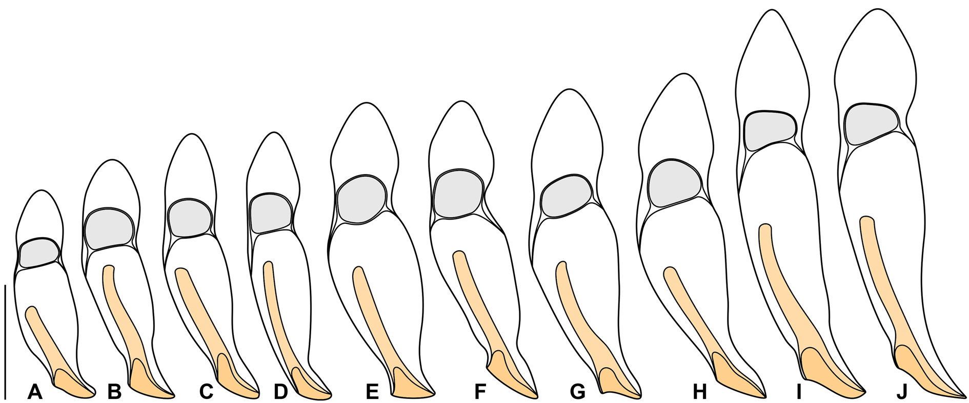
Notes on Thai I population
The Thai population matches very well the Vietnamese population. However, the Thai population shows a slightly wider range in the body length (95–175 μm vs 100–155 μm) and is slightly more slender than the Vietnamese population (length:width ratio 3.4–5.0:1 vs 3.0– 4.2:1). The variability of body size and shape of the Thai population is summarized in Table 1 View Table 1 and shown in Figs 2 View Fig A–J, 3B–C.
No known copyright restrictions apply. See Agosti, D., Egloff, W., 2009. Taxonomic information exchange and copyright: the Plazi approach. BMC Research Notes 2009, 2:53 for further explanation.