
Atopobathynella operculata, Reddy, Ranga, 2008
|
publication ID |
https://doi.org/ 10.5281/zenodo.183164 |
|
DOI |
https://doi.org/10.5281/zenodo.6229985 |
|
persistent identifier |
https://treatment.plazi.org/id/456687E0-FFB3-301A-35B5-391CFF027C73 |
|
treatment provided by |
Plazi |
|
scientific name |
Atopobathynella operculata |
| status |
sp. nov. |
Atopobathynella operculata sp. n.
( Figs 1–4 View FIGURE 1 View FIGURE 2 View FIGURE 3 View FIGURE 4 )
Type locality and material examined. The River Godavari at Rajahmundry town (16o9’ N 81 o47’ E), South India. The sampling site is located almost in the middle of the river basin wherefrom sand is being regularly mined and transported ashore. Here the riverbed has a deposit of fine sand and detritus particles, but with little or no clay, and is devoid of any macrophytic vegetation. Tidal influence from the nearby Bay of Bengal is non-existent, hence freshwater conditions prevail throughout the year.
Holotype female (dissected and mounted on 4 slides, catalogue no. SMF 32211). Allotype male (undissected, catalogue no. SMF 32212). Female paratype 1 (undissected, catalogue no. SMF 32212). Female paratype 2 (dissected on one slide, catalogue no. SMF 32213). Female paratype 3 (undissected, catalogue no. SMF 32214). Male paratype (dissected on one slide, catalogue no. SMF 32215). All are kept in the collection of the Deutsches Zentrum für Marine Biodiversitätsforschung ( DZMB) at Wilhelmshaven ( Germany), being a department of the Senckenberg Museum und Forschungsinstitut, Frankfurt ( SMF). Leg. Y. Ranga Reddy, 24 December 2002.
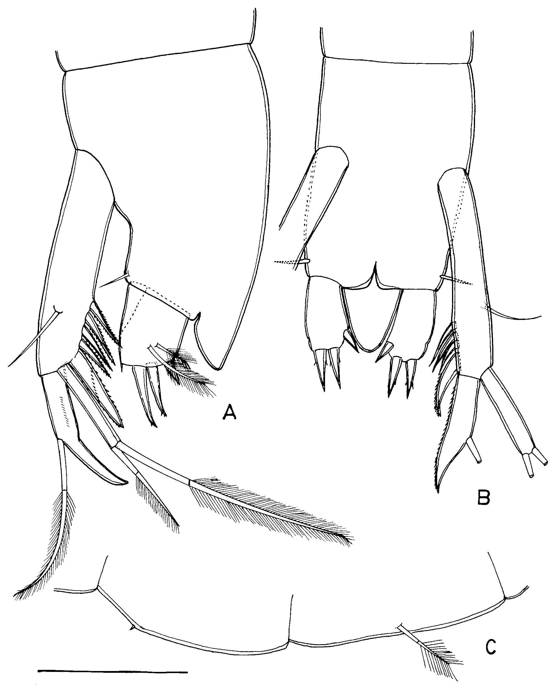
Description of adult female. Total body length of holotype 1.41 mm, of paratypes 1.34–1.56 mm. Body elongated, almost cyclindrical, segments progressively widening and lengthening towards posterior end. All body segments including head with numerous perforations. Head only about 16% longer than wide. Anal operculum massive, plate-like, somewhat subtriangular in outline, producing backwards and almost reaching the end of caudal furca ( Fig. 1 View FIGURE 1 A, B). Pleotelson with 1 seta on either side; seta smooth and much shorter than caudal furca. Furca elongately oval in ventral view ( Fig. 1 View FIGURE 1 B), but almost subquadrate in lateral view ( Fig. 1 View FIGURE 1 A), nearly 1.5 times longer than wide, with 2 apical spines of similar size, 1 small inner spine, and 2 unequal plumose setae. Furcal organ small and ventral.
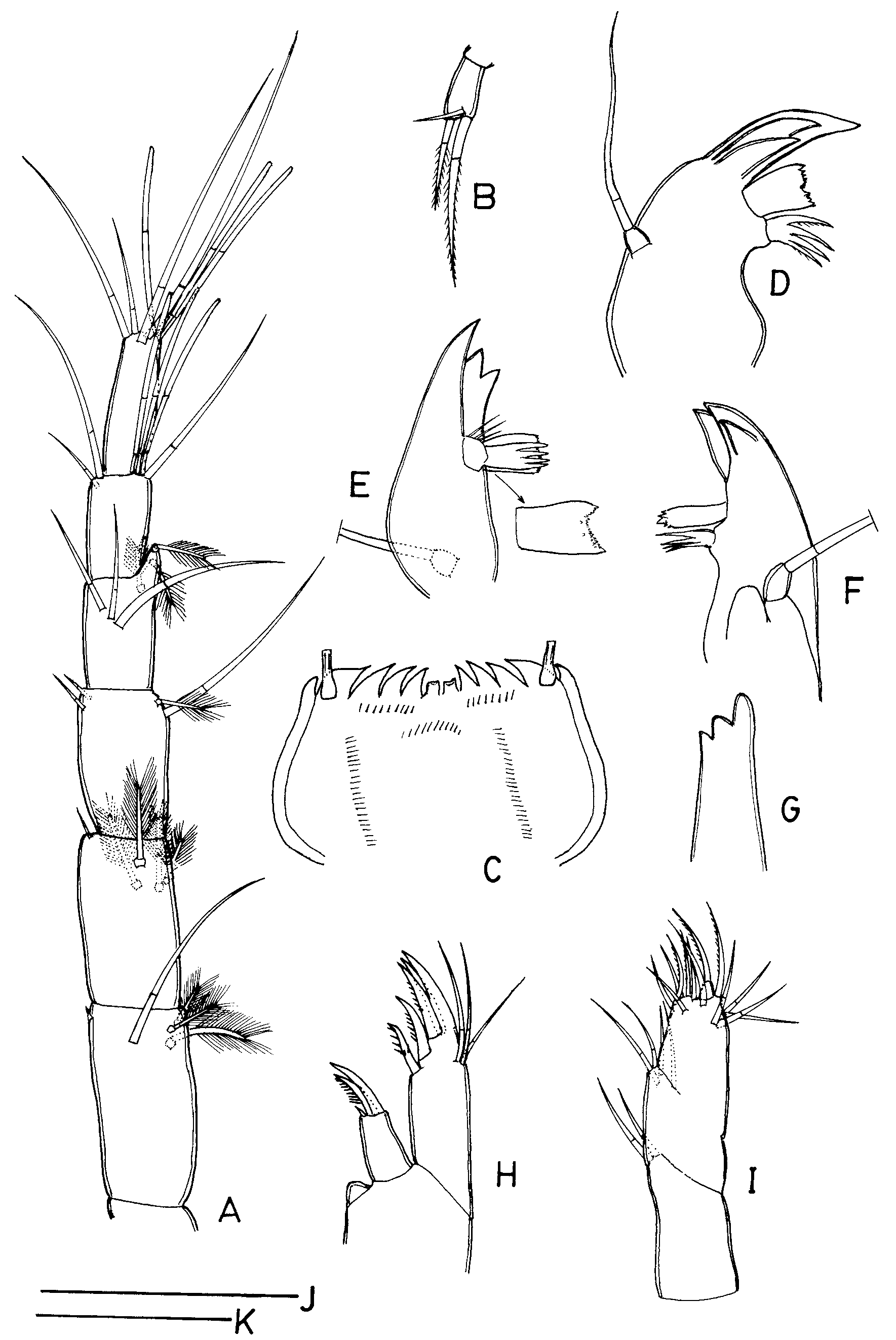
Antennule ( Fig. 2 View FIGURE 2 A): with 3-segmented peduncle and with 3-segmented inner flagellum, outer flagellum reduced. Whole antennule slender (9 times longer than maximum width), 42% longer than head. Length of the 3 segments of the stem greater than that of the remainder of the outer flagellum. First segment of peduncle longest, twice as long as its own width, with a group of 2 ventral and 1 dorsal plumose setae at distal outer corner, 1 long dorsal seta in the distal half, and 1 tiny seta at distal inner corner. Second segment slightly shorter than first segment, with a group of 3 ventral and 1 dorsal plumose setae near distal outer corner and 1 short, slender seta at distal inner corner. Third segment with 1 long seta on outer distal margin, 1 dorsal plumose seta at distal outer corner; 2 short setae, 1 dorsal and 1 ventral, near distal inner corner. First segment of inner flagellum with a group of 3 unequal dorsal setae in the distal half (these setae seem to be the rest of the reduced and fused outer flagellum), 1 ventral plumose seta below apophysis; apophysis distinct but slender, reaching proximal fourth of next segment, with 1 apical and 1 ventral, subapical plumose setae. Second segment of inner flagellum with a group of 1 simple seta and 3 aesthetascs, which are longer than next segment, at distal outer corner; 1 long apical and 1 short ventral setae at distal inner angle. Terminal segment slenderest, with 4 setae and 3 aesthetascs.
Antenna ( Fig. 2 View FIGURE 2 B): small, 1-segmented, only slightly dilated distally, with 2 terminal unequal plumose setae and 1 short, subterminal, simple seta; lateral seta absent.
Labrum ( Fig. 2 View FIGURE 2 C): flat, symmetrical in ventral view and characteristic in shape. Free margin straight, bearing 2 small median teeth, flanked on either side by 4 acuminate curved teeth, gradually increasing in size laterally, and with 1 smaller somewhat blunt tooth on either side; tubular pore with long, thick-walled, proximally dilated tube, occurring ventrally on either side at base of lateral main tooth. Also, fine spinules, in rows, discernible on ventral surface, as illustrated.
Mandible ( Fig. 2 View FIGURE 2 D–G): distal part of pars incisiva with 3 teeth, proximal tooth small; all teeth pointed in lateral view ( Fig. 2 View FIGURE 2 D–F), but blunt in frontal view ( Fig. 2 View FIGURE 2 G). Pars molaris (“Borstenlobus”) articulate, consisting of 5 claws; distal one defined at base, strongly developed into plate-like structure ( Fig. 2 View FIGURE 2 D, E) (almost cylindrical in a different view as in Fig. 2 View FIGURE 2 F), apical margin concave and finely denticulate; 2 additional denticles also occurring below denticulate margin ( Fig. 2 View FIGURE 2 E), 3 spinules seen at base of pars molaris on distal margin. Palp 1-segmented, only slightly longer than wide, bearing a long terminal seta.
Maxillule ( Fig. 2 View FIGURE 2 H): with 2 endites. Proximal endite small, subquadrate, twice as long as wide, carrying 2 unequal, serrulate, claw-like spines, and 1 tiny spinule; also, a simple, triangular lobe lying at base of endite. Distal endite straight, slender, 2.4 times as long as wide, armed with 3 terminal claws of similar size, 2 subterminal claws, all claws with serrulate inner margin, and 3 subterminal setae on distal outer margin.
Maxilla ( Fig. 2 View FIGURE 2 I): 4-segmented, second and third segments half fused, with 2, (4, 12), and 1 setae, respectively; distalmost segment tiny.
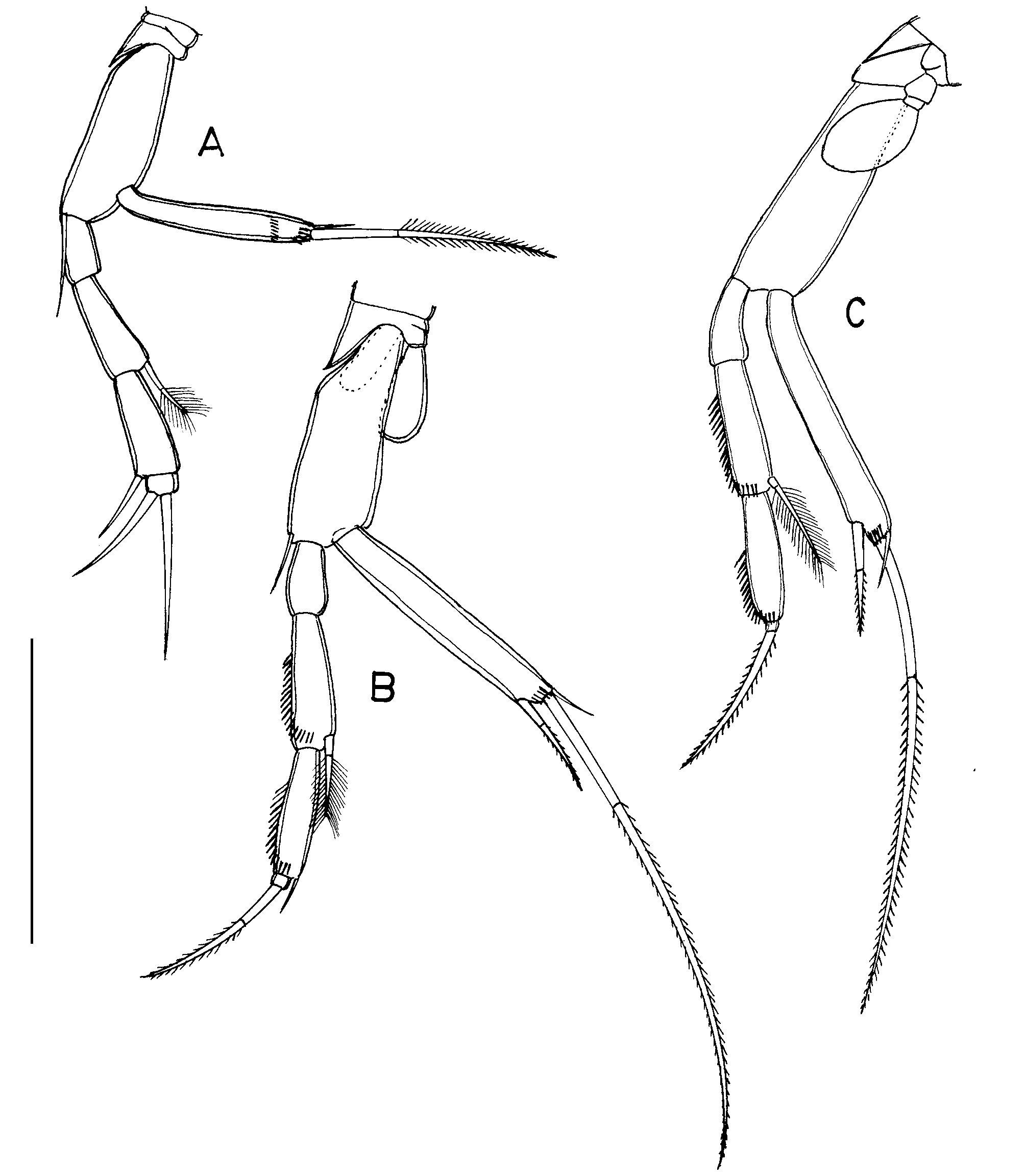
Thoracopods I–VII ( Fig. 3 View FIGURE 3 A–C): well developed, length gradually increasing from pairs I–III, last 4 pairs almost similar in size; thoracopods II–VII with 1-segmented epipod each. Coxa with distinct, pointed projection at distal inner border; basis of thoracopods II–VI alone with inner marginal seta. Exopod 1-segmented, with 2 very unequal terminal setae and 1short subterminal seta on ventral side; subterminal seta absent on thoracopod I alone. Endopod 4-segmented. Setal formulae: Th. I, 1+0/0+1/1+0/2(0); Th. II–IV, 0+0/0+1/0+1/ 1(0); Th. V–VII, 0+0/0+1/0+0/1(0).
Thoracopod VIII ( Fig. 1 View FIGURE 1 C): minute, sharply pointed, denticle-like structure.
Pleopod 1 ( Fig. 1 View FIGURE 1 C): represented by 1 strong, plumose seta.
Uropod ( Fig. 1 View FIGURE 1 A–B): sympod slender, 4.4 times as long as wide, bearing 4 spines in a row on distal inner margin and 1 short, simple seta on dorso-lateral surface almost opposite to spine row; distalmost spine stout, straight, 31% longer than other spines, which are acutely pointed and almost equal in size. Exopod cylindrical, nearly 6 times as long as wide, measuring 42% of sympod length and bearing 2 apical, unequal setae, outer seta spiniform, unipinnate and less than half the length of inner, bipinnate seta. Endopod sickle-shaped, reaching 62% of sympod length, lateral margins smooth, but a longitudinal of fine spinules at about the middle of proximal part, in lateral view; a single long, plumose seta at about midlength of outer margin.
Description of adult male ( Fig. 4 View FIGURE 4 A–C). Total body length of allotype 1.52 mm, of paratype 1.46 mm. Body and all appendages except antennule and thoracopod VIII as in female.
Antennule ( Fig. 4 View FIGURE 4 A): same as in female except for antennal organ on second segment; second segment 1.5 times as long as wide, antennal organ seen as a massive protuberance at distal inner angle, tip rounded with an opening.
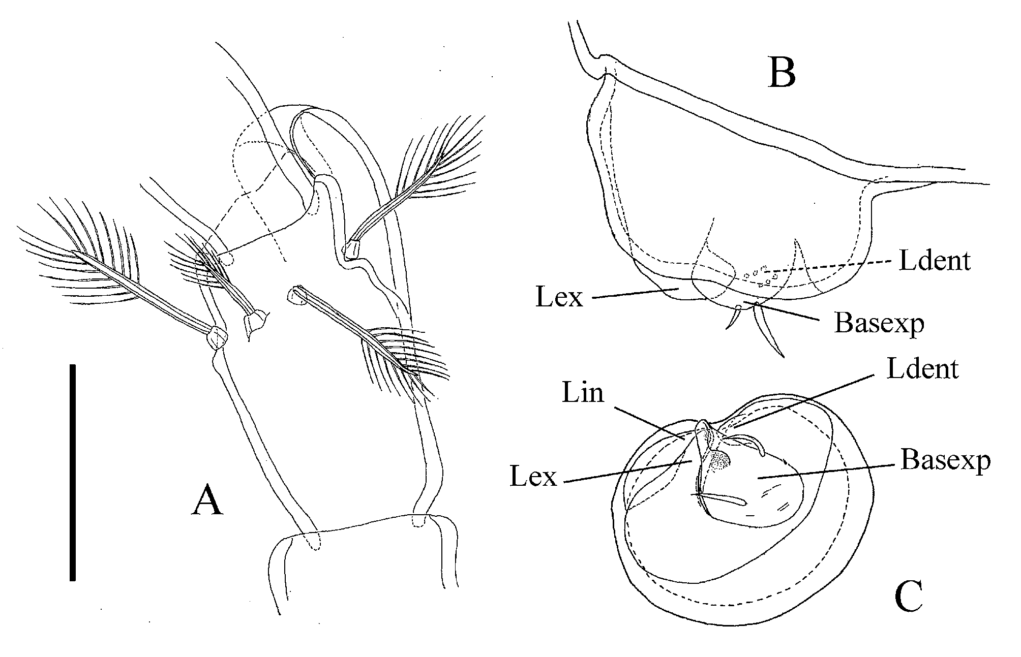
Thoracopod VIII ( Fig. 4 View FIGURE 4 B–C) rather small so that the constituent parts are difficult to make out and interpret. Protopod massive but without prominent penial region. In latero-external view ( Fig. 4 View FIGURE 4 B) two structures are discernible, one with 2 and one without setae. In ventral view ( Fig. 4 View FIGURE 4 C) the structure with 2 setae is extended into a spoon-like projection and is interpreted here as a basexopod with a basipodal seta (the bigger one) and an exopodal seta (the smaller one). The other structure without setae can be seen in ventral view ( Fig. 4 View FIGURE 4 C) as a conical projection reaching the spoon-like projection of the basexopod. This structure is interpreted here as the external lobe. Where both meet, the internal and the dentate lobe also converge. Dentate lobe with a few denticles can be seen in latero-external view ( Fig. 4 View FIGURE 4 B). Comparison with other species will reveal whether the interpretation presented here is tenable.
Intraspecific variation. The number of spines borne by the sympod of uropod is either 4 or 5. Etymology. The specific name refers to the prominent anal operculum (Latin adjective operculatus, operire = to close, cover); gender feminine.
Ecology. At the type locality, the new species was found as strays in the surficial sediments of knee-deep waters of an exposed island at the middle of the river bed. It was greatly dominated by Habrobathynella . The fauna that co-occurred with the new species was diverse but not rich, and included the following:
Bathynellacea View in CoL : Habrobathynella schminkei Ranga Reddy, 2004 , and Habrobathynella sp. Copepoda: Bryocyclops sp., Paracyclops sp., Parastenocaris curvispinus Enckell, 1970 , Parastenocaris gayatri Ranga Reddy, 2001 , Elaphoidella sp., Mesochra wolskii Jakubisiak, 1933 , Nitocra ?lacustris (Schmankevitsch, 1875), Folioquinpes chathamensis ( G.O. Sars, 1905) , and Phyllognathopus viguieri ( Maupas, 1892) . Cladocera: Macrothrix View in CoL sp., chydorids. Other taxa were: unidentified oligochaetes, nematodes, mites, and insect larvae.
| SMF |
Forschungsinstitut und Natur-Museum Senckenberg |
No known copyright restrictions apply. See Agosti, D., Egloff, W., 2009. Taxonomic information exchange and copyright: the Plazi approach. BMC Research Notes 2009, 2:53 for further explanation.
|
Kingdom |
|
|
Phylum |
|
|
Class |
|
|
Order |
|
|
Family |
|
|
Genus |
Atopobathynella operculata
| Reddy, Ranga 2008 |
Habrobathynella schminkei
| Ranga Reddy 2004 |
Parastenocaris gayatri
| Ranga Reddy 2001 |
Parastenocaris curvispinus
| Enckell 1970 |
Mesochra wolskii
| Jakubisiak 1933 |
Folioquinpes chathamensis (
| G.O. Sars 1905 |
Phyllognathopus viguieri (
| Maupas 1892 |