
Vorticella veloxiiforme, Pereira & Brito & Eizirik & Utz, 2018
|
publication ID |
https://doi.org/ 10.11646/zootaxa.4508.2.4 |
|
publication LSID |
lsid:zoobank.org:pub:294C2462-ADF9-4C03-ABA0-81FC5B641EBA |
|
DOI |
https://doi.org/10.5281/zenodo.5962964 |
|
persistent identifier |
https://treatment.plazi.org/id/49766545-5A63-FF9E-FF36-F9F7FE4345DC |
|
treatment provided by |
Plazi |
|
scientific name |
Vorticella veloxiiforme |
| status |
sp. nov. |
Vorticella veloxiiforme n.sp.
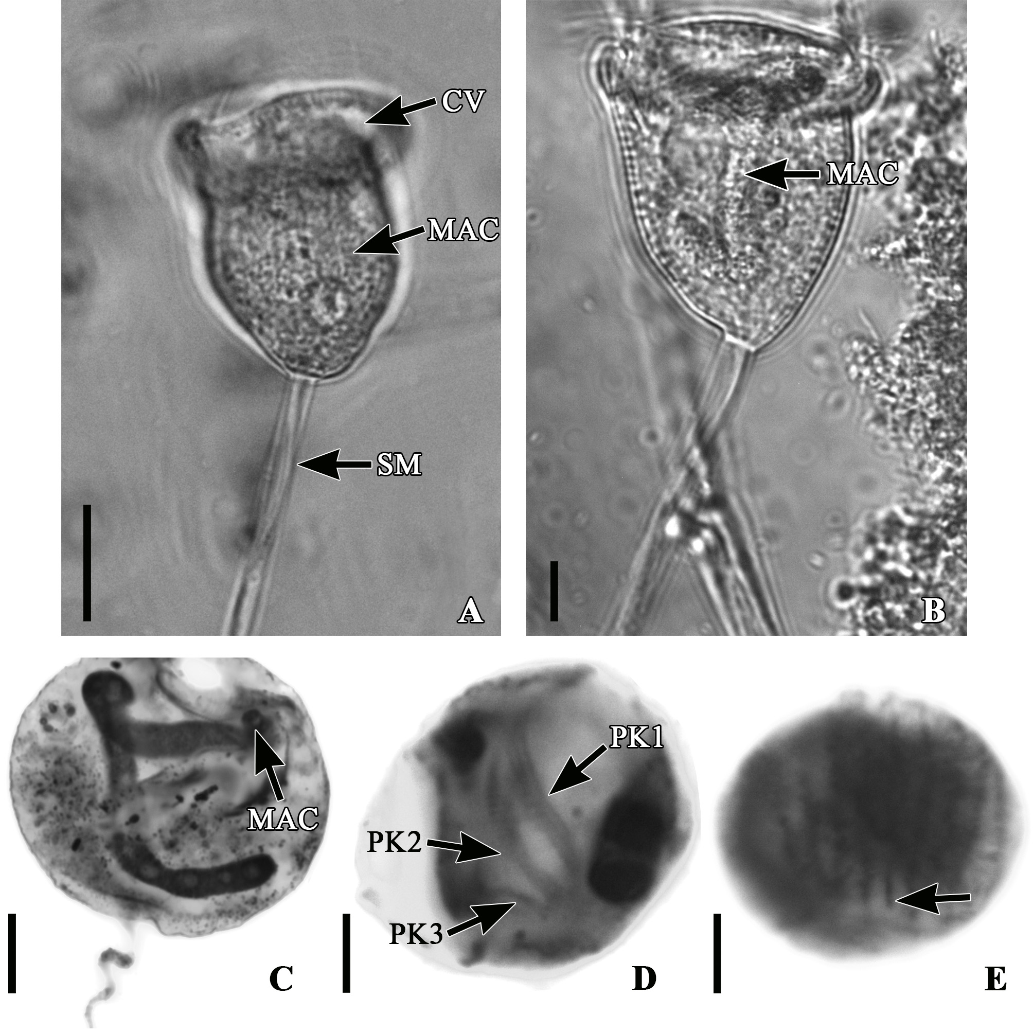
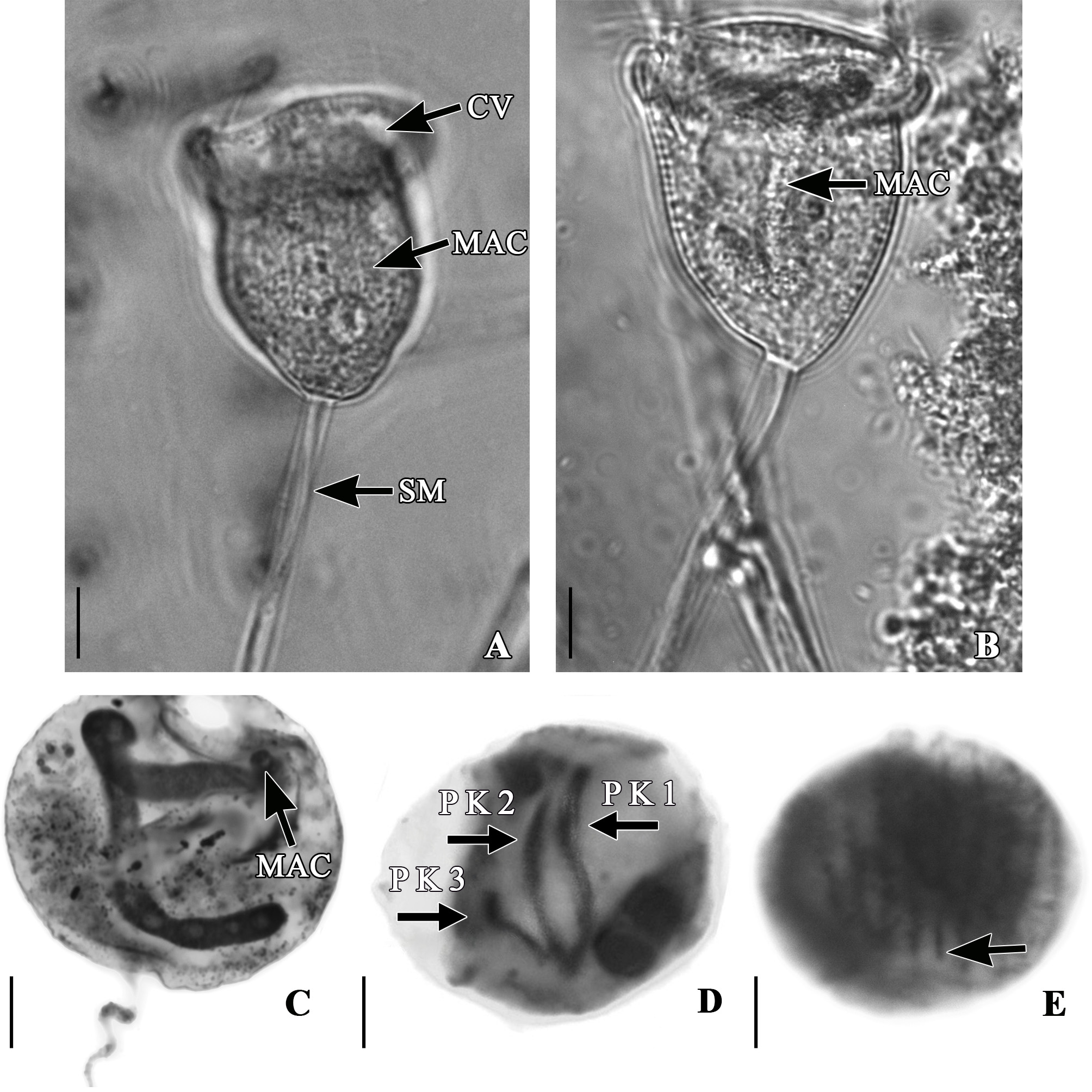
Diagnosis. Freshwater peritrich, with a cup-shaped zooid measuring 57 X 41 µm on average. Conspicuous silver line system on pellicle. Horizontal, J-shaped macronucleus occupying 2/3 of the cell volume. One contractile vacuole next to the peristomial lip. Stalk with a strongly contracting a helical spasmoneme, and green endoplasmic granules. Three infundibular polykineties, each comprised of three rows of kinetosomes: P2 and P3 terminate at the same level adstomally. P1 has one longer row of kinetosomes, terminating below the level of P2 and P3, which is hook-like shaped abstomally.
Type Locality. Patos Lagoon, Viamão (30° 19’70”S; 50°50’47”W), Rio Grande do Sul, Brazil.
Etymology. The specific epithet refers to the fast contraction mode of the trophont.
Deposition of slide. Type material: A slide with a protargol-stained colony (holotype) was deposited in the Protist Collection of the Museum of Sciences and Technology of the Pontifícia Universidade Católica do Rio Grande do Sul (PUCRS), under the number MCTP05. The paratype, a slide with a marked protargol-stained colony, was deposited in the Protist Collection of the Natural History Museum in London.
Morphology of live specimens. Zooids of V. veloxiiforme were inverted bell-shaped and measured on average 57.4 µm long and 41 µm wide ( Table 1). They also presented faint and closely spaced striae on the pellicle that, a thick peristomial lip, and a slightly elevated epistomial disk ( Figs. 1A View FIGURE 1 and 2A View FIGURE 2 ). A constriction below the peritomial lip was observed on the zooid body. A single contractile vacuole was found in the upper part of the zooid, close to the peristome ( Figs. 1A View FIGURE 1 and 2A View FIGURE 2 ). A J-shaped macronucleus was oriented along the oral-aboral axis and occupied approximately 2/3 of the volume of the cell body ( Figs. 1A View FIGURE 1 , 2A and 2B View FIGURE 2 ). The cytoplasm presented grayish and greenish food granules. The stalk measured on average 149.3µm in length ( Table 1). A spasmoneme was observed inside the stalk ( Figs. 1A View FIGURE 1 and 2A View FIGURE 2 ) and had several green endoplasmic granules throughout. Both the zooid and the stalk were very contractile.
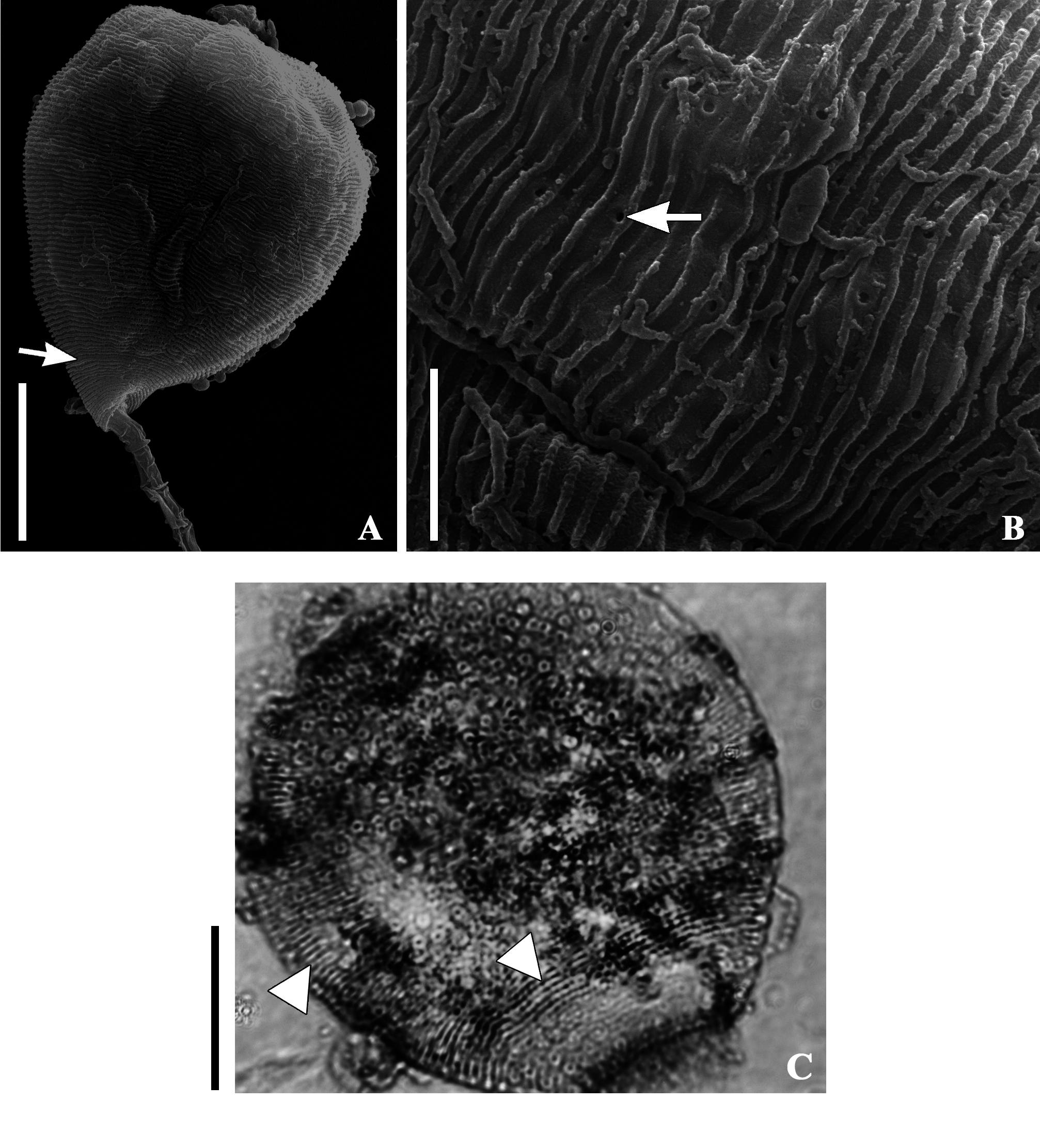
Morphology of stained specimens. The infraciliature and macronucleus of V. veloxiiforme was easily revealed by silver staining ( Figs. 1C and D View FIGURE 1 , 2D View FIGURE 2 ). Somatic myonemes extended from the peristomial lip to the scopula ( Fig. 1E View FIGURE 1 and 3C View FIGURE 3 ). The oral infraciliature was typical of peritrich ciliates, with all infundibular polykinetids composed by three rows of kinetosomes each ( Figs. 1D View FIGURE 1 and 2B View FIGURE 2 ). Rows of P1 were different in length: the adstomal end of the row closer to P2 was longer than the other two. The end of middle row was longer than distal rows in P1 ( Figs. 1D View FIGURE 1 and 2B View FIGURE 2 ). All rows of P2 showed different lengths abstomally, but all terminated at the same level adstomally ( Figs. 1D View FIGURE 1 and 2B View FIGURE 2 ). P3 consisted of three rows of kinetosomes forming a hook-like projection at the abstomal end ( Figs. 1D View FIGURE 1 and 2B View FIGURE 2 ). Adstomally, P3 terminated at the same level of P1 and P2 ( Figs. 1D View FIGURE 1 and 2B View FIGURE 2 ). The silver nitrate technique, and the scanning electron microscopy revealed a horizontal striation pattern and the presence of pellicular pores regularly spaced on the zooid ( Figs.3A and B View FIGURE 3 ). On average, 62 horizontal sileverlines were present between the peristome and the trochal band, and 20 were counted between the trochal band and scopula ( Table 2; Fig. 3C View FIGURE 3 ).
No known copyright restrictions apply. See Agosti, D., Egloff, W., 2009. Taxonomic information exchange and copyright: the Plazi approach. BMC Research Notes 2009, 2:53 for further explanation.