
Vorticella ampullaria, Pereira & Brito & Eizirik & Utz, 2018
|
publication ID |
https://doi.org/ 10.11646/zootaxa.4508.2.4 |
|
publication LSID |
lsid:zoobank.org:pub:294C2462-ADF9-4C03-ABA0-81FC5B641EBA |
|
DOI |
https://doi.org/10.5281/zenodo.5962966 |
|
persistent identifier |
https://treatment.plazi.org/id/49766545-5A65-FF95-FF36-F8DAFCA143CE |
|
treatment provided by |
Plazi |
|
scientific name |
Vorticella ampullaria |
| status |
sp. nov. |
Vorticella ampullaria n.sp.
Diagnosis. Freshwater peritrich with an elongate zooid measuring in vivo 70 X 25 µm on average. Presence of smooth silver line system on pellicle and narrow peristomial lip. A C-shaped macronucleus lies in the middle of the cell. Two contractile vacuoles are present: in oral and aboral regions. All infundibular polykinetids have three rows of kinetosomes each. The middle row of P1 is longer than others; the distal row of P1 is longer than row next to P2, which has different lengths adstomally and terminates at the adstomal curvature of P1. P3 terminates at the same level of P1 adstomally, with the middle row being longer at the abstomal end.
Type Locality. Patos Lagoon, Viamão (30°19’70”S; 50°50’47”W), Rio Grande do Sul, Brazil.
Etymology. The specific epithet refers to the family of the gastropod host.
Deposition of slide. Type material: A slide with a protargol-stained colony (holotype) was deposited in the Protist Collection of the Museum of Science and Technology of the Pontifícia Universidade Católica do Rio Grande do Sul (PUCRS), under the number MCTP06. The paratype, a slide with a marked protargol-stained colony was deposited in the Protist Collection of the Natural History Museum in London.
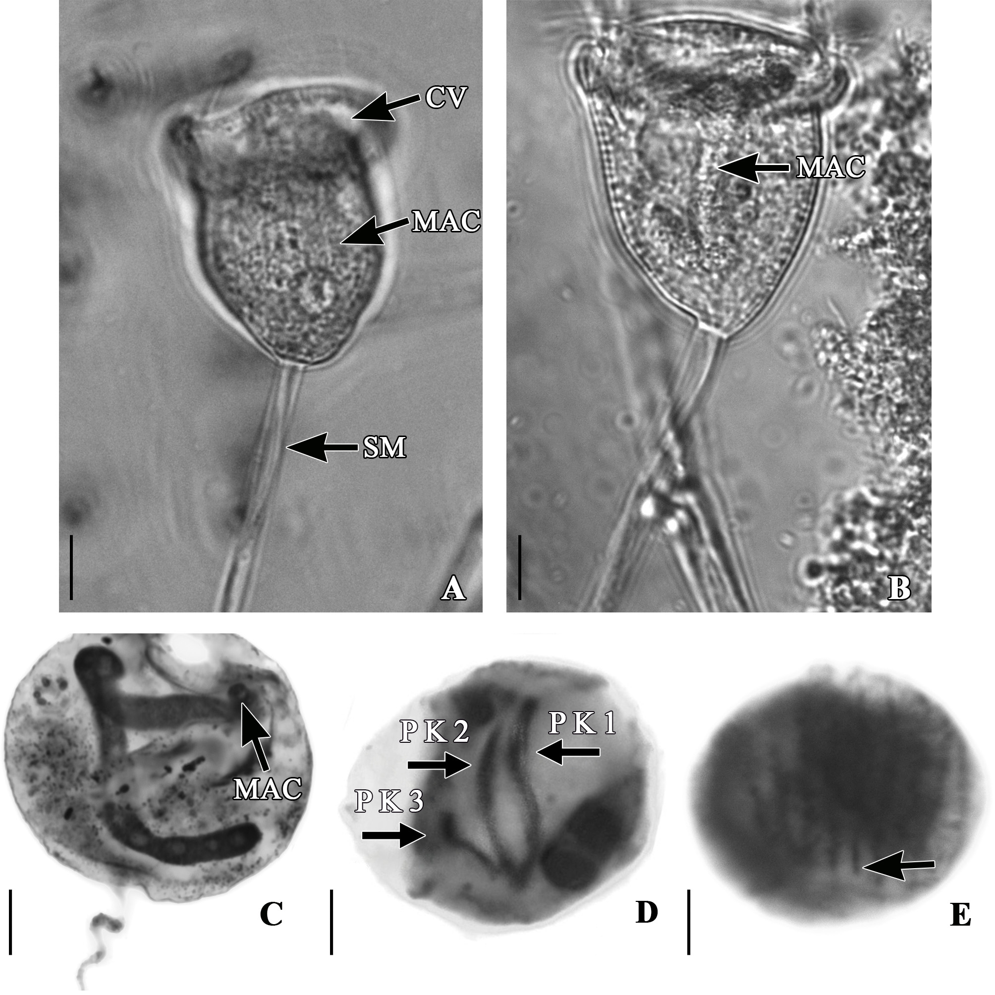
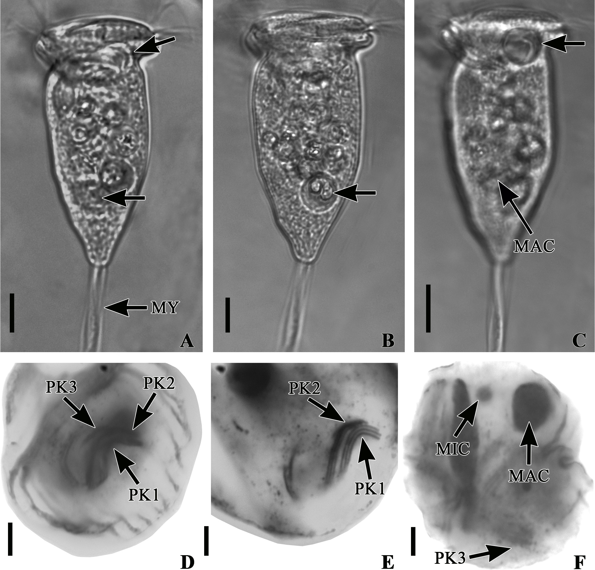
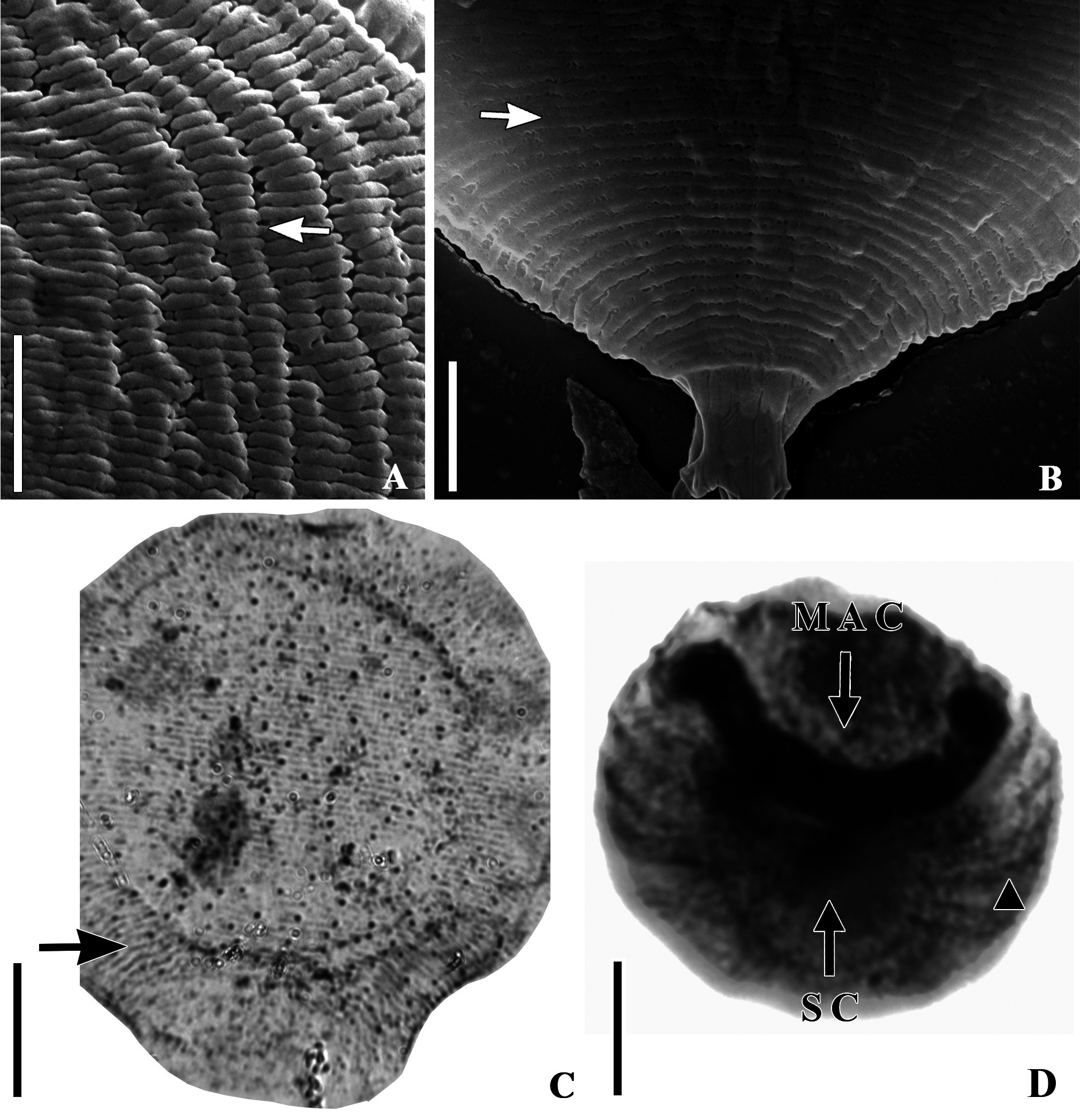
Morphology of live specimens. Elongate zooids, 69.8 µm long and 24.9 µm wide on average ( Table 1), presenting a smooth striated pellicle (visible by high magnification and silver nitrate technique), narrow peristomial lip, and slightly elevated epistomial disk ( Figs. 2C View FIGURE 2 and 4A View FIGURE 4 ). The proportion between upper and middle of the body are similar. A peculiar characteristic for peritrich ciliates was observed: the presence of two contractile vacuoles (one close to the peristome and the other close to the scopula) ( Figs. 2C View FIGURE 2 , 4A, B and C View FIGURE 4 ). The cytoplasm was colorless or slight greenish with many food vacuoles. A C-shape, elongate macronucleus was located vertically in the middle of the cell ( Figs. 2C View FIGURE 2 , and 4C View FIGURE 4 ). The scopula was rectangular-shaped, and the stalk measured 114.5 µm in length on average ( Table 1). A spasmoneme with endoplasmic granules was observed inside the stalk ( Fig. 4A View FIGURE 4 ).
Morphology of stained specimens. The infraciliature and the nuclear apparatus of V. ampullaria were easily revealed by silver staining ( Figs. 2D View FIGURE 2 and 4D and E View FIGURE 4 ). All infundibular polykinetids presented three rows of kinetosomes ( Figs. 2D View FIGURE 2 and 4D and E View FIGURE 4 ). P1 had three rows of different lengths in the adstomal end (the middle row is longer than the others; the distal row is longer than row next to P2) ( Figs. 2D View FIGURE 2 and 4E View FIGURE 4 ); P2 also had rows of different lengths adstomally that terminated at the curvature of P1 ( Figs. 2D View FIGURE 2 and 4E View FIGURE 4 ). P3 presented rows of same length that terminate at the level of P1 adstomally ( Figs. 2D View FIGURE 2 and 4F View FIGURE 4 ). Abstomally, rows of P3 had different lengths with the middle row being longer than the other two ( Figs. 2D View FIGURE 2 and 4F View FIGURE 4 ). The micro and macronucleus are visible in silver stained specimens ( Fig. 4F View FIGURE 4 ). The silver nitrate technique, and scanning electron micrographs revealed a pattern of horizontal smooth silver lines in the pellicle (there were 54 between the peristomial lip and the trochal band, on average, and 14 between the trochal band and scopula, on average; Table 2), a diagnostic character of the genus Vorticella ( Figs. 5B, C and D View FIGURE 5 ). The presence of pores on the membrane was also observed with SEM. These pores were regularly organized on the pellicle as well as a horizontal pattern of silver lines ( Figs. 4A and B View FIGURE 4 ).
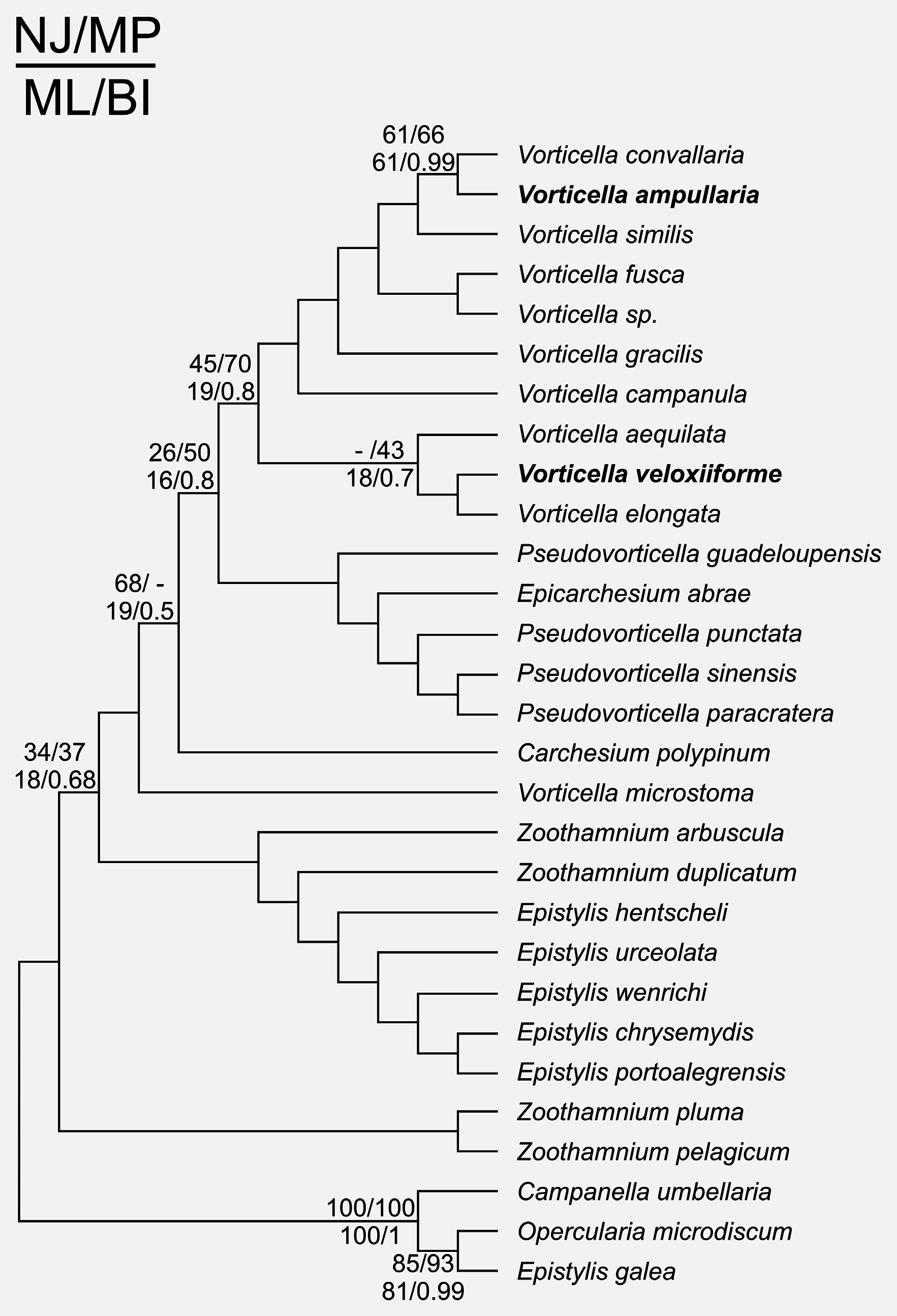
Molecular analyses Vorticella veloxiiforme n.sp. and V. ampullaria n.sp. clustered with other Vorticella species within the peritrich order Vorticellida ( Utz et al. 2010) ( Fig. 6 View FIGURE 6 ). V. ampullaria consistently clustered with V. convallaria with all methods, a relationship that received moderate to high support (61–66% bootstrap support [BS] in ML, NJ and MP; 0.99 posterior probability [PP] in the BI). The position of V. veloxiiforme was less stable, with most methods indicating a closer relationship with V. aequilata and V. elongata , albeit with limited support [18 and 43% BS in ML and MP, respectively; 0.74 PP in BI]).
No known copyright restrictions apply. See Agosti, D., Egloff, W., 2009. Taxonomic information exchange and copyright: the Plazi approach. BMC Research Notes 2009, 2:53 for further explanation.