
Stylopoma magnovicellata Silén, 1954
|
publication ID |
https://doi.org/ 10.11646/zootaxa.5379.1.1 |
|
publication LSID |
lsid:zoobank.org:pub:430102D2-4EAA-41B3-B57F-CC532F929DA3 |
|
DOI |
https://doi.org/10.5281/zenodo.10248931 |
|
persistent identifier |
https://treatment.plazi.org/id/4B6E902E-FF94-FFD5-FF46-FEFA1824FD0B |
|
treatment provided by |
Plazi |
|
scientific name |
Stylopoma magnovicellata Silén, 1954 |
| status |
|
Stylopoma magnovicellata Silén, 1954 View in CoL
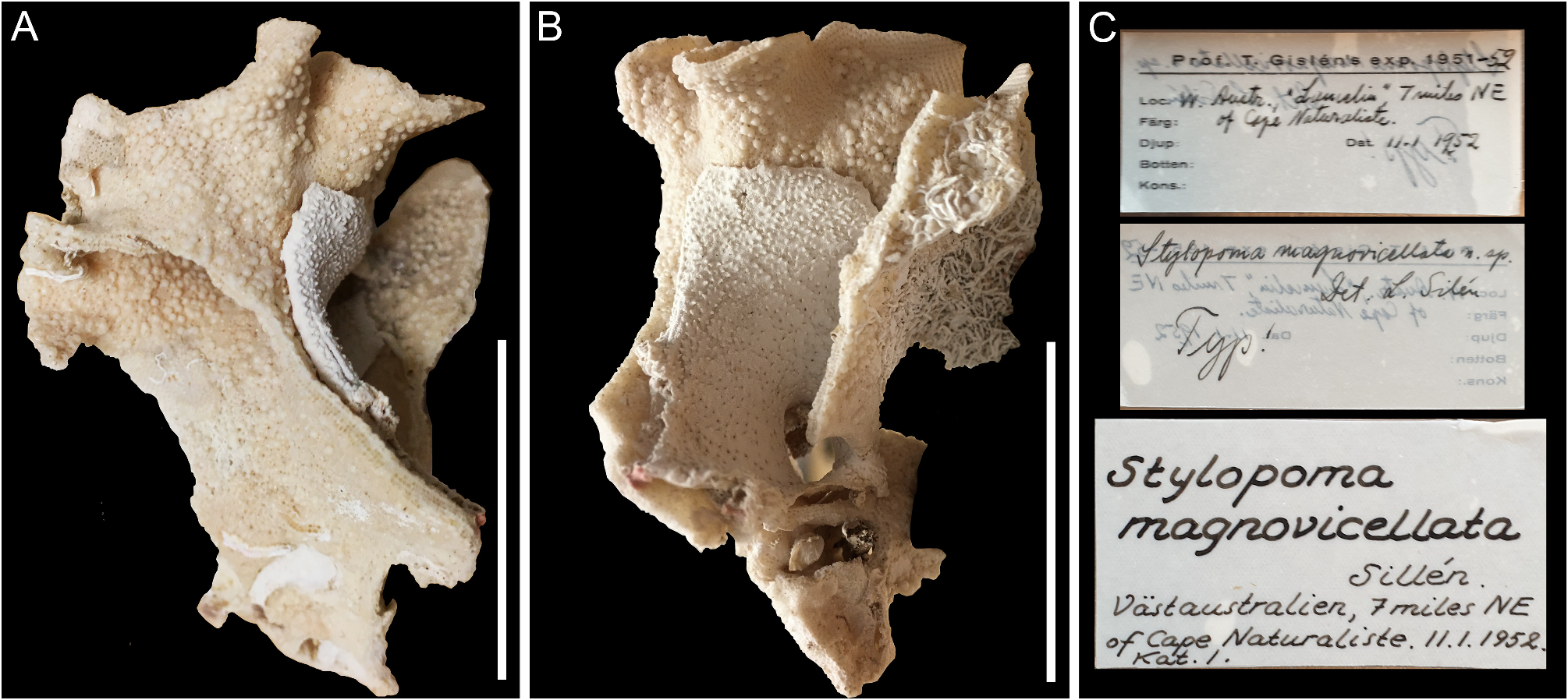
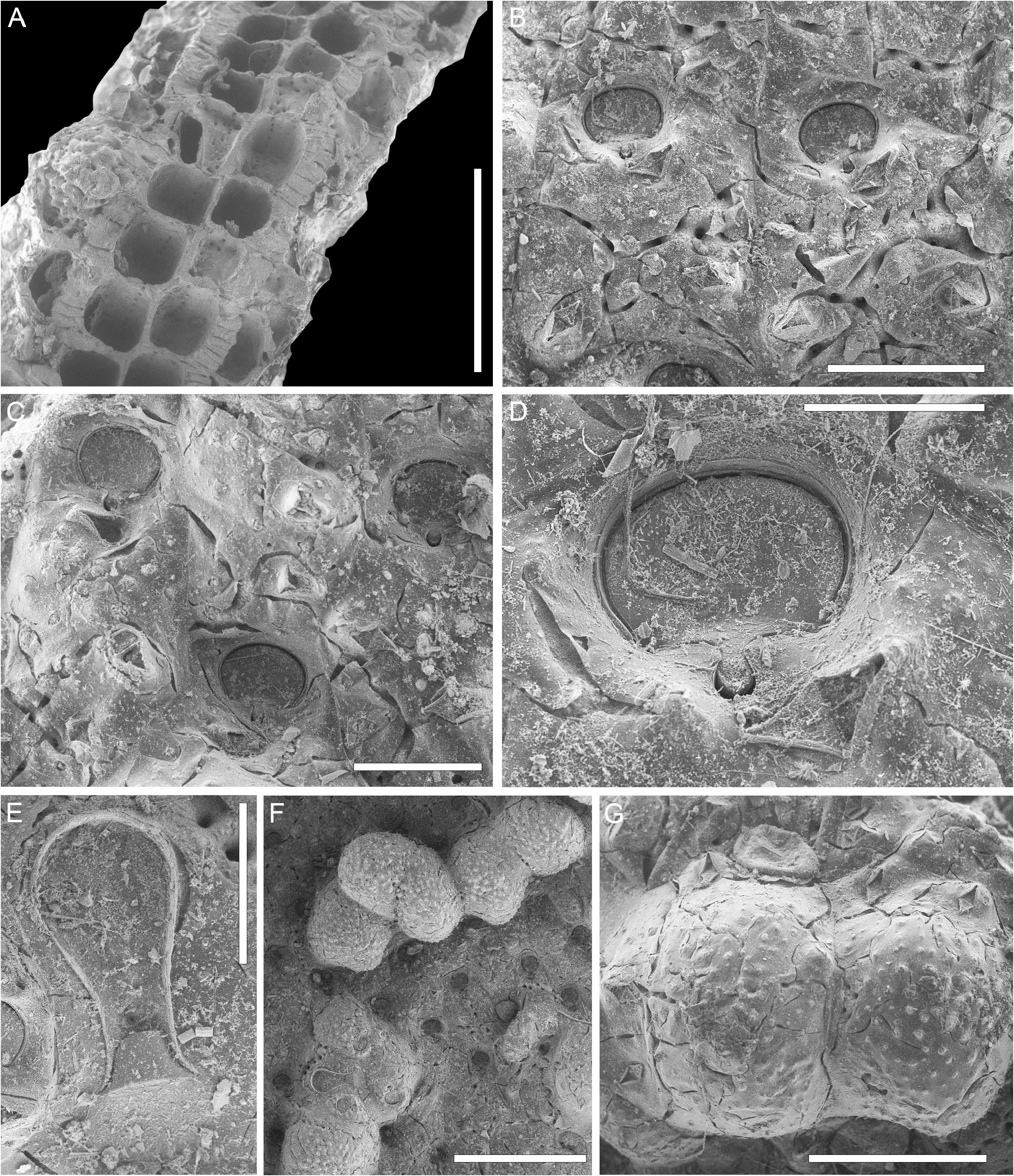
( Figs 33 View FIGURE 33 , 34 View FIGURE 34 ; Table 31)
Stylopoma magnovicellata Silén, 1954: 18 View in CoL , fig. 5, pl. 1, figs 2, 3.
Material examined. Holotype by monotypy LUZM 52 , south-western region of Western Australia, 7 miles NE of Cape Naturaliste. Leg. Prof. T. Gislén , Australia Expedition 1951–1952, collected 11.1.1952.
Description. Colony erect, rigid formed by bifoliate or multilayered, convoluted fronds; the holotype specimen 5.8 × 4 × 3 cm in size, with bifoliate fronds developing an additional layer per side, about 1.5 mm thick ( Figs 33 View FIGURE 33 , 34A View FIGURE 34 ).
Autozooids rectangular, hexagonal or irregularly polygonal, longer than wide (mean L/ W 1.45), distinct with boundaries traced by thin furrows, initially quincuncially arranged but the arrangement becoming irregular as a consequence of frontal budding ( Fig. 34A, B View FIGURE 34 ); interzooidal communications through a continuous series of small, circular pores visible on the inner vertical walls, 12–15 µm in diameter; in transverse section ( Fig. 34A View FIGURE 34 ), autozooids on the first two layers arranged back-to-back but irregularly arranged on subsequent layers; basal wall separating the two layers of autozooids generally thin, 25–55 µm. Frontal shield about 150 µm thick ( Fig. 34A View FIGURE 34 ), flat to slightly convex, granular with granules arranged in radial ridges and circular pseudopores, 10–15 µm in diameter, occupying furrows between the ridges ( Fig. 34B, C View FIGURE 34 ).
Orifice wider than long with horseshoe-shaped anter and drop-shaped sinus ( Fig. 34D View FIGURE 34 ).
Adventitious avicularia teardrop-shaped with raised, acutely triangular rostrum and mandible, and complete crossbar, 2–3 per zooid: one avicularium constantly placed below the orifice, medially adjacent to the sinus or laterally on either side, directed distolaterally ( Fig. 34C, D View FIGURE 34 ); 1–2 avicularia on the frontal shield variously arranged, either both placed proximally with one at each corner directed proximolaterally, or one placed at the lateral corner at about zooidal mid-length directed laterally and the other at the proximal corner directed proximolaterally, either both on the same side or one on each side of the zooid ( Fig. 34B, C View FIGURE 34 ).
Vicarious avicularia rare (only one observed), spatulate, 405 × 195 µm, crossbar complete, cystid polygonal having the same size of an autozooid (520 µm long by 380 µm wide) ( Fig. 34E, F View FIGURE 34 ).
Ovicell globular, large, covering the orifice of the maternal zooid and the frontal shield of the distal and the two lateral zooids; often ovicells of neighbouring zooids fusing together along the lateral margins ( Fig. 34F View FIGURE 34 ). Ooecium coarsely granular (granules about 25–40 µm in diameter), with sparse circular pseudopores (10 µm in diameter), and a row of slightly larger marginal pores (15 µm in diameter); avicularia similar in size and shape to those observed on the frontal shield present along the margins of the ooecium ( Fig. 34G View FIGURE 34 ).
Remarks. Thirteen species of Stylopoma are known from Australian waters ( Cook et al. 2018). In the most recent and complete revision of Stylopoma species from the Indo-West Pacific, Tilbrook (2001) did not include Stylopoma magnovicellata because no material of this species was available in the zoological bryozoan collection of the Natural History Museum, London. However, Tilbrook (2001) mentioned the species, although with the specific name misspelled as S. magniovicellata , with the invitation to clarify its exact identity. Bock (2023) illustrates the species (see http://bryozoa.net/cheilostomata/schizoporellidae/stylopoma _magnovicellata .html accessed 26.05.2023) and specifies that the figured specimens was previously misidentified as S. duboisii ( Audouin, 1826) . This suggests that the lack of additional records for this species might be due to its misidentification. The morphological characters on the SEM image of Bock (2023) match perfectly with those observed in the type specimen.
A fossil species described from the Miocene (Burdigalian) Quilon Formation (Kerala, India) as Stylopoma aff. magniovicellata Silén (again with the specific name misspelled) is likely a different species. Sonar & Badve (2019) pointed out that the Miocene taxon has a more convex frontal and a higher number of pseudopores compared with the nominal species. Additional differences are the number and arrangement of adventitious avicularia on the frontal (1–2 avicularia in S. aff. magniovicellata , both placed lateral to the orifice and distolaterally directed), as well as the size of the ovicell, much smaller in the fossil species (OvL 0.32–0.33, OvW 0.31–0.32).
| T |
Tavera, Department of Geology and Geophysics |
No known copyright restrictions apply. See Agosti, D., Egloff, W., 2009. Taxonomic information exchange and copyright: the Plazi approach. BMC Research Notes 2009, 2:53 for further explanation.
|
Kingdom |
|
|
Phylum |
|
|
Class |
|
|
Order |
|
|
Family |
|
|
Genus |
Stylopoma magnovicellata Silén, 1954
| Martino, Emanuela Di 2023 |
Stylopoma magnovicellata Silén, 1954: 18
| Silen, L. 1954: 18 |