
Liphistius gracilis, Peter J. Schwendinger, 2017
|
publication ID |
https://doi.org/10.5281/zenodo.893555 |
|
DOI |
https://doi.org/10.5281/zenodo.6042355 |
|
persistent identifier |
https://treatment.plazi.org/id/4C30A452-FFEA-FFE4-BB5A-F8773AF2FD9E |
|
treatment provided by |
Plazi (2017-10-18 21:12:42, last updated 2024-11-26 07:45:54) |
|
scientific name |
Liphistius gracilis |
| status |
sp. nov. |
Liphistius gracilis View in CoL sp. nov.
Figs 8-9 View Fig. 8 View Fig. 9
Types : MHNG, sample SIM-01/07; male holotype (matured 13.VIII.2001) , 5 male paratypes (matured 18.IX., 19.IX.2001, 5.I., 24.III., 3.VI.2002), 5 female paratypes; Malaysia, Johor, Kota Tinggi Waterfalls at foot of Gunung Muntahak , 170 m (evergreen rain forest); 24.-26.VI.2001, leg. P.J. Schwendinger. – SMF, sample TM-14/02; 1 male paratype (matured 5.XI.2016); same locality; 21.-22.VI.2014; leg. P.J. Schwendinger. – MHNG, SMF, sample Sum-00/01; 3 female paratypes [moulted 3.V. (allotype); early VIII.; 26.XII.2000; 16.V., 6.X.2001]; same locality; 4.II.2000; leg. P.J. Schwendinger.
Etymology: The species epithet is a Latin adjective meaning “gracile”, “slender”, “slim”, referring to the small body size of this species in comparison with L. malayanus and L. endau .
Diagnosis: Similar to L. malayanus and L. endau but much smaller in body size, with lighter body colouration and annulated legs and palps. Males distinguished from those of L. malayanus ( Fig. 4 View Fig. 4 ) and L. endau ( Fig. 6 View Fig. 6 ) by tibial apophysis of palp longer and directed more distad ( Fig. 8A, C View Fig. 8 ), carrying shorter megaspines, the dorsal one sitting clearly lower than the rest ( Fig. 8B View Fig. 8 , H-J); paracymbium relatively deeper than in L. malayanus and shallower than in L. endau ( Fig. 8 View Fig. 8 A-C cf. Fig. 4A View Fig. 4 and Fig. 6B View Fig. 6 , respectively); distal edge of contrategulum serrate ( Fig. 8A View Fig. 8 , D-G), its apex distinctly smaller ( Fig. 8 View Fig. 8 F-G cf. Fig. 4 View Fig. 4 G-L and Fig. 6F View Fig. 6 , H-O); dorsal wall of sclerotised part of embolus proper ending in a sharply prodorsad-bent lobe ( Fig. 8 View Fig. 8 D-G) [both walls equally wide in L. malayanus ( Fig. 4 View Fig. 4 G-H); dorsal wall prodorsal-curved (not sharply bent) in L. endau ( Fig. 6F, H View Fig. 6 )]. Vulva of L. gracilis sp. nov. ( Fig. 9 View Fig. 9 ) different from vulvae of L. malayanus ( Fig. 5 View Fig. 5 ) and L. endau ( Fig. 7 View Fig. 7 ) by ventral side of poreplate without bulging lateral and posterolateral margins, thus transition between poreplate and posterior stalk much less marked; anterior margin of poreplate with less distinct anterolateral lobes; receptacular cluster wider than long, more distinctly protruding beyond anterior margin of poreplate. Vulva different from that of L. johore ( Fig. 10A View Fig. 10 ) by lacking pronounced anterolateral invatinations in the margin of the poreplate, by having a wide and indistinct transition between poreplate and posterior stalk (narrow and distinct in L. johore ), any by possessing hairs in the genital atrium.
Description of male (holotype): Colour in alcohol (darker in life): Carapace mostly light brown, with darker patches along margins (medially broken on posterior margin), extending between coxal elevations; dark anterior margin enclosing very dark eye mound; dark W-shaped pattern behind eye mound indistinct; a few small dark spots on pars cephalica; a paramedian pair of dark patches anterior to fovea; chelicerae creamcoloured proximally, grey-brown distally; legs and palps mostly light brown, with darker zone proximally on patellae and tibiae of legs III-IV, and in proximal half of all metatarsi. Opisthosomal tergites cream-coloured, with dark marginal and central patches on tergite I, other tergites only with lateral and anterolateral patches; membranous part of opisthosoma cream-coloured. Sclerotised parts of ventral side of body light brown.
Bristles on carapace: Short bristles along all margins (strongest on posterior margin, longest behind, on and in front of eye mound), on coxal elevations and in area behind fovea, none anterior to fovea.
Cheliceral teeth: Ten and eleven small ones on promargin of cheliceral groove of right and left chelicera, respectively.
Scopula: Tarsus I with very thin scopula confined to distal fifth of ventral side, divided for its entire length by narrow pale, glabrous longitudinal median stripe and by some bristles; tarsus II with very thin scopula in distal quarter, only distally divided by median stripe; tarsus III with dense scopula covering distal half, only distally divided by median stripe; tarsus IV with dense scopula covering distal three-fifths, only distally divided by median stripe.
Claws: Paired tarsal claws on anterior legs with 4-6 denticles, on posterior legs with 5-6 denticles; unpaired claws in most cases with a single denticle on anterior legs, none on posterior legs.
Palp: Tibial apophysis relatively long, situated at distal margin of tibia, inclined distad [ Fig. 8A View Fig. 8 (showing paratype matured 5.I.2002), C], carrying four quite short megaspines, the dorsal one rising from a clearly more proximal position than the others [ Fig. 8B, H View Fig. 8 (showing paratype matured 5.I.2002), I-J]. Both apical lobes of cymbium indistinct, prodorsal one more rounded than retrodorsal one ( Fig. 8D View Fig. 8 ). Paracymbium relatively large and basally deep [ Fig. 8 View Fig. 8 A-B (showing paratype matured 5.I.2002), C-D], its cumulus indistinctly elevated, carrying moderately long stiff bristles [ Fig. 8 View Fig. 8 A-B (showing paratype matured 5.I.2002), C]. Subtegulum without apophysis. Tegulum short and wide, coarsely dentate along entire proximal margin, not or only indistinctly connected to contrategulum on retrodorsal side ( Fig. 8E View Fig. 8 ). Contrategulum without ventral process; distal edge finely serrate, proventrally ending in a U-shaped row of denticles ( Fig. 8D View Fig. 8 ), dorsally ending in a spatulate asymmetrical apex ( Fig. 8F View Fig. 8 ). Para-embolic plate only little elevated ( Fig. 8B View Fig. 8 , showing paratype matured 5.I.2002); sclerotized part of embolus proper without longitudinal keels or ribs and not carrying denticles ( Fig. 8 View Fig. 8 A-B, showing paratype matured 5.I.2002), its dorsal wall distinctly wider than its ventral wall, abruptly bent prodorsad approximately at a right angle ( Fig. 8F View Fig. 8 ) and ending in a pronounced angular lobe ( Fig. 8 View Fig. 8 D-E); membranous part of embolus proper apically widened and lying on lobe of dorsal wall of sclerotised part ( Fig. 8D View Fig. 8 ).
Measurements: Total length 11.32; carapace 4.67 long, 3.96 wide; opisthosoma 4.83 long, 3.64 wide; eye mound 0.68 long, 0.80 wide, AME well-developed; palpal coxae 1.38 long, 0.95 wide; labium 0.40 long, 0.79 wide; sternum 2.37 long, 1.50 wide (0.83 on ventral surface); palp 7.99 long (2.53 + 1.34 + 2.81 + 1.31); leg I 15.97 long (4.35 + 1.90 + 3.40 + 4.03 + 2.29); leg II 17.01 long (4.43 + 1.90 + 3.56 + 4.63 + 2.49); leg III 18.92 long (4.55 + 1.90 + 4.00 + 5.78 + 2.69); leg IV 23.89 long (5.38 + 2.06 + 5.10 + 8.03 + 3.32).
Description of female (allotype): Colour in alcohol (darker in life): As in male but with a more pronounced dark pattern on carapace, resulting in a more distinct W-shaped pattern behind eye mound, and with a pronounced, light brown, flower-shaped pattern on pars thoracica; all opisthosomal tergites with larger dark patches, tergites II-V additionally with dark anteromedian patches. Legs light brown, with dark proximal and subapical annulations on femora (also on palpal femur), tibiae, metatarsi (indistinct on posterior legs) and on tarsi (indistinct on posterior legs). Palpal tibia with dark proximal and apical annulation; tarsus mostly dark, proventral-distal area lighter.
Bristles on carapace: Slightly stronger and longer (especially in front of eye mound) than in male.
Cheliceral teeth: Eleven strong cheliceral teeth on promargin of cheliceral groove on both sides.
Claws: Paired tarsal claws on legs with mostly 4-5 denticles (one claw with only two); unpaired claws with two denticles on legs I-III, 1-2 on leg IV. Tarsi without scopula.
Vulva: Vulval plate ( Fig. 9 View Fig. 9 A-B) distinctly sclerotised and pigmented, wider than long. Genital atrium uniformly flat, its lateral parts not bulging on dorsal side, with several lateral and median hairs. Posterior stalk wide, indistinctly perforated with tiny micropores; transition to poreplate indistinct, only slightly constricted. Anterior margin of poreplate slightly to distinctly invaginated, with indistinct anterolateral lobes, without anterolateral invaginations; ventral side of poreplate without bulging lateral and posterolateral margins. CDO large, undivided, irregularly quadrangular, close to anterior margin of poreplate. Receptacular cluster clearly wider than long, deep, distinctly projecting beyond anterior margin of poreplate.
Measurements: Total length 13.53; carapace 5.34 long, 4.19 wide; opisthosoma 5.70 long, 4.15 wide; eye mound 0.74 long, 0.83 wide; palpal coxae 1.74 long, 1.27 wide; labium 0.63 long, 1.27 wide; sternum 2.53 long, 1.86 wide (0.91 on ventral surface); palp 8.98 long (3.01 + 1.58 + 2.14 + 2.25); leg I 11.82 long (3.56 + 1.94 + 2.73 + 2.25 + 1.34); leg II 12.22 long (3.60 + 1.98 + 2.73 + 2.49 + 1.42); leg III 12.74 long (3.60 + 1.98 + 2.45 + 3.05 + 1.66); leg IV 16.41 long (4.27 + 2.10 + 3.24 + 4.55 + 2.25).
Variation: Carapace lengths in males (n=7) 4.36-4.75, in females with a well-developed vulval plate (n=9) 4.29-5.34, in the smallest female with an egg case 4.66. Carapace widths 3.64-4.06, 3.11-4.22 and 3.51, respectively.
In four males (including the holotype) the scopula extends over the distal half of the ventral side of tarsus III and over the distal three-fifths of tarsus IV, in the other three males it extends over the distal three-fifths of tarsus III and over the distal two-thirds of tarsus IV; on legs I-II the extent of the scopula is the same in all males. Variation in the shape of the tibial apophysis of the male palp is shown in Fig. 8 View Fig. 8 A-C, H-J; variation in the shape of the distal edge of the contrategulum in Fig. 8 View Fig. 8 F-G; variation in the shape of vulval plates of the females examined in Fig. 9 View Fig. 9 . Two female paratypes ( Fig. 9C, E View Fig. 9 ), but not the allotype ( Fig. 9A View Fig. 9 ), have a moderately pronounced anterolateral lobe followed by a slight anterolateral invagination on each side of the poreplate. The largest (oldest) female ( Fig. 9A View Fig. 9 ) has more hairs in the genital atrium than smaller (younger) females ( Fig. 9E View Fig. 9 ). The posterior margin of the genital sternite of females is almost straight to distinctly invaginated ( Fig. 9 View Fig. 9 A-F).
Distribution: The types were collected from the surroundings of the Kota Tinggi Waterfall, at the foot of Gunung Muntahak, north of Kota Tinggi, in Johor State ( Fig. 1 View Fig. 1 , locality 8). Burrows of probably the same species were seen along the trail to the nearby Pelepah Waterfall. Two juveniles (don J. Kral) from Gunung Belumut, about 32 km further northwest, may also belong to this species.
Biology: At the Kota Tinggi Waterfall L. gracilis sp. nov. occurs together with the much larger L. endau which belongs to the same species-group. Burrows of the two species were seen only a few centimetres from each other. Liphistius endau was exclusively found very close to the waterfall and to the stream, L. gracilis sp. nov. also further away, but both were absent from high earthbanks (old road sides) in a palm oil plantation adjacent to the rain forest. Most burrows were simple, equipped with a single trapdoor and dug into the soil; a few small ones were sac-like nests with two trapdoors, constructed on the moss-covered surface of rock (as also known from cave species and from juveniles of forest-dwelling species; see Schwendinger, 1987). The trapdoors of seven penultimate males were 1.1-1.3 cm long and 1.6-1.9 cm wide; that of the largest female 1.3 and 2.0, respectively. All burrows had a maximum of eight, relatively long signal lines running over soil, stones and tree roots; the longest signal line (of the largest female) was 21 cm long.
Four males collected in June became adult in August to November of the same year; two males reached maturity in January and February after over 1.5 years in captivity (thus probably not corresponding to conditions in nature). In early July and early August two females built egg cases in captivity, 1.7-2.0 cm in diameter and 1.1-1.4 cm high, containing 30 and 36 eggs suspended on a thin layer of fine silk strands. Adult females moulted twice per year, in May and again in August to November.
lateral and posterolateral margins on ventral side ( Platnick & Sedgwick, 1984: fig. 80), with relatively large pores on dorsal side, with a large CDO (at its posterior margin level with the poreplate), and with a large receptacular cluster projecting far beyond anterior margin of poreplate; genital atrium without hairs, including a strongly trapezium-shaped (posteriorly wide, anteriorly very narrow, posterior margin almost straight) posterior stalk ( Fig. 10A View Fig. 10 ; Platnick & Sedgwick, 1984: figs 79-80).
Male: Unknown.
Female: This species was described on the basis of only the female holotype. For an easy comparison with other species treated here, the dorsal aspect of the vulval plate was re-drawn from Platnick & Sedgwick (1984: fig. 79) and is shown in Fig. 10A View Fig. 10 .
Relationships: The vulval plate (with a large CDO, with a huge receptacular cluster projecting far beyond the anterior margin of the well-sclerotised poreplate and with a well-sclerotised posterior stalk fully connected to the poreplate) indicates that L. johore is a member of the malayanus -group. Relatively small size, the presence of anterolateral invaginations on the poreplate (also present, but less distinct, in some females of L. gracilis sp. nov.; Fig. 9C, E View Fig. 9 ) and the apparent absence of bulged lateral and posterolateral margins on the ventral side of the poreplate (not clearly evident from original illustrations of the vulval plate of the holotype, see Platnick & Sedgwick, 1984: fig. 80) indicate that L. johore and L. gracilis sp. nov. are more closely related to each other than to L. malayanus and L. endau .
Distribution and remarks: The type locality is given as “Sungai Rengit, Pengarang” ( Fig. 1 View Fig. 1 , locality 9). Kampung Sungai Rengit and the Pengarang ferry Liphistius johore Platnick & Sedgwick, 1984
Fig. 10A View Fig. 10
Liphistius johore Platnick & Sedgwick, 1984: 30 View in CoL , figs 79-80
(description of female).
Type material: Bernice Pauahi Bishop Museum, Honolulu, USA; female holotype (not examined); Malaysia, Johore [sic], Sungai Rengit, Pengarang ; 19.XII.1961; leg. K.J. Kunchuna.
Material examined: None.
Diagnosis: Seemingly a fairly small species (carapace length of female holotype 5.15, carapace width 4.00). Female with annulated legs and yellow-coloured to orange-coloured carapace and opisthosomal tergites ( Platnick & Sedgwick, 1984: 30); vulva with a wide poreplate with a distinct anterolateral invagination on each side of anterior margin, seemingly without bulging terminal and immigration post (with nearby remains of the “Pengarang Battery”, a British defence post overrun by the Japanese Army during the fall of Singapore in World War II) are about 15 km apart. I spent a full day searching in both the fairly undisturbed forest on the publicly accessible little hill next to the Pengarang ferry terminal (the nearby, distinctly higher and nicely forested Bukit Pengarang lies on the land of a large Malaysian naval base and is off limits to civilians and foreigners) and in the strongly disturbed forest of Bukit Pelali, about 5 km north of Kampung Sungai Rengit, but found no traces (not even empty burrows) of Liphistius .
Platnick N. I., Sedgwick W. C. 1984. A revision of the spider genus Liphistius (Araneae, Mesothelae). American Museum Novitates 2781: 1 - 31.
Schwendinger P. J. 1987. On the male of Liphistius trang (Araneae: Mesothelae), with notes on the natural history of the species. Natural History Bulletin of the Siam Society 35: 19 - 25.
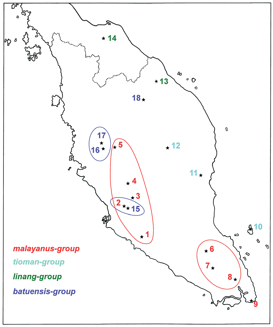
Fig. 1. Localities of Liphistius species of the malayanus - group, tioman - group, linang - group and batuenis - group in peninsular Malaysia and southern Thailand (coast of Sumatra omitted): 1 - Gunung Angsi (type locality of L. malayanus); 2 - Templer Park and Gua Anak Takun (L. malayanus, L. batuensis); 3 - Genting Highlands (L. malayanus); 4 - Fraser’s Hill (L. malayanus); 5 - Cameron Highlands (type locality of L. malayanus cameroni); 6 - Sungai Jasin in Endau Rompin National Park (type locality of L. endau); 7 - Gunung Belumut (L. endau); 8 - Gunung Muntahak (L. endau; type locality of L. gracilis sp. nov.); 9 - Sungai Rengit (type locality of L. johore); 10 - Gunung Kajang on Tioman Island (type locality of L. tioman); 11 - Gua Charas in Bukit Charas (type locality of L. panching); 12 - Nusa Camp in Taman Negara (type locality of L. negara sp. nov.); 13 - Jeram Linang Waterfall (type locality of L. linang sp. nov.); 14 - Sankalakhierie Mountains (type locality of L. indra); 15 - Batu Caves (type locality of L. batuensis); 16 - Gua Tempurung (type locality of L. tempurung); 17 - Gua Cicak (L. tempurung); 18 - Gua Keris (type locality of L. priceae sp. nov). Localities with conspecific populations are encircled. Colours distinguish species groups.

Fig. 4. Liphistius malayanus, details of palp of four males: Templer Park (A, C-D, H-I); holotype of L. malayanus cameroni (B, E, G); Cameron Highlands, ZRC (F, J), Fraser’s Hill, MHNG (K-L). (A) Paracymbium and tibial apophysis of left palp, retroventral view. (B) Tibial apophysis of right palp, ventral view. (C) Distal part of left palpal organ, proventral view (arrow indicating V-shaped row of denticles at proventral end of distal edge of contrategulum). (D, F) Same, retrodorsal and slightly proximal view. (E) Same, retrodorsal view. (G-H) Left palpal organ, distal view (dorsal side up). (I, K) Distal edge of contrategulum of right palp, distal view (dorsal side to the left). (J) Same, distal and slighly prolateral view. (L) Same of left palp, distal and slighly prolateral view (dorsal side to the right). Abbreviations: a - dorsal apex of contrategulum; de - distal edge of contrategulum. Scale lines: 1.0 mm (A; B; C-D; E, G; F; H-L).

Fig. 5. Liphistius malayanus, vulval plate of eight females: paratype of L. m. cameroni from Berinchang, Cameron Highlands (A- B); exuvia of female from Ringlet, Cameron Highlands (C); female (SMF 64093) from Fraser’s Hill (D); female (moulted 24. VI. 2002) from Fraser’s Hill; poreplate slightly deformed by brief immersion in cold KOH (E); female (SMF 56206) from Fraser’s Hill (F); female (SMF 60037) from Genting Highlands (G); female (moulted 8. XII. 2005) from Genting Highlands (H); exuvia of female from Templer Park (I-J). (A, C-I) Dorsal view. (B, J) Ventral view. Scale lines: 1.0 mm (A-B; C, F; D; E, G; H; I-J).
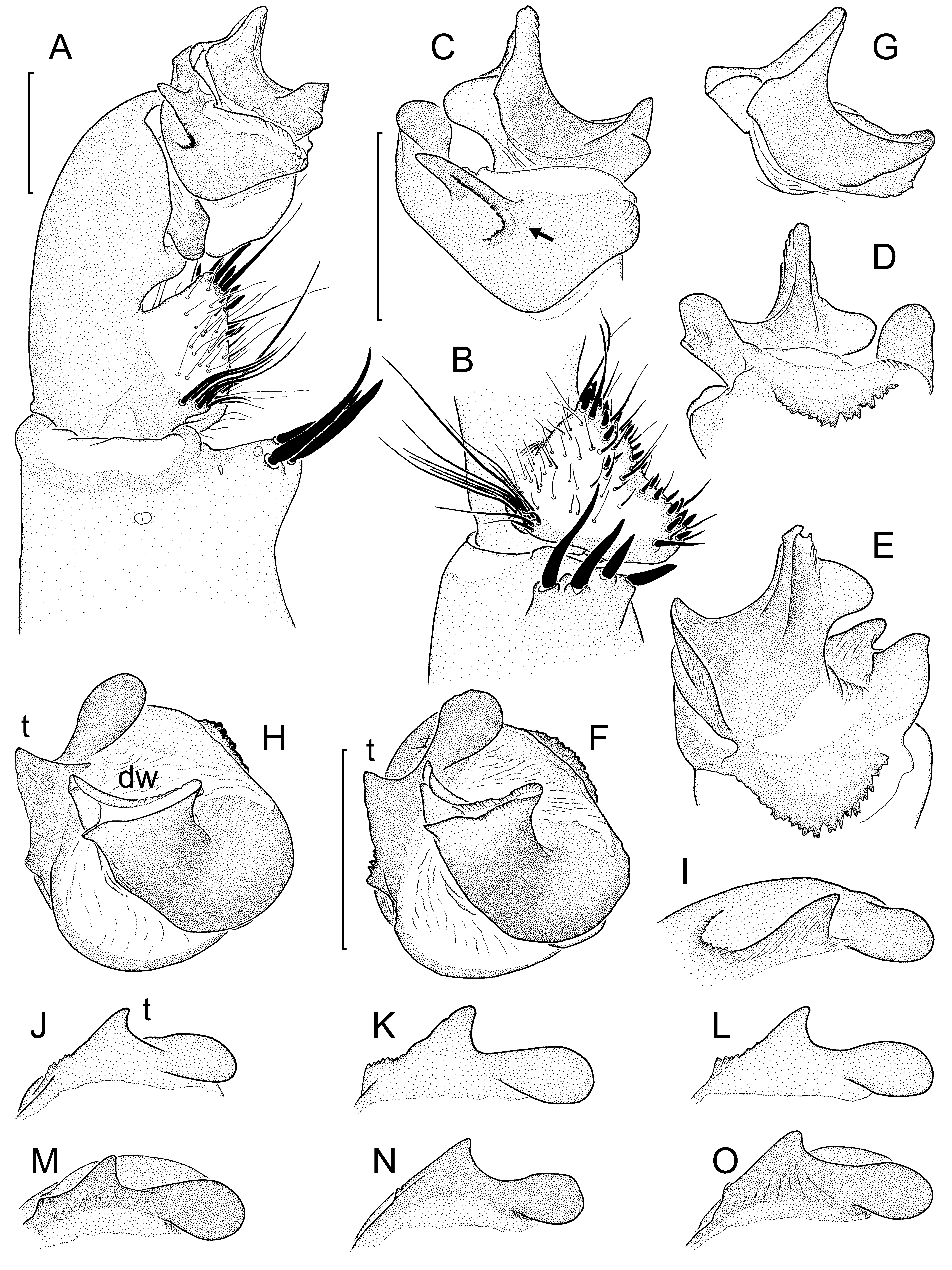
Fig. 6. Liphistius endau, details of left palp of eight males: from type locality, matured 27. II. 2002 (A-F); from type locality, matured 1. II. 2002 (G-I); from unknown locality, died 22. III. 2012 (J); from unknown locality, killed 30. IX. 2010 (K); from Gunung Belumut (L); from Kota Tinggi, matured 22. X. 2014 (M); same place, matured 20. VIII. 2014 (N); same place, matured 6. XI. 2014 (O). (A) Distal part of palp, ventral view. (B) Paracymbium and tibial apophysis, retroventral view. (C) Palpal organ, proventral view (arrow indicating U-shaped row of denticles at proventral end of distal edge of contrategulum). (D) Same, retrodorsal and slightly proximal view. (E) Same, retrodorsal view. (F, H) Same, distal view (dorsal side up). (G) Embolus complex, proventral and slightly distal view. (I) Distal edge of contrategulum, distal and slighly prolateral view (dorsal side to the right). (J-O) Same, distal view. Abbreviations: dw - dorsal wall of sclerotized part of embolus proper; t - tooth on distal edge of contrategulum. Scale lines: 1.0 mm (A-B; C-E, G-H; F, I-O).

Fig. 7. Liphistius endau, vulval plate of six females: adult from unknown locality (A-B); largest female from Kota Tinggi (C); exuvia of reproductive female (moulted 27. IV. 2002) from type locality (D-E); exuvia of reproductive female (moulted 28. IV. 2002) from type locality (F-G); medium-sized juvenile from type locality (H); small juvenile from type locality (I). (A, C-D, F, H-I) Entire vulval plate, dorsal view. (B) Poreplate, ventral view. (E, G) Entire vulval plate, ventral view. Scale lines: 1.0 mm (A-B; C; D-E; F-G; H-I).
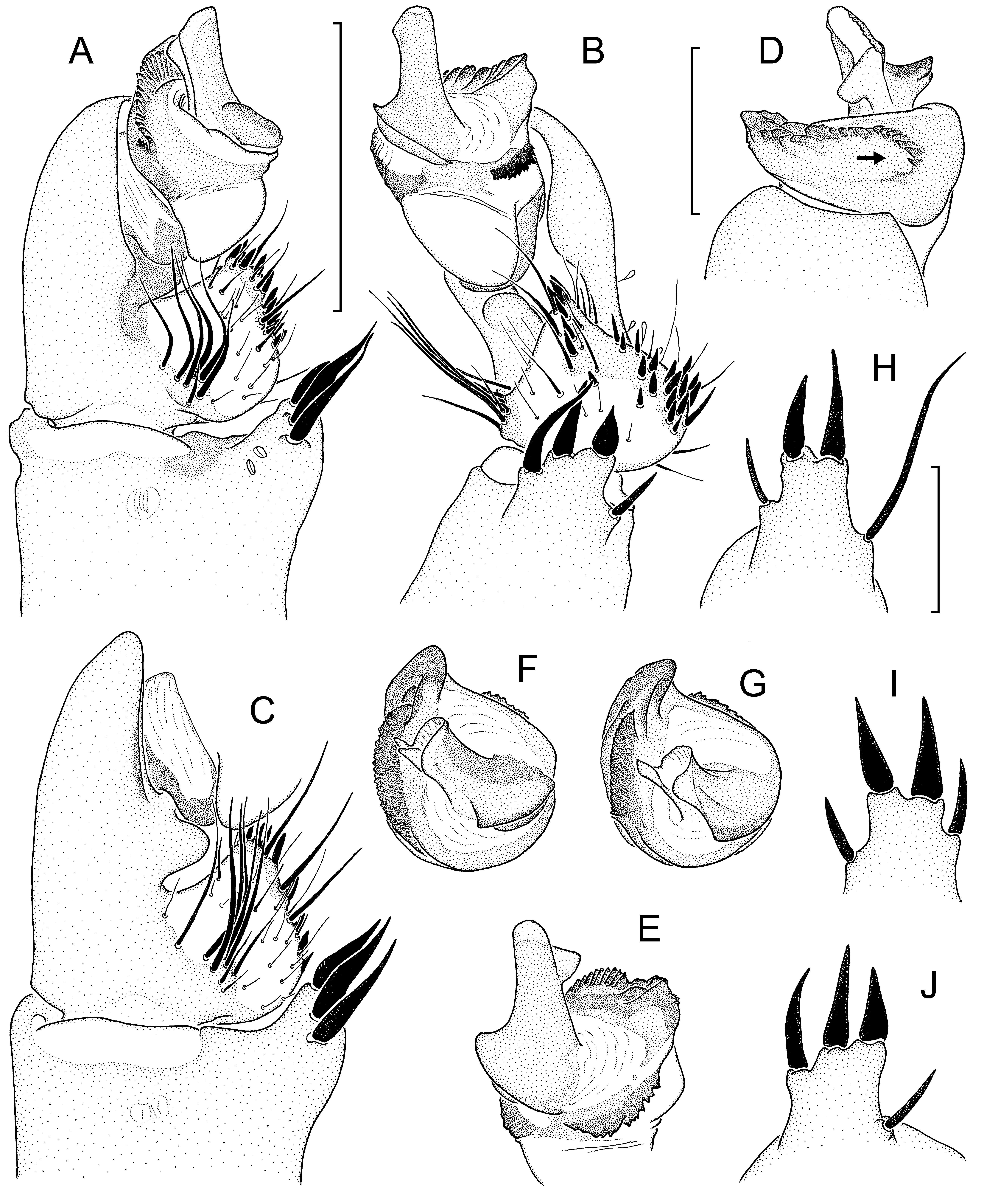
Fig. 8. Liphistius gracilis sp. nov., details of palp of three males: paratype, matured 5. I. 2002 (A-B, H); holotype (C-F, I-J); paratype, matured 19. IX. 2001 (G). (A) Distal part of palp, ventral view. (B) Same, retrolateral view. (C) Subtegulum, cymbium and distal part of palpal tibia, ventral view. (D) Distal part of cymbium and palpal organ, proventral view (arrow indicating U-shaped row of denticles at proventral end of distal edge of contrategulum). (E) Same, retrodorsal view. (F-G) Same, distal view (dorsal side up). (H-I) Right tibial apophysis, retrolateral and slightly proximal view. (J) Left tibial apophysis, same view. Scale lines: 1.0 mm (A-C), 0.5 mm (D-G; H-J).
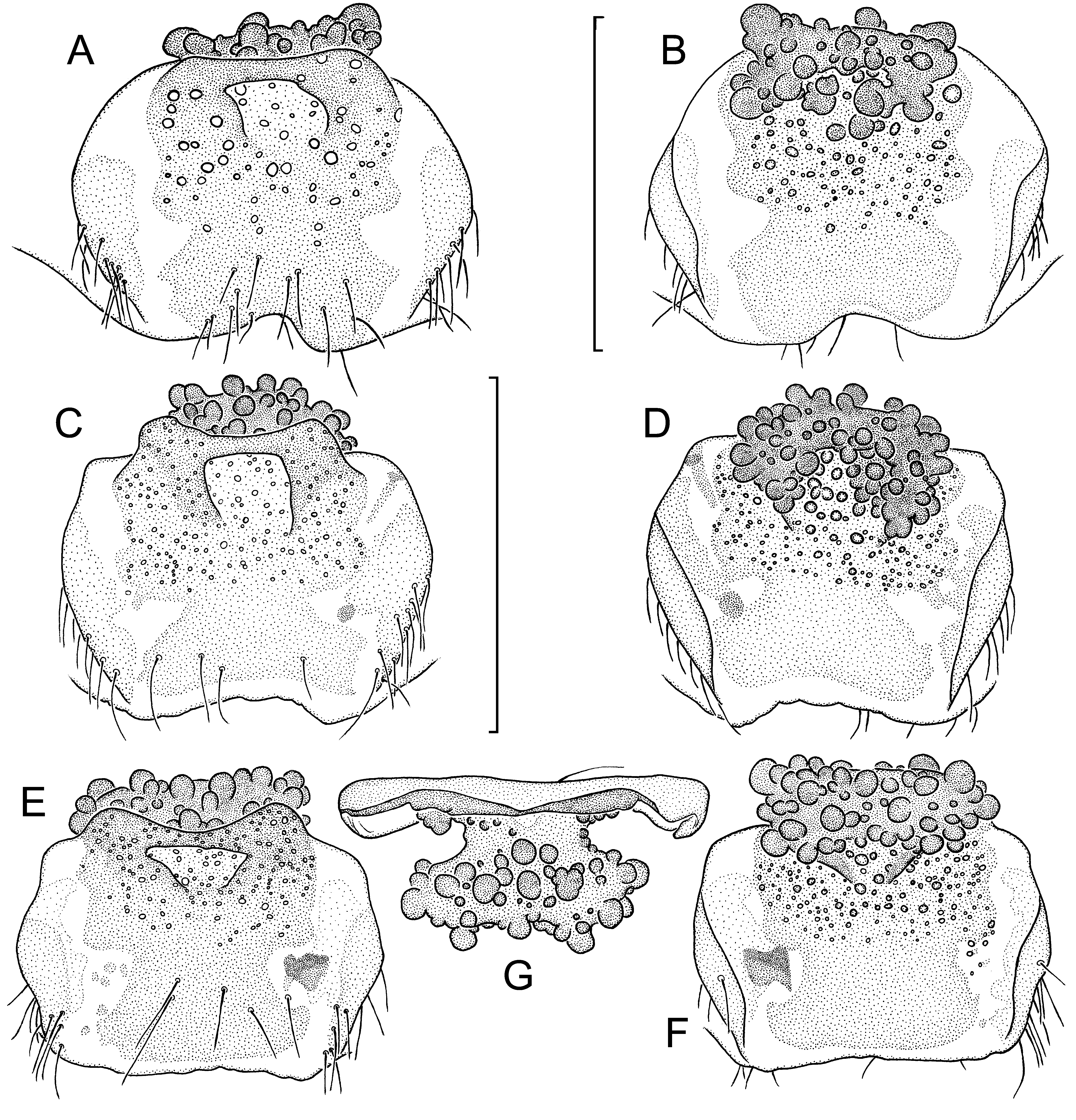
Fig. 9. Liphistius gracilis sp. nov., vulval plate of three females (all from exuviae): allotype (A-B); paratype, moult 31. VIII. 2001 (C- D); paratype, moult 27. IX. 2001 (E-G). (A, C, E) Vulval plate, dorsal view. (B, D, F) Same, ventral view. (G) Same, anterior view. Scale lines: 1.0 mm (A-B; C-G).

Fig. 10. Liphistius johore, female holotype (A); Liphistius panching, male from type locality (B). (A) Dorsal view of vulval plate (redrawn from Platnick & Sedgwick, 1984: fig. 79) (arrows indicating anterolateral invaginations in margin of poreplate). (B) Distal view of left palpal organ (dorsal side up) (redrawn from Sedgwick & Platnick, 1986: fig. 5).
| MHNG |
Museum d'Histoire Naturelle |
No known copyright restrictions apply. See Agosti, D., Egloff, W., 2009. Taxonomic information exchange and copyright: the Plazi approach. BMC Research Notes 2009, 2:53 for further explanation.
|
Kingdom |
|
|
Phylum |
|
|
Class |
|
|
Order |
|
|
Family |
|
|
Genus |
Liphistius gracilis
| Peter J. Schwendinger 2017 |
Liphistius johore
| Platnick N. & Sedgwick W. C. 1984: 30 |