
Phyllium (Phyllium) gantungense Hennemann, Conle, Gottardo & Bresseel, 2009
|
publication ID |
https://doi.org/ 10.11646/zootaxa.2322.1.1 |
|
persistent identifier |
https://treatment.plazi.org/id/4C724261-6C62-3A54-FF39-FB633772C2E7 |
|
treatment provided by |
Felipe |
|
scientific name |
Phyllium (Phyllium) gantungense Hennemann, Conle, Gottardo & Bresseel |
| status |
sp. nov. |
Phyllium (Phyllium) gantungense Hennemann, Conle, Gottardo & Bresseel View in CoL n. sp.
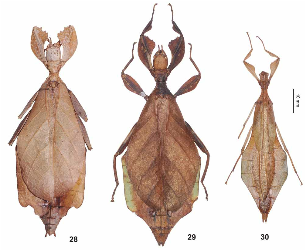
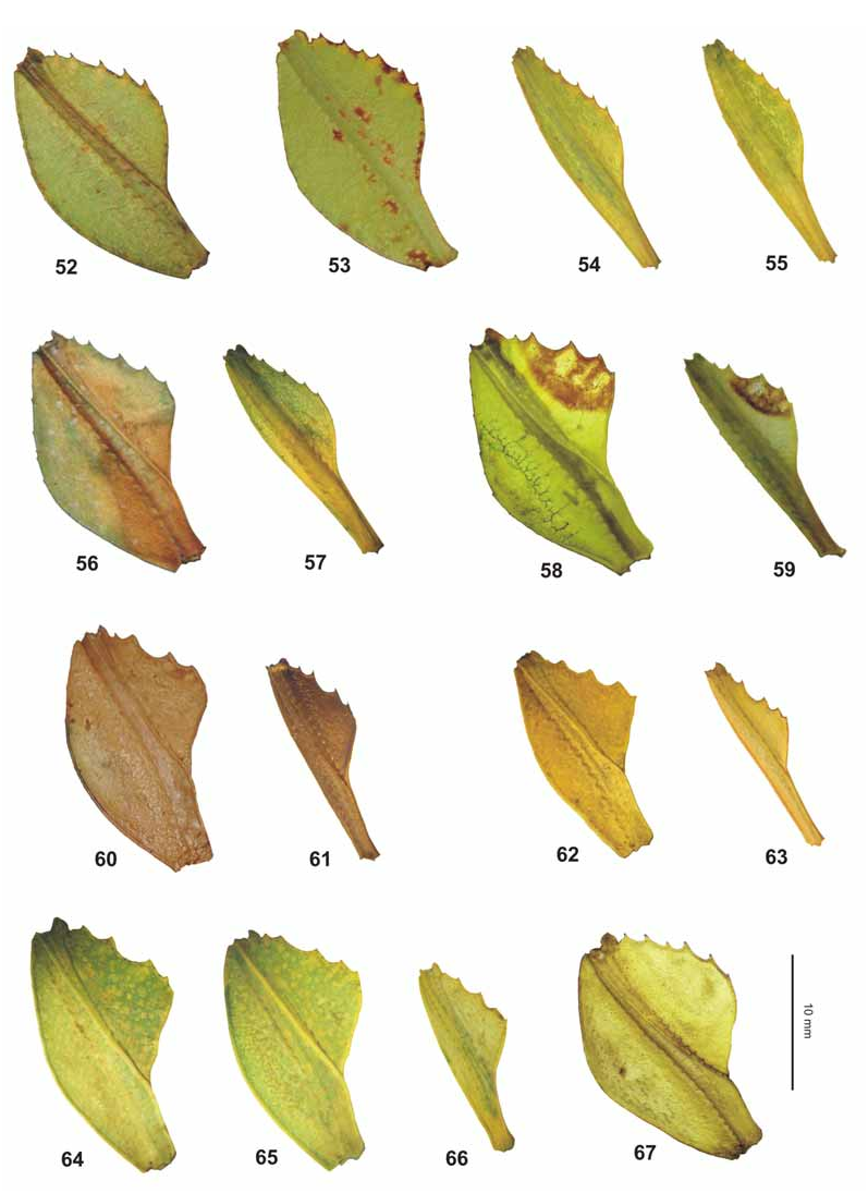
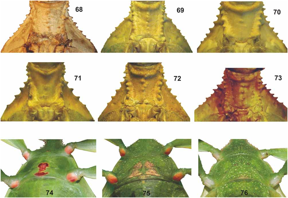
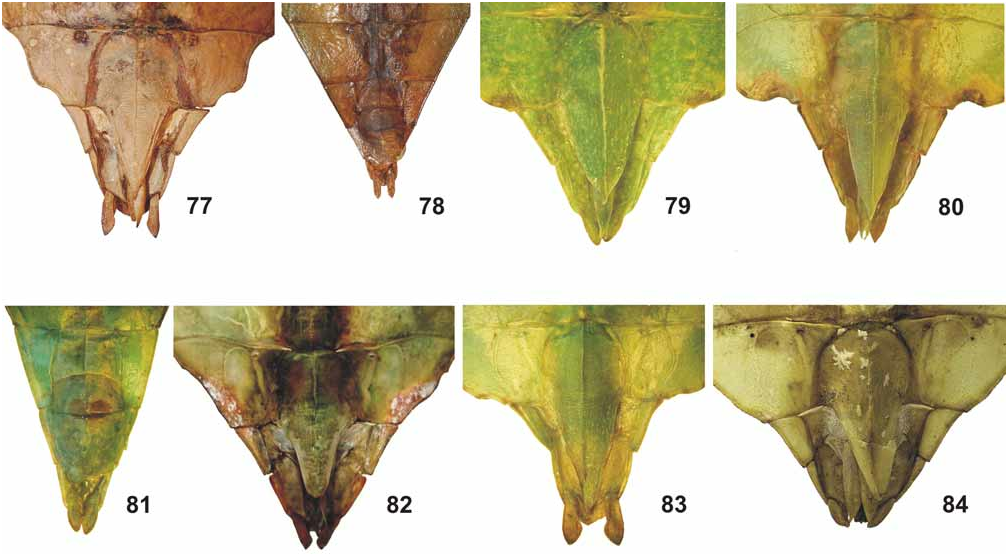
( Figs. 28–30 View FIGURES 28–30 , 60–61 View FIGURES 52–67 , 68 View FIGURES 68–76 , 77–78 View FIGURES 77–84 , 88 View FIGURES 85–94 , 101–102, 116)
HT, ♀: Philippines, Palawan, Brooke’s Point, Mount Gantung ( MSNG) .
PT, 1 egg (ex abdomen of HT): Philippines, Palawan, Brooke’s Point, Mount Gantung ( MSNG) .
PT, ♂: Philippines, Palawan, Brooke’s Point, Mount Gantung , IX.2008 ( MSNG) .
PT, ♀ + 2 eggs (ex abdomen): Philippines, Palawan, Brooke’s Point, Mount Gantung , IX.2008 (coll. MG) .
PT, ♀: Nord-Palawan, 20.08.1997, ex coll. Stumpe (coll. OC).
Comparison: A combination of morphological characters of both sexes of Ph. gantungense n. sp., including the granulose vertex and distinct longitudinal median row of tubercles on the meso-praescutum, indicate a close relation to Ph. bilobatum Gray, 1843 , Ph. hausleithneri Brock, 1999 , Ph. mindorense n. sp., and Ph. woodi Rehn & Rehn, 1933 . However, it differs from all these species by the straight and ± parallel-sided outer margins of abdominal segments V and VI (♀) or V only (♂♂), while these segments are more tapering in the other species.
From the Philippine Ph. bilobatum ♀ also differ by: the larger size; antennae with nine segments (ten in bilobatum ); slightly shorter tegmina, which reach about half the way along abdominal segment VII (VIII in bilobatum ) and having only four teeth on the interior lobe of the profemora (five in bilobatum ).
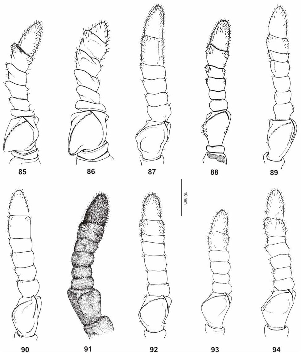
It is also well distinguished from the Malayan Ph. hausleithneri by: the relatively longer profemora, which are> 2x longer than the head; more distinct spination along the lateral margins of the mesopleurae ( Fig. 68 View FIGURES 68–76 ) and dark grey instead of blue marking on the interior surface of the meso- and metacoxae of both sexes. ♀ also differ by having only 33–35 teeth on the pars stridens of antennomere III (44–48 in hausleithneri , Fig. 88 View FIGURES 85–94 ) and ♂♂ can also be distinguished by the longer antennae, which reach as far back as half way along abdominal segment V, and consist of 26 segments (23–24 in hausleithneri ).
31: ♀ HT: Captive reared from Philippines (E-Luzon) [ ZSMC]
32: ♀ PT: Captive reared from Philippines (E-Luzon) [coll. FH, No. 0688-7]
33: ♀ PT: Captive reared from Philippines (E-Luzon) [coll. FH, No. 0688-8]
34: ♀ PT: Captive reared from Philippines (E-Luzon) [coll. FH, No. 0688-30]
35: ♂ PT: Captive reared from Philippines (E-Luzon) [coll. FH, No. 0688-31]
36: ♂ PT: Captive reared from Philippines (E-Luzon) [coll. FH, No. 0688-23]
From the Philippine Ph. mindorense n. sp. it can also be distinguished by: the shape of the profemora of both sexes ( Figs. 60–61 View FIGURES 52–67 ), which have the outer margin of the profemora almost unarmed and only have four teeth on the interior lobe (five in mindorense ) and the presence of a median spine on the transverse anterior ridge of the meso-praescutum (absent in mindorense ). ♂♂ can additionally be distinguished by the antennae consisting of 26 segments (only 22 in mindorense ).
From the Philippine Ph. woodi ♀ also differ by: the almost smooth outer margin of the exterior lobe of the profemora (acutely serrate in woodi ) and only four teeth on the interior lobe (six in woodi ); relatively longer and more slender meso-praescutum; slightly longer tegmina, which reach about half way along abdominal segment VII (to posterior margin of VI in woodi ) and slightly more elongate and triangular anal segment.
The distinctive egg of Ph. gantungense n. sp. (Figs. 101–102) is characterized by a set of peculiar features, which include: the reduction of the appendages which are mainly developed along the outer margin of the operculum and on the polar area of the capsule; structure of the appendages, which are comparatively smaller and resemble small palm-trees rather than being feather-like; the presence of well-organized, deep and broad depressions on the lateral capsule surfaces and operculum. These features might represent specializations of Ph. gantungense n. sp. indicating a possible isolated position within the genus.
Etymology: The specific name refers to the type-locality, Mount Gantung in the Philippine Province of Palawan.
Diagnosis: ♀ ( Figs. 28–29 View FIGURES 28–30 ). Moderate to large for the subgenus (body length 76.4–89.5 mm); profemora with a moderately broad and rounded exterior lobe; abdomen rather slender (maximum width 30.7–36.0 mm), with straight and parallel-sided outer margins of abdominal segments V and VI. Dorsal and lateral surfaces of the body and legs mainly pale apple green, ventral surfaces distinctly darker, tarsi greyish brown. Eyes light yellowish brown; antennae reddish-bronze, with a hint of green colouration on antennomere VIII. Interior surfaces of meso- and metacoxae with a conspicuous dark grey marking. Preserved specimens ranging from pale to dark brown. Head 1.3x longer than wide, vertex sparingly granulose, with two minute, very shallowly impressed elliptical areas in the centre, and a prominent conical tubercle medioposteriorly. Antennae slender, consisting of nine antennomeres; apical antennomere (IX) conical, 1.6x longer than wide, and about 1.6x longer than VIII, apex rounded. Pars stridens on antennomere III with 33–35 teeth ( Fig. 88 View FIGURES 85–94 ). Pronotum trapezoidal, gradually narrowed towards posterior portion, with anterior margin about 2.4x broader than posterior margin, the latter straight. Meso-praescutum about as long as wide with lateral margins slightly convex and very slightly narrowing towards the posterior; armed with a row of six spiniform tubercles and 2– 3 much smaller intercalated nodes. Median longitudinal row formed by six prominent tubercles. Anterior margin of mesonotum with a swollen transverse ridge, medially armed with a prominent spiniform, more or less composite tubercle. Mesopleurae not distinctly expanded in anterior quarter, then increasingly diverging towards the posterior; their lateral margins armed with five prominent spiniform teeth and several small intercalated nodes ( Fig. 68 View FIGURES 68–76 ). Pro- and metasternum irregularly granulose; mesosternum granulose-tuberculate and strongly convex longitudinally. Tegmina oval-oblong, reaching half way along abdominal segment VII. Alae rudimentary (3.5–4.0 mm). Abdominal segments II–IV gradually widened; IV widest segment and strongly angulate medially; V–VII very moderately narrowing, with the outer margins of V and VI straight and ± parallel-sided. Outer margin of VII angulate medially and converging posteriorly, occasionally may be protruded into a prominent posteriorly extending lobe (HT, Fig. 28 View FIGURES 28–30 ). VIII much narrower than VII, with lateral margins tapered to roundly lobed ( Figs. 28–29 View FIGURES 28–30 ). IX and X convergent caudad. Anal segment (X) roundly triangular, slightly wider than long, with the apex rather rounded. Subgenital plate projecting over posterior margin of tergite IX, with the longitudinal median carina reaching the apex of plate; outer margins slightly convex in posterior portion ( Fig. 77 View FIGURES 77–84 ). Profemora with a moderately broad and rounded exterior lobe, outer margin smooth or with a few very minute and indistinct dentations. Interior lobe slightly wider than exterior lobe, the outer margin armed with four triangular teeth and occasionally with a smaller one proximally ( Fig. 60 View FIGURES 52–67 ). Interior lobe of protibiae roundly triangular. Protarsi slender and> ¾ the length of corresponding tibia; probasitarsus about 6–7x longer than wide and shorter than combined length of remaining tarsomeres, except claws; tarsal claws simple. Lower margin of tarsal claws in meso- and metatarsi pectinate (4–5 teeth, examined at 50x magnification).
37: ♀ PT: Philippines (Mindoro Id., Mount Halcon ) [coll. FH, No. 0088-1]
38: ♂ HT: Philippines (Mindoro Id., Mount Halcon ) [ ZSMC]
♁♁ ( Fig. 30 View FIGURES 28–30 ). Medium-sized for the subgenus (body length 61.8 mm) with a rather broad and conspicuously angulate, medially parallel-sided abdomen (maximum width 19.6 mm). General colour of the preserved specimen strongly darkened due to preservation being mid brown with slight shades of green on the tegmina and outer margins of the abdomen. Head and thorax generally structured like in ♀. Antennae consisting of 26 segments and reaching half way along abdominal segment V. Pronotum with anterior margin 2x broader than posterior margin. Meso-praescutum 1.5x longer than wide, lateral margins with a row of 5–6 tubercles, longitudinal median row with four prominent tubercles. Lateral margins of mesopleurae with 4–5 small teeth and 8–9 intercalated nodes. Tegmina ovate and slightly constricted apically, projecting beyond abdominal segment III. Alae reaching half way along abdominal segment VIII. Abdominal segments II and III slightly diverging. IV and VI sharply angular (ca. 45°), V with outer margins straight and parallel-sided; VI widest segment. VII–IX gradually narrowing. Anal segment (X) about 1.2x longer than wide with outer margins roughly parallel-sided in anterior 2/3, posterior margin slightly indented medially; dorsal surface tectiform with a longitudinal median keel. Vomer broadly triangular, with a narrow and slightly up-curving terminal hook. Poculum 1.2x longer than wide, moderately convex and with a faint longitudinal median carina in anterior third; posterior margin rounded and slightly projecting over tergite IX ( Fig. 78 View FIGURES 77–84 ). Exterior lobe of profemora very narrow with the broadest point much slender than the shaft of the femur itself; outer margin very gently rounded and smooth. Interior lobe about 3.6x wider than exterior expansion and armed with four acute teeth ( Figs. 61 View FIGURES 52–67 ). Interior lobe of protibiae as in ♀. Protarsus elongate and just a little shorter than corresponding tibia, probasitarsus 9.3x longer than wide. Tarsal claws pectinate as in ♀.
Eggs (Figs. 101–102). The following description is based on eggs extracted from the abdomen of the ♀ paratypes, and on the examination of photographs of fully developed eggs laid by the ♀ paratypes, which however could not be preserved.
Of moderate size for the genus. Capsule roughly rectangular, about 1.9x longer than wide, distinctly laterally flattened, with the lateral surfaces sub-parallel, and both dorsal and ventral surfaces slightly convex. Lateral and ventral surfaces each with eight large circular depressions arranged in two sub-parallel longitudinal rows. Outer margins of the depressions bearing short and erect extensions resembling minute palm-trees. Interior surface of depressions rugulose and rather glossy. Surface between the depressions velvetlike and with a weakly developed network of narrow hairy ridges. Anterior third of dorsal surface with two conspicuous circular depressions of equal size, but occasionally oval-oblong in shape. Micropylar plate elongate-oval, well impressed and positioned in posterior 2/3 of dorsal egg surface; covering approximately half length of capsule and outer margin bearing short hairy structures. Micropylar cup placed just below the centre of plate and set with a tuft of hairy extensions. Posterior portion of dorsal surface with a central depression just behind the micropylar plate; two further similar depressions developed also on the polar area. Operculum slightly oval and with two semicircular depressions; space between the depressions slightly raised. Surface densely covered with hairy extensions, including a row of long appendages resembling small palmtrees along the outer margin. General colour mid brown.
Measurements [mm]: length 3.6 mm, width 1.9–2.0 mm, height 2.3–2.5 mm, length of micropylar plate 1.6–1.8 mm.
Variation: As with other members of the subgenus, ♀ of Ph. gantungense n. sp. show considerable variation concerning to the shape of the abdomen, which may have segments VII and VIII simple ( Fig. 29 View FIGURES 28–30 ) or protruded into more or less posteriorly extending lobes ( Fig. 28 View FIGURES 28–30 ). The lobes of segment VII are rather narrow and resemble the HT of Ph. bilobatum Gray, 1843 . It also appears that the lobed abdomen appears to be associated with more prominent teeth of the interior lobe of the profemora. Nothing is known about the variation in ♂♂.
Comments: Ph. gantungense n. sp. represents the second species of leaf insect recorded from Palawan, where it appears to be sympatric with Phyllium (Phyllium) palawanense Grösser, 2001 .
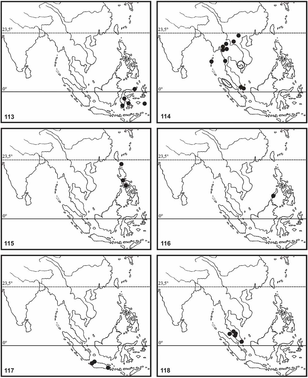
Distribution ( Fig. 116 View FIGURES 113–118 ): So far known only from Mount Gantung, Palawan Island, Philippines.
| MSNG |
Museo Civico di Storia Naturale di Genova 'Giacomo Doria' |
| MG |
Museum of Zoology |
| ZSMC |
Zoologische Staatssammlung |
No known copyright restrictions apply. See Agosti, D., Egloff, W., 2009. Taxonomic information exchange and copyright: the Plazi approach. BMC Research Notes 2009, 2:53 for further explanation.
|
Kingdom |
|
|
Phylum |
|
|
Class |
|
|
Order |
|
|
Family |
|
|
Genus |