
Theloderma bicolor ( Bourret, 1937 )
|
publication ID |
https://doi.org/ 10.5281/zenodo.212140 |
|
DOI |
https://doi.org/10.5281/zenodo.6169162 |
|
persistent identifier |
https://treatment.plazi.org/id/4D4D87EB-5F04-5505-FF35-FE49FBCAFF51 |
|
treatment provided by |
Plazi |
|
scientific name |
Theloderma bicolor ( Bourret, 1937 ) |
| status |
|
Theloderma bicolor ( Bourret, 1937) View in CoL
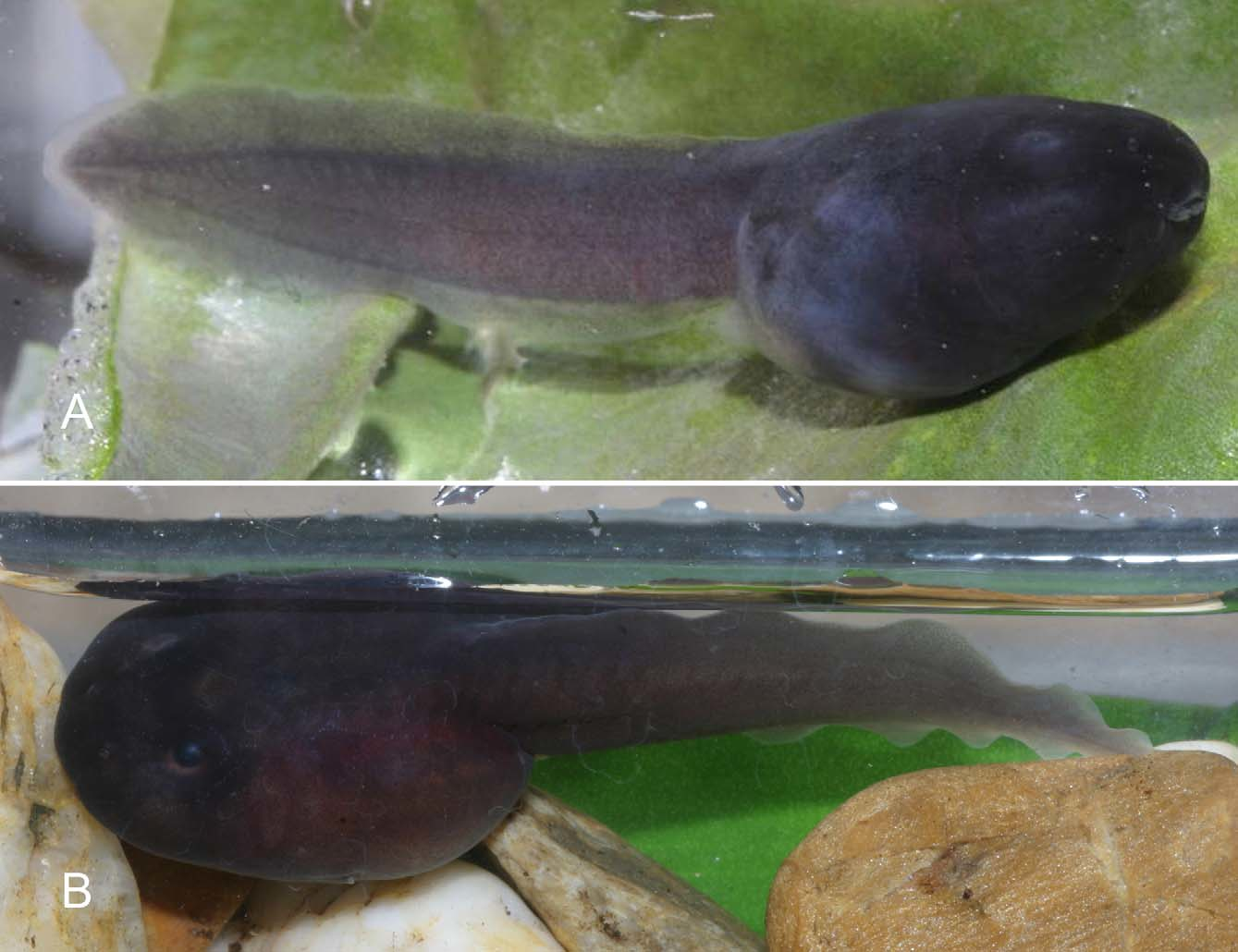
The following larval description of T. bicolor is based on a single captive bred specimen in Gosner stage 25 (IEBR A.2011.4, GenBank accesssion number JX046474 View Materials ) preserved at the Amphibian Breeding Station in Hanoi ( Fig.1 View FIGURE 1 A), which derived from parents from Sa Pa, Lao Cai Province, northern Vietnam.
Tadpole matching: For a specific assignment of the tadpole IEBR A.2011.4, we applied a DNA barcoding approach for an unambiguous genetic assignment of the tadpole used for our description with an adult male (VNMN 1394, GenBank accesssion number JX046475 View Materials ) from Sa Pa, Lao Cai Province, northern Vietnam, which largely agreed with the original description for T. bicolor provided by Bourret (1937). We used the mitochondrial 16S rRNA gene (using the primers 16sar-L and 16sbr-H of Palumbi et al. 1991); for the exact methods used see Schmitz et al. (2005) and Wildenhues et al. (2011).
Comparison of the resulting 568 bp long fragment of the 16S rRNA between the adult male and the tadpole showed only a negligible single base pair difference (corresponding to 0.18%) therefore, an unambiguous assignment of this tadpole to the species T. bicolor is guaranteed.
Colour pattern. Colouration in life (description of the colouration in life of tadpole is based on photographs taken in the Amphibian Breeding Station in Hanoi in 2009): body dorsally and laterally black to dark blue; gills and heart not visible; tail musculature lighter coloured, with reddish brown to grey pigments; upper and lower tail fin transparent with brown to grey pigments; vent tube unpigmented; iris black ( Fig. 1 View FIGURE 1 A).
Colouration in preservative: body predominantly dark pigmented from snout to tip of tail, except for a narrow transparent margin without pigments along last third of tail fin; pigmentation of body denser than tail pigmentation; body colouration varies from dark reddish brown, in first half, to dark bluish grey in remaining body; skin around nares and oral disc unpigmented and whitish; ventral body side slightly paler, also reddish brown to greyish brown; last quarter of ventral body side up to vent tube beige and unpigmented; tail musculature brown to grey with brown pigments; pigments form a dark brown stripe along apex and ventral side of myotomes; tail fin almost transparent with brown pigments; second half of tail denser pigmented, intensified around V-shaped myotomes.
Description in dorsal view. Body somewhat ovoid with a round, only slightly pointed snout, reaching a total length of 29.9 mm; mouth terminally located; maximum body width between spiracle and vent tube; eyes of small size (eye diameter 0.09 times of body length), situated dorsolaterally at end of first body third, and directed more dorsally; nares small rounded, slightly rimmed, anterodorsally positioned and directed, nearer to snout than to pupil (rostro-narial distance 0.75 times of narrow pupilar distance); internarial distance 0.63 of interpupilar distance; muscles of tail of moderate size, widest at base (width of tail musculature at base 0.13 of maximum body width); line organ present and well developed dorsally in first half of body, mostly around eyes and nares ( Fig. 2 View FIGURE 2 A).
Description in lateral view. Body slightly depressed to round-bellied (body height 0.82 times of body width) with a rounded, only slightly pointed snout; conical shaped spiracle sinistral, laterally positioned at beginning of second half of body (distance from tip of snout to opening of spiracle 0.73 times of body length), opening of the spiracle consists of an oval slit, positioned somewhat below longitudinal axis, oriented in posterodorsal direction, and connected with body and not visible in dorsal view; vent tube medially positioned at body end, ventrally located and adhered to lower tail fin by an outer wall; tail length 1.58 times of body length; myotomes of tail musculature of moderate development (height of tail musculature at base 0.60 times of maximum body height, and 0.71 times of maximum tail height); V-shaped myotomes of tail musculature gradually tapering from base of second half of tail towards end of tail, not reaching the tail tip; tail fin moderate, slightly pointed at the end; upper fin inserts at base of tail; highest point of upper fin at last third of tail ( Fig. 2 View FIGURE 2 B).
Oral disc. triangular-shaped, emarginated, anteroventrally positioned (oral disc width 0.30 of maximum body width); upper labium with large medial gap, marginal papillae confined to lateral corners; posterior labium with one row of marginal and one submarginal row of papillae, papillae small, finger-shaped to rounded; labial tooth row formula: 3(2-3)/3 with 34 keratodonts per 0.5 mm; upper and lower beak black and serrated; upper beak arched, lower beak V-shaped ( Fig. 2 View FIGURE 2 C).
Measurements (in mm). BH 6.1; BL 12.0; BW 7.5; ED 1.1; IND 2.0; IP 3.2; LF 0.9; MTH 5.1; NK 34; NP 77; NPD 2.2; ODW 2.3; RND 1.67; SS 8.8; TAL 19.0; TL 29.9; TMH 3.7; TMW 3.2; UF 1.5.

No known copyright restrictions apply. See Agosti, D., Egloff, W., 2009. Taxonomic information exchange and copyright: the Plazi approach. BMC Research Notes 2009, 2:53 for further explanation.
|
Kingdom |
|
|
Phylum |
|
|
Class |
|
|
Order |
|
|
Family |
|
|
Genus |