
Obama marmorata ( Schultze & Müller, 1857 ), Schultze & Muller, 1857
|
publication ID |
https://doi.org/ 10.1111/zoj.12358 |
|
DOI |
https://doi.org/10.5281/zenodo.5621676 |
|
persistent identifier |
https://treatment.plazi.org/id/4E2E87C6-212F-B622-A835-FAE7CC9616B2 |
|
treatment provided by |
Plazi |
|
scientific name |
Obama marmorata ( Schultze & Müller, 1857 ) |
| status |
|
Obama marmorata ( Schultze & Müller, 1857) View in CoL
Diagnosis: Species of Obama up to 100 mm long, 10 mm wide; dorsum covered with small longitudinal green brown dots anastomosed into longitudinal striae on a pastel orange background along the anterior 1/6th, ivory along the rest. Eyes occupying a lateral band on each side of the body, 1/5th of body width. Relative mouth/ body length c. 72%. Relative gonopore/body length c. 86%. Oesophagus present. Copulatory apparatus 5.43 ± 0.8 mm from anterior to posterior. Dorsal insertion of the penis dorsal, or even posterior to the level of the gonopore. Penis orientated backwards to the left side of the body. Male atrium not folded. Cyanophilic glands pierce a U-shaped surface of the male atrium, extending ventro-anteriorly and laterally onto its wall. Lining epithelium of female atrium with stratified appearance.
Molecular diagnosis: this species includes all populations that cluster with individuals MZUSP PL 1602,. site per substitutions indicates bar scale The. set data concatenated the from inferred tree phylogenetic ML. 1 Figure MZUSP PL 1601, MZUSP PL 1600 and MZUSP PL 1604 ( Table 1) from this study, with significant support in an adequate molecular delimitation model.
MATERIAL EXAMINED
Slides of sections of the copulatory apparatus of three specimens of Geoplana marmorata described and figured by C. G. Froehlich (1959), drawings by, and slides loaned by, E. M. Froehlich: Gm2: Blumenau, Santa Catarina, Brazil, E. M. Froehlich & Froehlich, coll. 2/Jun/1953. Copulatory apparatus (see fig. 4 in Froehlich, 1959): sagittal sections on two slides. Gm4: Pomerode (formerly Rio do Testo), Santa Catarina, Brazil, E. M. Froehlich & Froehlich, coll. 28/Jun/1953. Pharynx: sagittal sections on one slide; copulatory apparatus (see fig. 5 in Froehlich, 1959): sagittal sections on four slides. Gm6: Blumenau, Santa Catarina, Brazil, E. M. Froehlich & Froehlich, coll. 2/Jul/1953. Copulatory apparatus (see fig. 7 in Froehlich, 1959): sagittal sections on six slides. MZUSP PL 1600 (field number F3914): Parque Estadual da Serra do Tabuleiro, Paulo Lopes, Santa Catarina, Brazil (27°50′34″S, 48°55′33″W). F. Carbayo et al., coll., 15/Jul/2009. Anterior region end 7 mm long, sagittal sections on ten slides; a portion 7 mm behind, 19 mm long, horizontally sectioned on 47 slides; pre-pharyngeal region: tranverse sections on 27 slides; a portion 12 mm long including the pharynx, sagittal sections on 78 slides; a portion 10 mm long including the copulatory apparatus, sagittal sections on 26 slides; a 4-mm-long piece of tissue anterior to the pre-pharyngeal region was kept frozen in absolute ethanol. MZUSP PL 1601 (Field number F3351): Parque Estadual da Serra do Tabuleiro, São Bonifácio, Santa Catarina, Brazil (27°51′54.4″S, 48°55′0 9.8″W), F. Carbayo et al., coll. 19/Jan/2009. Anterior end 3 mm long transverse sections on three slides; a portion 9 mm long including the pharynx, sagittal sections on 23 slides; a portion 9 mm long including the copulatory apparatus, sagittal sections on 32 slides; a 7-mm-long piece of the posterior tip kept frozen in absolute ethanol. MZUSP PL 1602 (field number F3184): Parque Estadual da Serra do Tabuleiro, Paulo Lopes, Santa Catarina, Brazil, 27°57′31.3″S, 48°45′41.9″W), F. Carbayo et al., coll. 9/Jan/2009. Divided into two, preserved in 80% ethanol and absolute ethanol, respectively. MZUSP PL 1603 (field number F3234): Parque Estadual da Serra do Tabuleiro, Paulo Lopes, Santa Catarina, Brazil (27°56′51.5″S, 48°45′0 4.5″W). F. Carbayo et al., coll. 12/ Jan/2009. A portion 6 mm long including pharynx, sagittal sections on 12 slides; a portion 7 mm long including copulatory apparatus, sagittal sections on ten slides. Incompletely mature. MZUSP PL 1604 (field number F4620): Parque Nacional da Serra de Itajaí. Blumenau, Santa Catarina, Brazil (27°03′13.9″S, 49°05′0 9.8″W).
J. Pedroni et al, coll., 25/Jul/2010. Middle region of the body in absolute ethanol; remaining body parts in 80% ethanol.
Additional material studied
NHMW 2743, Series 1: four slides labelled ‘ Geoplana marmorata ’: No available locality. Serial sagittal sections including pharynx and copulatory apparatus.
Type locality
Blumenau, State of Santa Catarina, Brazil.
DESCRIPTION
Body up to 10 cm when crawling ( Fig. 3 View Figure 3 ); at rest, body 50–60 mm long and ∼ 6 mm wide. Body margins nearly parallel, except the anterior 1/4, and the posterior 1/6 of the body, rounded and pointed, respectively. Dorsum slightly convex, ventral side almost flat.
Dorsum ‘golden yellow’ along the anterior 1/6, ‘light ivory’ along the rest, richly ornamented with ‘green brown’ dots, anastomosed into longitudinal striae. These striae are longer and thinner in the anterior region than in the posterior. Striae are lacking at the margins of the body and in a thin medial stripe running from the anterior tip to the pharyngeal region. Posterior to pharynx, small dots occupy the medial region. Ventral side mostly ‘light ivory’, ‘nut brown’ at the anterior tip, whitish in the region of the pharynx and copulatory apparatus; margins of the anterior third of the body ‘orange brown’ ( Fig. 3 View Figure 3 , inset). After fixation, the dorsum colour became less brilliant and slightly faded.
Eyes marginally arranged in a single-to-triple row from the anterior end to 10 mm behind, then spreading dorsally in a band about 1/5 of the body width that becomes gradually narrower to posterior end ( Fig. 4 View Figure 4 ). Eyes of two types, a single-cup type, ∼ 30 μm in diameter, in the anterior 1/6 of the body, and a second type, 55 μm in diameter, consisting of three pigmented lobes, occurring from the second 1/6 behind. Eyes within clear halos around them. Sensory pits as simple invaginations 35 μm deep, located ventromarginally in a single row, from the very anterior end up to 15 mm posterior. Creeping sole 97.5% of body width.
In preserved specimens, the position of the pharyngeal aperture, on the ventral surface behind the anterior end, varies from 70% to 78% of total length, median 73% ( Table 2 View Table 2 ). The genital aperture (gonopore) varies from 82% to 88% of total body length, median 86% ( Table 2 View Table 2 ).
The sensory pits are simple invaginations 40 μm deep, ventromarginally distributed in a single row along approximately the anterior third of the body. The epidermis is ciliated just over the creeping sole. The glandular margin contains four types of glands, namely gross xanthophilic granules, fine erythrophilic granules, fine xanthophilic granules and cyanophilic granules.
The three typical geoplaninid cutaneous muscle layers are present. Their fibres are organized similarly throughout the body as follows: one circular followed by a diagonal with decussate fibres, and then a longitudinal layer with fibres arranged into poorly delimited bundles with 12–25 fibres each. The cutaneous musculature thickness relative to body height at the pre-pharyngeal region (CMI, see Froehlich, 1955) is 7.0%. Three parenchymal muscle layers are present: a dorsal layer of decussate diagonal fibres, a supraintestinal layer of transverse fibres and a subintestinal layer with transverse fibres.
The nervous system consists of a broad ventral plate, 80 μm in dorso-ventral thickness, extending almost the full width of the animal.
The mouth is nearly in the middle of the pharyngeal pouch ( Fig. 5 View Figure 5 ). The pharyngeal pouch is 1.6 times longer than the pharynx. An oesophagus is present. The pharynx is cylindrical-to-bell-shaped. Its outer epithelium is underlain by a one-fibre-thick layer of longitudinal muscle, followed by loose circular fibres. The inner pharyngeal epithelium is underlain by a thick layer (60–75 μm thick) of circular muscle fibres, with some longitudinal ones interspersed. The distal portion of the pharynx is pierced by gland cells of three types, producing cyanophilic, erythrophilic or xanthophilic granules.
Testes are dorsal to the digestive diverticula, roundedto-pyriform, 350 μm in larger diameter, located between the supraintestinal parenchymal muscle layer and intestine. They extend from the level of the ovaries to nearly the dorsal root of the pharynx. The sperm ducts are dorsal to the ovovitelline ducts. The distal portions of the sperm ducts bend forward and medially to communicate with the paired, extrabulbar branches of the prostatic vesicle ( Figs 6 View Figure 6 , 7 View Figure 7 ; Figs S2–S17). The unpaired portion is partially embraced by muscle fibres of the common muscle coat. The prostatic vesicle is C-shaped in lateral view. It is lined with a ciliated, columnar epithelium, pierced by glands producing erythrophilic granules and surrounded by a layer 20 μm thick of crisscrossed muscle fibres.
The ejaculatory duct runs through the penis. It is of more or less constant diameter throughout its length. It is lined with a cuboidal, ciliated epithelium underlain by a 10-μm-thick layer of circular muscle fibres. The penis is large, and wide (2.5–2.9 times longer than wide), occupying the entire male atrium. It projects backwards and to the left side of the body from the roof of the male atrium. The dorsal insertion of the penis is dorsal to the gonopore level, or even behind it. The penis is lined with a non-ciliated, cuboidal epithelium, and crossed by two types of glands, producing, respectively, erythrophilic and xanthophilic granules; xanthophilic glands are highly abundant and their cell bodies lie anterior and posterior to the penis bulb; their cell necks frequently join in small bundles each 20 μm in diameter until they open into the male atrium ( Figs 8, 9 View Figure 9 ). They pierce the entire epithelium of the penis except that of the tip. The underlying musculature is composed of a 12-μm-thick double layer of circular and longitudinal muscles, partially intermingled. The musculature of the stroma of the penis consists of scattered, diversely orientated fibres.
The male atrium is ample, with non-folded walls, lined with a columnar, non-ciliated epithelium. Numerous glands containing fine, weakly cyanophilic granules cross the U-shaped surface of the male atrium. The atrial surface pierced by these glands extends on the anterior ventral midline region ( Fig. 9 View Figure 9 ; Figs S5– S13) and the lateral walls. The cell bodies of these glands are located anterior and lateral to the penis bulb. In Froehlich’s specimens, probably stained with L, length; W, width; A-m and A-gp are distances from the anterior end to the mouth (pharyngeal aperture) and to the gonopore respectively. % values are for these as a percentage of total body length. *Sectioned specimens. Lcop, length of the copulatory apparatus from the junction of the sperm ducts to the oviduct-female genital canal
haematoxylin-eosin, the granules are pinkish. The atrial epithelium is surrounded by a musculature (10 μm thick) composed of a layer of circular muscles followed by a layer of longitudinal muscles partially intermingled with circular ones; the longitudinal muscle layer is absent where the epithelium is crossed by cyanophilic glands.
Paired ovaries are elongate, 400 μm in length and 30 μm in diameter, located at a distance from the anterior end about 30% of the body length. They lie above the ventral nerve plate, 2 mm apart. The vitellaria are distributed around the intestine. The ovovitelline ducts emerge from the latero-dorsal aspect of the ovaries.
Behind the region of the gonopore, they ascend laterally to the female atrium, and join each other above the posterior region of this atrium. The distal ascending portions of the ovovitelline ducts are pierced by shell glands. The common glandular ovovitelline duct is short and continuous with the female genital canal.
This canal is a dorso-anteriorly flexed diverticulum, projecting from the dorso-posterior region of the female atrium ( Fig. 10 View Figure 10 ; Figs S15, S16). The female atrium is ample, with lateral folds, and broadly communicates with the male atrium. It is lined with a non-ciliated epithelium with multilayered aspect, 40–150 μm thick. The epithelium is pierced by glands containing xanthophilic granules, and underlain by a 40-μmthick layer of crisscrossed muscle fibres. The ventral and lateral epithelium is additionally pierced by glands containing cyanophilic granules. The female atrium is about half the length of the male atrium. The gonopore canal is lined with a columnar ciliated epithelium, with erythrophilic granules apically.
The copulatory apparatus has an anterior–posterior extent of 5.6 ± 0.9 mm ( Table 2 View Table 2 ) from the junction of the sperm ducts to the junction of the oviducts.
Ecological note
We found specimens in the following environments: at the edge of a native wood partially open to stock grazing; a native forest partially logged decades before; a regrown native forest; and a wide cleared area opened in a native forest area for agriculture and grass grazing.


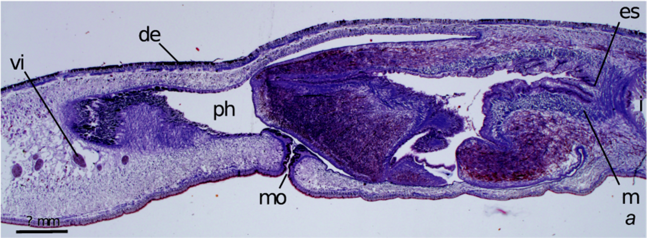
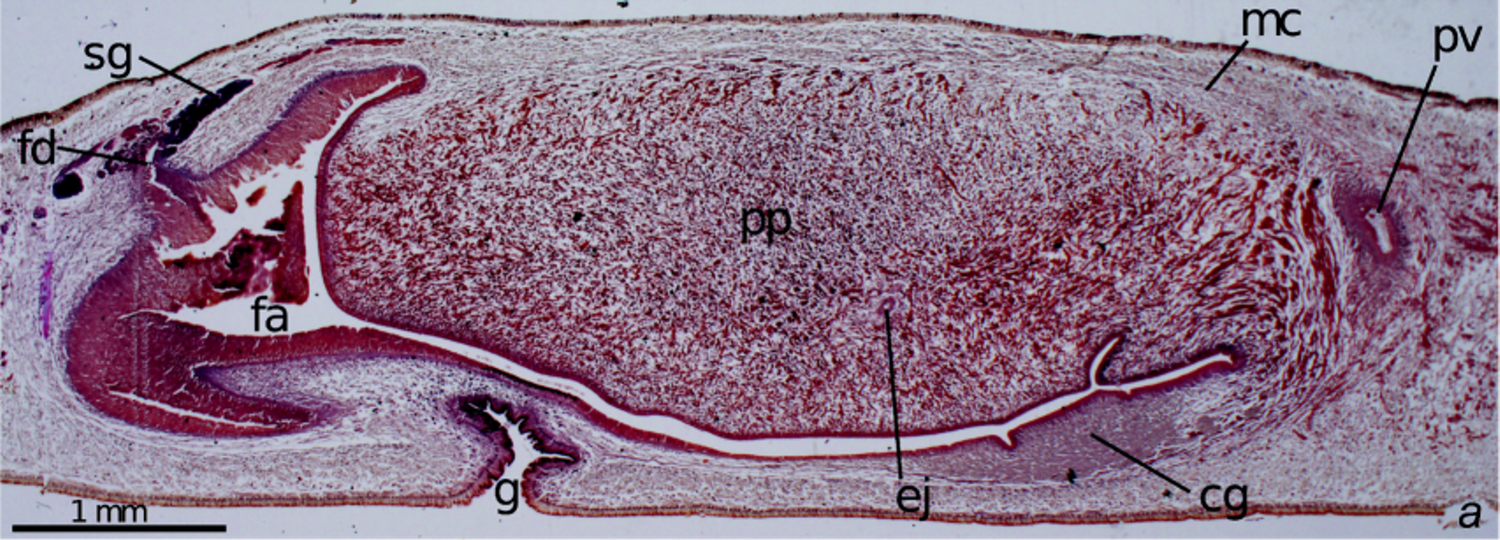

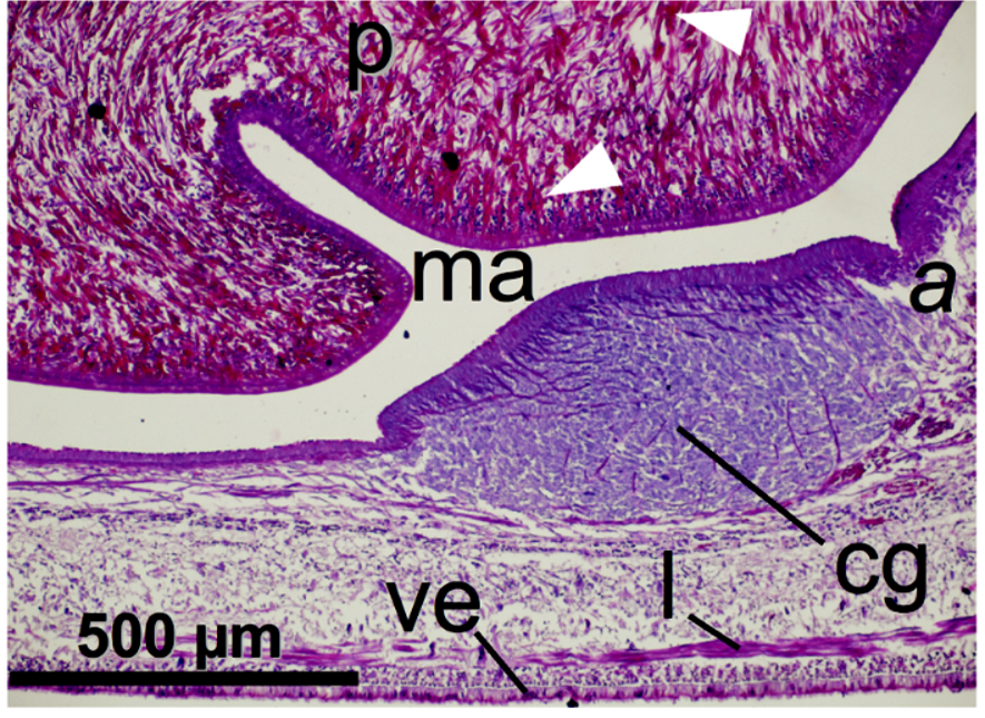
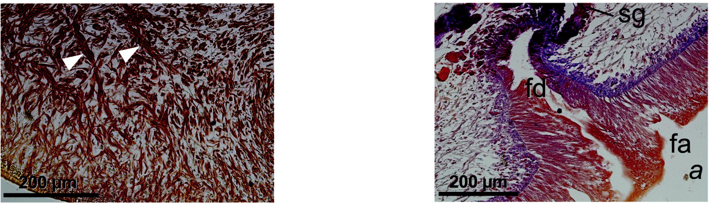
Table 2. Obama marmorata: dimensions (mm) of preserved specimens from Brazil
| L | W | A-m | A-m% | A-gp | A-gp% | Lcop | |
|---|---|---|---|---|---|---|---|
| Froehlich 1959* | 84 | 9.5 | 61 | 73 | 73 | 87 | 6.8 |
| Froehlich 1959* | 50 | 9 | 35.5 | 71 | 42 | 84 | 4.8 |
| Froehlich 1959* | 50 | 7 | 35 | 70 | 43 | 86 | 4.7 |
| F3914* | 66 | 10 | 48 | 73 | 58 | 88 | 6.2 |
| F3351* | 60 | 15 | 42 | 70 | 49 | 82 | 5.5 |
| F3184 | 30 | 5 | 23 | 77 | – | 85 | |
| F4260 | 41 | 5 | 30 | 73 | 35 | 86 | |
| Median | 73 | 86 | |||||
| Mean ± SD | 54 ± 17 | 8.6 ± 3.5 | 39 ± 12.5 | 50 ± 14 | 5.6 ± 0.9 | ||
| Müller (live) | 100 | 10 |
No known copyright restrictions apply. See Agosti, D., Egloff, W., 2009. Taxonomic information exchange and copyright: the Plazi approach. BMC Research Notes 2009, 2:53 for further explanation.
|
Kingdom |
|
|
Phylum |
|
|
Class |
|
|
Order |
|
|
Family |
|
|
SubFamily |
Geoplaninae |
|
Genus |