
Mutaparadoxipus duodigifinis, Gross, Vladimir, Miller, William R. & Hochberg, Rick, 2014
|
publication ID |
https://doi.org/ 10.11646/zootaxa.3835.2.6 |
|
publication LSID |
lsid:zoobank.org:pub:756F0BAC-8D78-48E3-9C07-FFA47FB9E2EA |
|
DOI |
https://doi.org/10.5281/zenodo.5671195 |
|
persistent identifier |
https://treatment.plazi.org/id/4E45BF75-BE0C-FFDB-FB8C-74E7FC05F801 |
|
treatment provided by |
Plazi |
|
scientific name |
Mutaparadoxipus duodigifinis |
| status |
sp. nov. |
Mutaparadoxipus duodigifinis View in CoL sp. nov.
Table 1 View TABLE 1 , Figures 1–3 View FIGURE 1 View FIGURE 2 View FIGURE 3
Diagnosis. Same as for the genus.
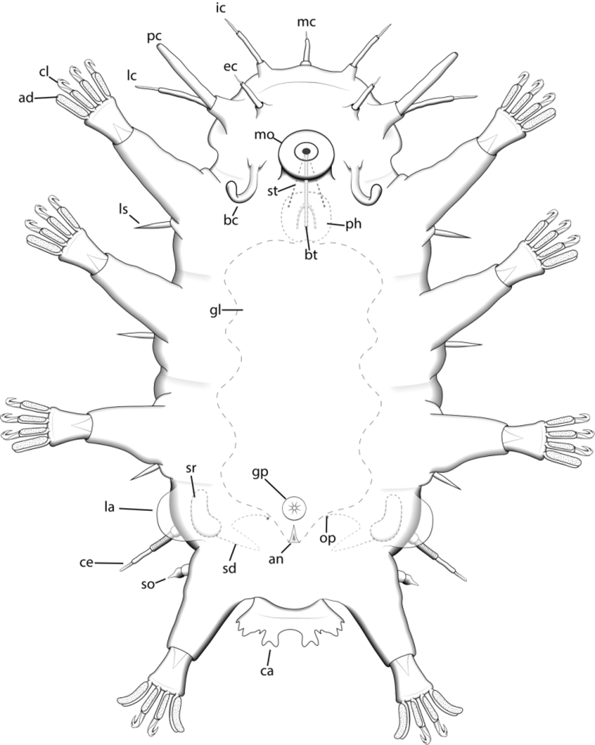
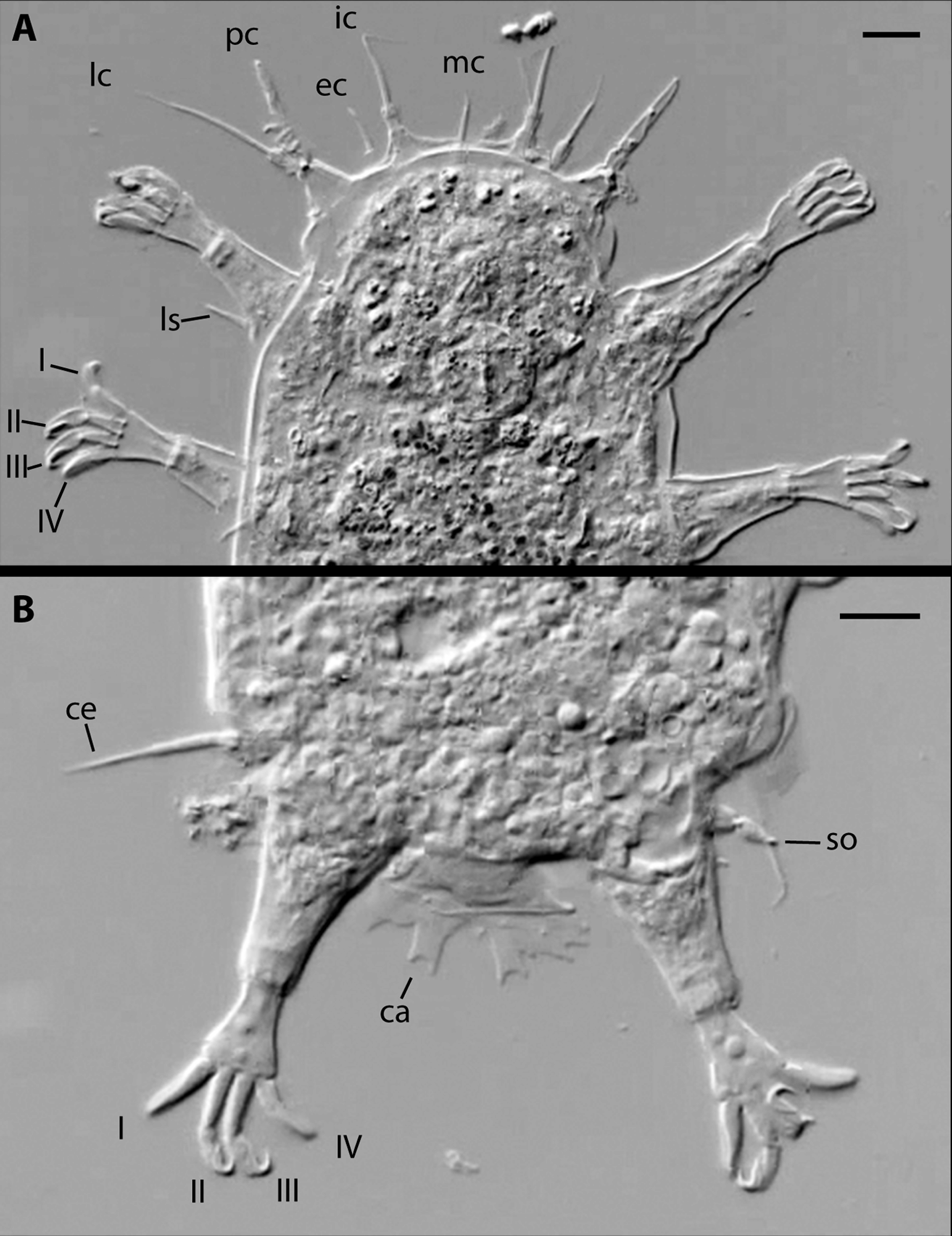
Description. Body length of 108–123 µm. Maximum width (between legs III–IV) of 50–53 µm. Cuticle without plates but with fine punctations, which are more prominent dorsally than ventrally. Indistinct segmentation is observable from both dorsal and ventral aspects. Caudal ala (12 µm long, 25 µm wide) is flap-like and extends posteriorly approximately one-third the length of legs IV ( Figs. 1 View FIGURE 1 , 2 View FIGURE 2 B, 3C). Lateral alae (9 µm long, 13 µm wide) are less distinct and attach slightly anterior to leg IV. The female gonopore consists of six rosette cells and is midventral, approximately halfway between legs III and IV ( Figs. 1 View FIGURE 1 , 3 View FIGURE 3 C). The male gonopore is U-shaped, opens posteriorly, and is positioned slightly more posterior than the corresponding female gonopore. Seminal receptacles are kidney-shaped with a coiled, S-shaped duct as in P. orzeliscoides ( Kristensen & Higgins 1989) , located adjacent to lateral ala. The seminal receptacle ducts open to the outside on either side of the gonopore.
The mouth cone extends ventrally and is positioned approximately halfway between the rostral margin of the animal and leg I ( Figs. 1 View FIGURE 1 , 3 View FIGURE 3 A). A typical heterotardigrade-type bucco-pharyngeal apparatus is present, with three placoids and a pair of straight stylets. Stylet supports could not be observed. The median cirrus (19 µm) is dorsal and, like all of the other cirri, consists of a scapus, a thick flagellum, and a thin flagellum ( Figs. 1 View FIGURE 1 , 2 View FIGURE 2 A). The thin flagellum is approximately one-third the length of the thick flagellum. The internal cirri (30 µm) are anteroventral and attach via a cirrophore at the base ( Figs. 1 View FIGURE 1 , 2 View FIGURE 2 A, 3A). The external cirri (14 µm) are ventral and attach directly posterior to the internal cirri, almost exactly lateral to the lateral cirri/clavae, which share a common pedestal. The lateral cirri (27 µm) are longer than and dorsal to the primary clavae (20 µm), which lack constrictions but have a terminal pore ( Figs. 1 View FIGURE 1 , 2 View FIGURE 2 A, 3A). The sausage-shaped secondary (buccal) clavae (20 µm) are lateral to the mouth cone and curve posteroventrally ( Figs. 1 View FIGURE 1 , 3 View FIGURE 3 A). There is no evidence of any buccal sensory plates at their bases.
The cirrus E (20 µm) is located on the posterior dorsal trunk and consists of a cirrophore, scapus, and 3-part flagellum ( Figs. 1 View FIGURE 1 , 2 View FIGURE 2 B). Each telescopic leg consists of a coxa, femur, tibia, and tarsus. Attached to the tarsus are four digits (14 µm long), each of which bears a ventral proximal paddle-shaped adhesive pad ( Figs. 1 View FIGURE 1 , 3 View FIGURE 3 B). The adhesive pads on the clawless digits are longer than those of the clawed digits (8 µm vs. 5 µm). Additionally, a distal sickle-shaped claw (5 µm) is present on digits I–III of legs I–III as well as digits II & III of leg IV (see Fig. 2 View FIGURE 2 A for digit numbering scheme). Accessory points are present only on the claws of digits II & III of each leg. A short sense organ consisting of two parts, a cylindrical base and a teardrop-shaped terminal structure (5 µm each, 10 µm total), is present on leg IV ( Fig. 2 View FIGURE 2 B). The coxae of legs I–III each bear a single spine (9–10 µm).
Type specimens. Holotype female ( USNM 1231540), 108 µm long, dorsoventral orientation, collected by V. Gross on 11 June 2012. Paratype male, 119 µm long. Digital photos and videos of the paratype were submitted to the Smithsonian Institution for archival. Second paratype male ( USNM 1231541) submitted on SEM stub.
Etymology. The species epithet, duodigifinis (“two toe ends”), is derived from duo, “two”; digitus, “toe”; and finis, “end,” referring to the two different types of toes present in the species; either with or without claws.
Type locality. Approximately 7 km east of Hutchinson Island near Fort Pierce, Florida (27° 28.90′N, 80° 13.72′W). Depth of 9.1–11.5 m. Collected in June 2012.
Comparisons. Additional species of Mutaparadoxipus gen. nov. have not been discovered. Thus, comparisons remain at the generic level. M. duodigifinis gen. nov., sp. nov. differs from P. orzeliscoides , which has claws and adhesive pads on all digits of all legs; Opydorscus fonsecae , which has adhesive pads and claws on all digits of legs I but lacks claws on all digits of legs II–IV; and Orzeliscus belopus , which has elongated adhesive pads on all digits but lacks claws altogether.
Additional meiofauna found in the same samples with the new species include: Tardigrada ( Batillipes , Tanarctus , Wingstrandarctus , Paradoxipus , Orzeliscus , and Halechiniscus ), Nematoda, Rotifera (Bdelloidea), Crustacea, Acoelomorpha, Kinorhyncha, Platyhelminthes (Kalyptorhynchia), and Gastrotricha (Chaetonotida and Macrodasyida).
LIST OF ABBREVIATIONS gp gonopore
ic internal cirrus la lateral ala
an anus lc lateral cirrus
ad adhesive pad ls leg spike
ap accessory point mc median cirrus bc buccal clavae mo mouth cone
bt buccal tube op seminal receptacle duct opening ca caudal ala pc primary clava ce cirrus E ph pharynx
cl claw sd seminal receptacle duct cs claw sheath so leg IV sense organ ec external cirrus sr seminal receptacle gl gut lumen st stylet

| USNM |
Smithsonian Institution, National Museum of Natural History |
No known copyright restrictions apply. See Agosti, D., Egloff, W., 2009. Taxonomic information exchange and copyright: the Plazi approach. BMC Research Notes 2009, 2:53 for further explanation.
|
Kingdom |
|
|
Phylum |
|
|
Class |
|
|
Order |
|
|
Family |
|
|
Genus |