
Rhabdoblattella alexeevi Anisyutkin
|
publication ID |
https://doi.org/ 10.11646/zootaxa.4236.1.2 |
|
publication LSID |
lsid:zoobank.org:pub:23412386-CB17-49CA-9C47-BD71DD9C5372 |
|
DOI |
https://doi.org/10.5281/zenodo.6015703 |
|
persistent identifier |
https://treatment.plazi.org/id/4F3A5135-FFB0-DC08-4FE3-FD146E5BFE82 |
|
treatment provided by |
Plazi |
|
scientific name |
Rhabdoblattella alexeevi Anisyutkin |
| status |
sp. nov. |
Rhabdoblattella alexeevi Anisyutkin , sp.nov.
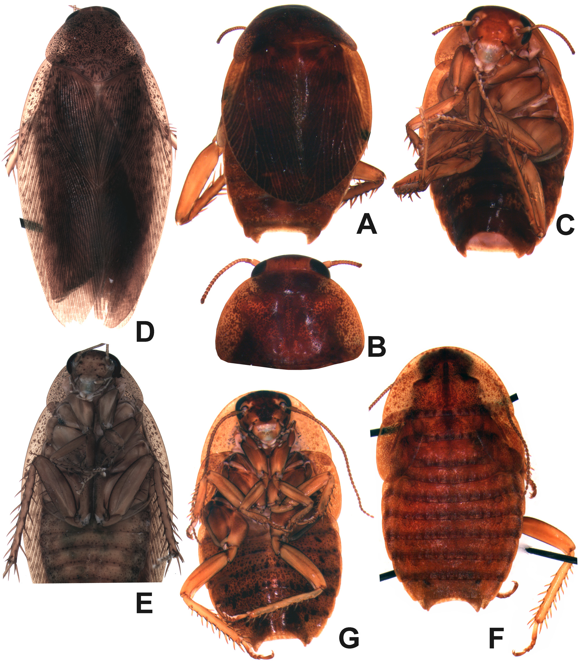
( Figs. 4A–C View FIGURE 4 , 5 View FIGURE 5 A–N)
Material. Holotype—male, SOUTH INDIA, state Kerala, Cardamon Hills, Muttapatti near Munnar , 1700 m, sieving in forest at the foot of a group of tree ferns, 22 November 1972, coll. C. Besuchet / I. Löbl (48) ( MHNG).
Etymology. This species is named in honour of the late Prof. Dr. Andrey N. Alexeev (1930–2015), a famous parasitologist and a true Russian intellectual, under whose supervision the authors of the present paper had a pleasure to work.
Description. Male (the holotype). General colour reddish brown ( Fig. 4A–C View FIGURE 4 ); facial part of head brown; eyes black; pronotum, tegmina and abdomen densely speckled, with small dark maculae; legs dirty yellowish. Surfaces smooth and lustrous. Head convex ( Figs. 4C View FIGURE 4 , 5 View FIGURE 5 A); epicranial sutures present; ocellar spots small, but distinct; weak transverse hollow located between antennal sockets; distance between eyes 1.2 times eye length; distance between antennal sockets 2.5 times of the scape length (about 0.4 mm); approximate length ratio of 3rd–5th segments of maxillary palps 1.3: 1.0: 1.8. Pronotum transverse; widely rounded along anterolateral margins, caudal margin very weakly angulate ( Figs. 4A, B View FIGURE 4 , 5 View FIGURE 5 B). Scutellum very small. Tegmina and wings shortened ( Fig. 4A View FIGURE 4 ), only reaching 7th abdominal tergite; rounded apically; venation distinct; all main veins present; Sc thickened (well visible on ventral side of tegmen). Wings completely hidden under tegmina, much shorter than tegmina, with a simplified venation. Anterior margin of fore femur armed as in the type B, with 6 spines, including 2 apical ones. Fore tibiae not thickened distally. Structure of hind tarsi as described for the genus (see above); arolium vestigial. Abdominal tergites 5th and 6th specialized ( Fig. 5 View FIGURE 5 C). 5th tergite with median membranous area and thin transverse sclerotized strip located in the centre of this area ( Fig. 5 View FIGURE 5 , s.str.). 6th tergite with larger membranous area, as compared with 5th tergite, membranous area separated by transverse sclerotized strip into two parts ( Fig. 5 View FIGURE 5 , s.str.), anterior part with membranous tubercle and small transverse sclerotized plate at base of tubercle. Posterolateral angles of tergites obtuse ( Fig. 5 View FIGURE 5 C). Anal plate (tergite X) with the widely rounded caudal margin and a distinct median incision ( Fig. 5 View FIGURE 5 D, E). Cerci short, flatten, with segments distinctly separated ( Fig. 5 View FIGURE 5 D, E). Paraprocts of the blaberid-type, right paraproct with a sclerotized outgrowth directed cranially ( Fig. 5 View FIGURE 5 E). Hypandrium weakly asymmetrical, with caudal margin rounded and projected caudally ( Fig. 5 View FIGURE 5 F); a median tooth present (Fig. F, G, m.t.); right stylus fusiform, bent at its base ( Fig. 5 View FIGURE 5 F), left stylus absent.
Male genitalia ( Fig. 5 View FIGURE 5 H–N). Right phallomere (R+N) with caudal part of sclerite R1T transverse ( Fig. 5 View FIGURE 5 H, I, c.p.R1T), cranial part of R1T and R2 rounded ( Fig. 5 View FIGURE 5 H, I, cr.p.R1T); R3 with branches of unequal length and width; R5 wide, weakly sclerotized; bristles absent. Sclerite L2D (L1) divided into basal and apical parts ( Fig. 5 View FIGURE 5 J); basal part rod-like, moderately widened cranially ( Fig. 5 View FIGURE 5 J, b.L2D); caudally with "curved upward subsclerite" ( Fig. 5 View FIGURE 5 J, K, u.s.); apical part in shape of sclerotized and folded plate ( Fig. 5 View FIGURE 5 J, K); bristles absent. Sclerite L3 (L2d) with distinct basal subsclerite ( Fig. 5 View FIGURE 5 L–N, b.L3); "folded structure" distinct, with bristles ( Fig. 5 View FIGURE 5 L–N, f.s.); groove hge present. Sclerite L4U (L3d) distinct.
Female unknown.
Measurements (mm). Head length: 1.9, width 1.9, pronotum length 2.5, width 3.7, tegmina length 4.8, width 2.3.
Comparison. The new species readily differs from all its congeners in having the distinctly shortened tegmina and wings. R alexeevi , sp.nov. is similar to R. euptera , sp.nov. in the presence of abdominal tergal glands, but can be readily distinguished from it by the location and structure of these glands (on the 5th and 6th tergites in R. alexeevi , sp.nov., contrary to the 6th and 7th ones in R. euptera , sp.nov.), and the presence of the apical part of sclerite L2D in the male genitalia.
Note. The shortening of tegmina, the reduction of ocelli and arolia are probably evidence of a shift to a more reclusive lifestyle. The well visible epicranial sutures and the reduced ocelli are rather larval characters, i.e. the modus of evolution R. alexeevi , sp.nov. seems to be the retardational paedomorphosis (retrogenesis) (sensu Iordansky 2005).

| MHNG |
Museum d'Histoire Naturelle |
No known copyright restrictions apply. See Agosti, D., Egloff, W., 2009. Taxonomic information exchange and copyright: the Plazi approach. BMC Research Notes 2009, 2:53 for further explanation.