
Elmohardyia cearensis, Marques, Dayse W. A. & Rafael, José A., 2015
|
publication ID |
https://doi.org/ 10.11646/zootaxa.3972.3.1 |
|
publication LSID |
lsid:zoobank.org:pub:D524C44A-6CAF-41B5-9461-89CF3C63DB1B |
|
DOI |
https://doi.org/10.5281/zenodo.6118778 |
|
persistent identifier |
https://treatment.plazi.org/id/4F7E878E-2969-FFCA-6BDD-FCD8E1EEE072 |
|
treatment provided by |
Plazi |
|
scientific name |
Elmohardyia cearensis |
| status |
sp. nov. |
Elmohardyia cearensis View in CoL sp. nov.
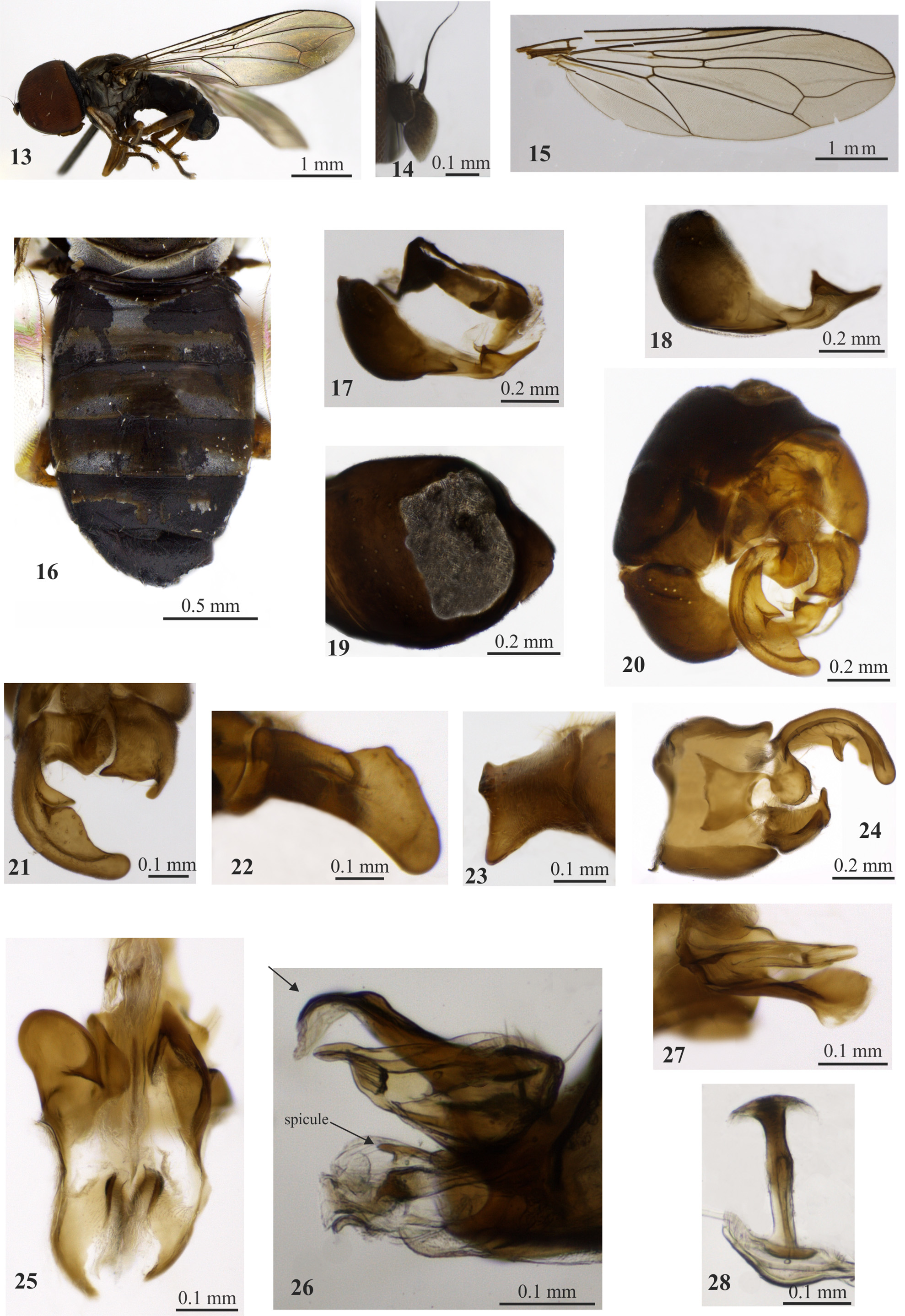
Figs 13–28 View FIGURES 13 – 28
Diagnosis. Tergite 2 with large basal gray pruinose band and two posterolateral gray pruinose spots. Sternite 6 with a subtriangular subapical projection and acute apex. Surstyli asymmetrical, the left one strongly developed, curved inward, about 2.2X longer than right surstylus. Left gonopod more developed than right one, with rounded apex. Phallic guide with one additional process that is enlarged distally.
Description of male holotype. ( Fig. 13 View FIGURES 13 – 28 ). Body length 4.5 mm. Head. Eyes contiguous for a distance of eighteen facets. F, EM, V = 0.4 mm, 0.4 mm, 0.3 mm. Frontal triangle and face gray pruinose. Postcranium dark, brown pruinose dorsally and gray pruinose laterally and ventrally. Antennae ( Fig. 14 View FIGURES 13 – 28 ) with scape dark brown; pedicel dark brown, with three dorsal and two ventral bristles; postpedicel dark brown at basal half and remaining light brown to yellow. LPP/WPP = 2.1. Labellum yellow. Thorax. Postpronotal lobe brown, gray-brown pruinose. Scutum dark brown to black, brown pruinose. Notopleuron brown, gray pruinose with twelve weak bristles. Scutellum black, gray pruinose anteriorly and brown pruinose along margins, with inconspicuous bristles. Mesopleuron and mediotergite dark brown to black, gray pruinose. Wing. ( Fig. 15 View FIGURES 13 – 28 ). Length 4.7 mm. LW/MWW = 3.4. LTC/LFC = 1.3. Membrane slightly light brown infuscated, almost entirely covered with microtrichia, except for cells bc, c, basal three quarters of sc, basal half of r1, small basal area of r2+3 and r4+5, br, bm, basal half of cup and basal one third of anal lobe without or with very sparse microtrichia. Vein r-m placed near basal third of cell dm. Vein dm-cu straight. Halter brown, except for yellow stem. Legs. ( Fig. 13 View FIGURES 13 – 28 ). Fore and mid coxae dark brown, hind coxa dark yellow to brown, all coxae gray pruinose. Trochanters yellow. Femora dark brown with base and apex yellow, entirely gray pruinose posteriorly. Tibiae yellow, gray pruinose posteriorly. Tarsi dark yellow, except fifth tarsomere dark brown. Pulvilli yellow. Abdomen. ( Fig. 16 View FIGURES 13 – 28 ). Dark brown to black, gray pruinose on tergite 1, on a large band at the base of tergite 2 and on posterolateral spots of tergites 2–5; tergite 1 with two stout black lateral bristles. Tergite and sternite 6 as in Fig. 17 View FIGURES 13 – 28 . Sternite 6 ( Fig. 18 View FIGURES 13 – 28 ) with a subtriangular subapical projection and acute apex. Syntergosternite 8 as long as tergite 5 ( Fig. 16 View FIGURES 13 – 28 ) and with large membranous area ( Fig. 19 View FIGURES 13 – 28 ). Terminalia. Epandrium and surstyli yellow ( Fig. 20 View FIGURES 13 – 28 ). Surstyli ( Figs 20–21 View FIGURES 13 – 28 ) asymmetrical. Left surstylus strongly developed, curved inward, about 2.2X longer than right surstylus, with basal lobe and a narrow sinus medially ( Fig. 21 View FIGURES 13 – 28 ); wider at distal third in lateral view ( Fig. 22 View FIGURES 13 – 28 ). Right surstylus with small projections apicolaterally ( Figs 21, 24 View FIGURES 13 – 28 ), subquadrangular, truncated in lateral view ( Fig. 23 View FIGURES 13 – 28 ). Subepandrial sclerite as in Fig. 24 View FIGURES 13 – 28 . Left gonopod more developed than right one, with rounded apex ( Fig. 25 View FIGURES 13 – 28 ). Phallic guide ( Figs 26, 27 View FIGURES 13 – 28 ) with one additional dorsal process which is enlarged distally and directed downward. Phallus with a small subapical spicule ( Fig. 26 View FIGURES 13 – 28 ). Ejaculatory apodeme as in Fig. 28 View FIGURES 13 – 28 . Female unknown.
Type material. HOLOTYPE ♂: “ BRASIL, CE[ará], Ubajara, Parque Nac.[ional] de Ubajara, Cachoeira do Cafundó, 03°50'13"S, 40°54'35"W ” “Armadilha Suspensa, 16–31.xii.2012, F. Limeira-de-Oliveira, J.S. Pinto - Júnior cols [collectors]” “ Holotype ♂, Elmohardyia cearensis Marques & Rafael " ( CZMA).
Holotype condition. Left wing detached, mounted on microslide, left hind leg glued on label. Terminalia placed in a microvial with glycerin.
Etymology. The specific name refers to the type locality, Ceará state.
Distribution. Brazil: Ceará (Caatinga Biome).
Discussion. Elmohardyia cearensis sp. nov. is close to E. galeata Rafael & Menezes due to the asymmetrical surstyli, inward curved left surstylus and shape of the phallic guide. Elmohardyia cearensis sp. nov. differs from E. galeata by tergites 2–5 being gray pruinose posterolaterally (only tergite 5 gray pruinose posterolaterally in E. galeata ) and by the subquadrangular right surstylus, with its small apicolateral projections (without apicolateral projections in E. galeata ).
No known copyright restrictions apply. See Agosti, D., Egloff, W., 2009. Taxonomic information exchange and copyright: the Plazi approach. BMC Research Notes 2009, 2:53 for further explanation.