
Heteromurus (Verhoeffiella) absoloni Kseneman, 1938
|
publication ID |
https://doi.org/ 10.11646/zootaxa.4039.2.3 |
|
publication LSID |
lsid:zoobank.org:pub:7DD7CB5E-E4E5-4424-A3CE-0D822B440512 |
|
DOI |
https://doi.org/10.5281/zenodo.5611549 |
|
persistent identifier |
https://treatment.plazi.org/id/504DC64D-FFEC-9413-FF25-FE84FB65FB9A |
|
treatment provided by |
Plazi |
|
scientific name |
Heteromurus (Verhoeffiella) absoloni Kseneman, 1938 |
| status |
|
Redescription of Heteromurus (Verhoeffiella) absoloni Kseneman, 1938
Designation of neotype. The type material of Heteromurus (Verhoeffiella) absoloni has not been retrieved in spite of thorough investigations in regional Museums and in Czech republic, including: Natural History Museum of Montenegro, Podgorica, National Museum of Bosnia and Herzegovina, Sarajevo, Croatian Natural History Museum, Zagreb, The Moravian Museum, Brno, Mendel University in Brno - Department of Zoology, Fisheries, Hydrobiology and Apiculture (formally Zoologicky ustav vysoke školy zemedelske v Brne where Kseneman was working, pers. com. Jan Bezdek). It was also not possible to locate Ksenemans collection (Rusek and Weiner, pers. com.). Type material can be therefore considered as lost. We propose here as neotype a specimen from the type locality.
Material examined. Montenegro, Boka kotorska, Risan, Golodražnica (cave) (coordinates WGS 84 X =18.69962°, Y =42.50282°, alt. 110 m.). Neotype adult female on slide, 04.xii.2014, M. Lukić leg.(CLL 4557); 5 ex. on slides and 19 ex. in 96% alcohol, same data (CLL 4555). Three ex. directly mounted on slides and one ex. mounted on slide after DNA extraction ( BOLD Sample ID:4028C01_MNECLL2278), 24 ex. in 96% alcohol, 24.iv.2010, J. Bedek leg.(CLL 2278); 3 ex. on slides, 8 ex. in 96% alcohol, 24.iv.2010, A. Kirin leg.(CLL 2279); 2 ex. on slides, 4 ex. in 96% alcohol, 24.iv.2010, A. Komerički leg.(CLL 2280); 3 ex. on slides, 34 ex. in 96% alcohol, 31.iii.2012, J. Bedek leg.(CLL 4000); 2 ex. on slides, 77 ex. in 96% alcohol, 31.iii.2012, M. Lukić leg. (CLL 4001); 17 ex. in 96% alcohol, 31.iii.2012, A. Komerički leg. (CLL 4185).
Material deposition. All material in 96% alcohol and following ex. on slides, including neotype, deposited in collection of the Croatian Biospeleological Society, Zagreb, Croatia: 2 ex. (CLL 2278), 3 ex. (CLL 2279), 1 ex. (CLL 2280), 1 ex. (CLL 4000), 1 ex. (CLL 4001), 6 ex. (CLL 4555). Following ex. on slides deposited in the collection of the Muséum National d’Histoire Naturelle de Paris: 1 ex. (CLL 2278), 1 ex. (CLL 2280), 2 ex. (CLL 4000), 1 ex. (CLL 4001).
Diagnosis. Color pale orange, pigment as orange dots. Scales present on dorsal and ventral side of head and body, on Ant. Ib and on Ant. II, on legs, ventrally and dorsally on manubrium, ventrally on dens. Antennae 0.5 to 0.7 as long as (head + body) length, Ant. III and IV annulated, often fused. Ant. segment ratio as Ia:Ib:II:III:IV = 0.15:0.61:1:1.5:2.0. Tita of leg III longer than those of legs I and II; length 0.40, 0.49, 0.58 mm for Tita I, II, III. Labial triangle with 9–12 smooth chaetae, and no scales. Dorsal macrochaetotaxy as (3+4),(3+3)/0,2,3,(3+0+4+4).
S-chaetotaxy: ms formula 1,?1/1,0,1,0,0; sens formula, 2,2/1,3,3,3,3, with S-like chaetae at least 10+10 on Abd. IV. Unguis thin and elongated, with one pair of inner basal teeth at 25% from its base and one unpaired inner tooth at 50% from its base; unguiculus pointed, 75% as long as inner edge of unguis; tenent hair acuminate, smooth. Manubrium with about 15 smooth pointed mac dorsally, unpaired ventro-basal smooth mac and 2+2 ventro-apical serrated mes; dens with one short internal basal spine, mucro bidentate with one basal spine.
Redescription. Head and body length combined without antennae: 2.7 to 3.2 mm (N=8). Habitus as in Fig. 1 View FIGURE 1 . Abd. IV 1.75 – 2 times as long as Abd. III. Color pale orange, pigment scattered as orange dots ventrally and dorsally on body, manubrium, coxa, paler on trochanter and femur. Intersegment area, mouth parts, antennae, other leg segments, and dens unpigmented. Body dorsally and laterally with white regular patches on an orange background ( Fig. 1 View FIGURE 1 ). Eyes absent, no pigment patch at their place. Body, mouthparts and appendages with a large diversity of phaneres. Most can be assigned to the traditional four types of chaetae: ordinary chaetae, S-chaetae, trichobothria and scales; several other kinds of specialized chaetae occur on mouth and antennae. Pseudopores of same size as mac socket, observed on antennae, tergites, legs and manubrium.
Antennae ( Figs 2–14 View FIGURES 2 – 8 View FIGURES 9 – 14 ). Antennae 0.54 to 0.68 as long as (head + body) length (N=8). Antennae 3 times longer than head diagonal. Ant. segment ratio as Ia:Ib:II:III:IV = 0.15:0.61:1:1.5:2.0 (mean values, N=9). Ant. III and IV annulated, except the proximal part of Ant. III and the distal part of Ant. IV. Antennae often asymmetrical, because of the fusion of Ant. III and IV. Pseudopores present ventro-apically on Ant. Ib (2–5), Ant. II (8–14) and Ant. III (5), located in the intersegment just above sensorial areas.
Antennal phaneres very diversified (ordinary chaetae, S-chaetae, scales and papillae). The following categories have been recognized ( Fig. 2 View FIGURES 2 – 8 ):
– Type a—serrated mac, dorsally on Ant. Ib and II, shorter than those of head and body.
– Type b—thick serrated mes, on all antennal segments ( Fig. 9 View FIGURES 9 – 14 ).
– Type c—thin serrated mes, less ciliated than type b, on Ant. III and IV ( Fig. 9 View FIGURES 9 – 14 ).
– Type d—pointed smooth mac, ventrally on Ant. Ib and II.
– Type e—pointed smooth mes, ventrally on Ant. Ia, Ib and II.
– Type f—acuminate smooth mes, thin, basally thinner than e-type mes, straight and inserted perpendicular to integument, on all antennal segments but only ventrally on Ant. Ia, Ib ( Fig. 9 View FIGURES 9 – 14 ).
– Type g—pointed smooth mic similar in shape to e-type mes but shorter, basally on Ant. Ia and II ( Fig. 10 View FIGURES 9 – 14 ).
– Type h—swollen S-chaetae ( Fig. 11 View FIGURES 9 – 14 ), dorso-externo-apically on Ant. Ib (2), II (4–6) and III (2) ( Figs 4, 5, 6 View FIGURES 2 – 8 ).
– Type i—thick subcylindrical S-chaetae, on all antennal segments ( Fig. 12 View FIGURES 9 – 14 ): 3–4 ventrally on Ant. Ia ( Fig. 3 View FIGURES 2 – 8 ), several grouped in ventro-externo-distal sensorial fields on Ant. Ib, II and III ( Fig. 4 View FIGURES 2 – 8 ), and 2–7 distally on Ant. IV ( Fig. 7 View FIGURES 2 – 8 ).
– Type j—short, pointed, bent S-chaetae ( Fig. 13 View FIGURES 9 – 14 ), 2–8 distally on Ant. II and 3–4 on Ant. III, located near sensorial area; not seen on Ant. Ib.
– Type k—thin, short, hyaline S-chaetae ( Fig. 14 View FIGURES 9 – 14 ), on the whole length of Ant. III and IV and a few ventrally on Ant. Ib and II, some larger and intermediate with type i, especially on Ant. IV.
– Type l—thin, long S-chaetae ( Fig. 10 View FIGURES 9 – 14 ), similar in shape and size to f-type mes, but blunt at the tip, thinner proximally and not straight, present ventrally on Ant. Ia and Ib.
Scales rounded, of medium size, their surface covered with numerous short spicules arranged longitudinally;
numerous dorsally on Ant. Ib and II, few ventrally on Ant. Ib; absent on Ant. Ia, III and IV. Subapical organite of Ant. IV long, thick and slightly bifurcate ( Fig. 7 View FIGURES 2 – 8 ).
Subapical papilla of Ant. IV, devoid of "pin seta" ( Fig. 7 View FIGURES 2 – 8 ).
The different categories of phaneres are distributed as follows on each antennal segment: Ant. Ia dorsally with 2 basal mic (type g) and thick serrated mes (type b); ventrally with 3 basal mic (type g),
pointed smooth mes (type e), acuminate smooth mes (type f) and sensorial area constituted of 10–12 S-chaetae of
type l and 3–4 S-chaetae of type i ( Fig. 3 View FIGURES 2 – 8 ). No scales.
Ant. Ib dorsally with dense cover of scales, without chaetae except apical row of serrated mac (type a), thick
serrated mes (type b), and two externo-apical small swollen S-chaetae (type h); ventrally with very few scales,
large number of pointed smooth mes (type e), thick serrated mes (type b), few acuminate smooth mes (type f),
distal row of pointed smooth mac (type d) or large pointed smooth mes (type e), ventro-external sensorial field
constituted of several S-chaetae of type i, few short S-chaetae of type k externo-apically to sensorial field, and
several S-chaetae of types i, k and l proximally to field. No basal mic.
Ant. II dorsally with dense cover of scales mixed with sparse serrated mac and mes (types a and b), distally a few acuminate smooth mes (type f), apical row of small mac (type d) or large straight mes (type e) and externoapical group of 4–7 swollen S-chaetae of type h (AIIO, Fig. 5 View FIGURES 2 – 8 ); ventrally without scales, with many short thick serrated mes (type b), pointed smooth mes (type e, less numerous), acuminate smooth mes (type f), distal row of small mac (type d) or large smooth mes (type e) and externo-apical sensorial field constituted of 25–30 S-chaetae of type i, similar to that of Ant. Ib, and 2–8 chaetae of type j; 2–3 short S-chaetae of type k present externo-distally between sensorial field and AIIO; longer S-chaetae of type k present laterally to and mostly proximally to sensorial field. Basal mic present (1 dorsal, 2 ventral, 1 latero-external, 1 latero-internal).
Ant. III–IV chaetotaxy with tendency to form whorls of chaetae, sometimes spiraling, without clear serial pattern across successive whorls ( Fig. 8 View FIGURES 2 – 8 ) that are composed of 4 types of chaetae variously interspersed (b, c, f and k, Fig. 9 View FIGURES 9 – 14 ); dorsally with less c-type chaetae and all chaetae smaller in size compared to the ventral ones. Ant. IV with several thick subcylindrical S-chaetae (type i) in distal part ( Fig. 7 View FIGURES 2 – 8 ).
AIIIO located dorso-externo-apically, with 2 swollen S-chaetae side by side (type h), surrounded internally by one thin short S-chaeta (type k) and externally by three short external S-chaetae, of which 2 pointed and bent (type j), and 1 thin (type k); few additional S-chaetae of type k present below ( Fig. 6 View FIGURES 2 – 8 ). Ant. III ventro-externo-distally with sensorial field constituted of about 20–30 i-type chaetae, and 1–3 small pointed j-type chaetae. Basal mic absent or undifferentiated on Ant. III and absent on Ant. IV. Scales absent on Ant. III–IV. Ant. IV with large slightly bifurcate subapical organite and a subapical papilla ( Fig. 7 View FIGURES 2 – 8 ).
When Ant. III and IV are fused, the described pattern of Ant. III distal chaetae is strongly modified.
Mouthparts ( Figs 15–20 View FIGURES 15 – 20 ). Labral formula 4/5,5,4. No labral papillae. Prelabral and labral chaetae smooth, acuminate, of similar size. Labrum margin with inverted medial U-form intrusion devoid of primary granules, distal smooth area large ( Fig. 15 View FIGURES 15 – 20 ). Ventro-distally on labrum two unequal combs with one apical row of about 10 and 15 teeth respectively, and a pair of sinuous rods ( Fig. 15 View FIGURES 15 – 20 ). Labial triangle with 9–12 chaetae, all smooth (N=9) and no scales ( Fig. 20 View FIGURES 15 – 20 ); lateral area (submentum) with 5 smooth chaetae. Labial palp with 5 proximal chaetae and 5 papillae (A,B,C,D,E); guard chaeta of papilla A integrated in papilla B complex which thus appears to have 5 guard chaetae ( Fjellberg 1999: 325); papilla C without guard chaeta; papilla D with 4 guard chaetae; papilla E with 4 guard chaetae and well developed labial process ( Fig. 16 View FIGURES 15 – 20 ). Maxillary outer lobe with one basal chaeta, simple maxillary palp, three sublobal hairs and one smaller chaeta ( Fig. 17 View FIGURES 15 – 20 ); basal fold with one chaeta. Maxilla with strong tridentate claw, 5 lamellae and a thin rod, long and bent ( Fig. 18 View FIGURES 15 – 20 , after Fjellberg 2007: 160). Mandible asymmetrical with 5 teeth on right and 4 on left on all examined specimens ( Fig. 19 View FIGURES 15 – 20 ). Clypeus with many pointed smooth and serrated mesochaetae. About 10+10 smooth post-labial chaetae (PLQ of Mari-Mutt 1985 pro parte) interspersed with scales.
Head and body ( Figs 21–30 View FIGURES 21 – 22 View FIGURES 23 – 30 ). Chaetae are rather diverse on head ( Fig. 27 View FIGURES 23 – 30 ) and body ( Fig. 28 View FIGURES 23 – 30 ), with the following categories usually relatively easy to recognize:
– serrated mac, either clubbed (sensu Stach 1960: plate I) or subcylindrical-pointed ( Figs 27 View FIGURES 23 – 30 a1–a2 and 28a1–a2);
– serrated mes on head and body ( Figs 27 View FIGURES 23 – 30 b1–b2 and 28b);
– thin mes slightly serrated on its distal half and associated with thin S-chaetae ( Figs 25, 26 and 28 View FIGURES 23 – 30 c);
– smooth mes, only on head ( Fig. 27 View FIGURES 23 – 30 c);
– trichobothria on head ( Fig. 27 View FIGURES 23 – 30 d) and tergites, often fallen in our specimens; those of head (1+1) located laterally and rather short (50 µm), those of tergites longer;
– S-chaetae, limited to tergites, smooth, very short, thick, pointed (ms, Figs 24, 28 View FIGURES 23 – 30 d);
– thin S-chaetae, limited to tergites, smooth, short and hyaline, subcylindrical, blunt (sens, Figs 24–26, 28 View FIGURES 23 – 30 e);
– smooth, thin, blunt, short to long S-like chaetae, longer, less transparent and slightly thicker than thin Schaetae, only on Abd. IV ( Fig. 28 View FIGURES 23 – 30 f1 and f2);
– mic, 2 minute on each anal valve, and 2+2 on female genital plate ( Fig. 28 View FIGURES 23 – 30 g);
Scales on dorsal and ventral side of head and body, rounded to oval-elongated, sometimes truncated, hyaline, overlapping, of various size (from 15 to 100 µm long), with a dense cover of short spicules arranged in more or less regular lines ( Figs 21, 22 View FIGURES 21 – 22 ).
Chaetae, scales and trichobothria of dorsal side of head and body usually detached in microscopic preparations or on skins retrieved after DNA extraction. Chaetae have circular or oval sockets, those of mac and large mes easily recognizable by their size, but those of tergite S-chaetae not distinguishable from those of small mes. Scale sockets are characterized by cupuliform morphology of their socket. Trichobothria have small round socket often with minute point in the middle, but often indistinguishable from mes sockets.
Head dorsally with macrochaetae arranged as in Fig. 23 View FIGURES 23 – 30 , and dense cover of small to large scales and short serrated mes; laterally with scales mixed with medium size serrated mes; between antennae with few smooth mes; posteriorly with row of serrated mes regularly interspersed with large scales; ventrally with smooth and serrated mes and small to medium-size scales; 1+1 trichobothria posterior to ocular area.
Dorsal chaetotaxy illustrated in Fig. 23 View FIGURES 23 – 30 . Pseudopore formula 1,1/1,1,1,1. Trichobothrial formula 1/0,0/0,2,3,2. Macrochaeta formula (3+4),(3+3)/0,2,3,(3+0+4+4). Macrochaeta arrangement stable; multiplets sensu Szeptycki 1979 only present anteriorly on Th. II; Th. II with 3 anterior and line of 4 posterior mac; Th. III with 3 posterior and 3 lateral mac; Abd. IV with 3 medial, 4 antero-lateral and 4 postero-lateral mac; Abd. V with 12 to 16 mac; Abd. VI with 11 mac in two rows, including an uneven one in the anterior row ( Fig. 23 View FIGURES 23 – 30 ). Serrated mes present on anterior part on Th. II, dorsally on Abd. V and VI ( Fig. 22 View FIGURES 21 – 22 ) and very few laterally on all tergites. Many rather long S-like chaetae on Abd. IV, 50% longer than mes of other tergites, arranged symmetrically ( Fig. 21 View FIGURES 21 – 22 ). Sens difficult to observe, often detached or hidden by scales, 3–4 times longer than mac socket; ms very short, about the length of mac socket. S-chaetotaxy given in Tab. 2 and Figs 23–26 View FIGURES 23 – 30 . Sens associated with thin serrated mes in tandem on Abd. II–III (also possibly in other locations). S-chaetotaxy: ms formula 1,?1/1,0,1,0,0 (ms on Th. III observed in two specimens, possibly an aberration as it would be unique in Entomobryoidea); sens formula, 2,2/1,3,3,3,3, with S-like chaetae at least 10+10 on Abd. IV.
Scales rounded, of various sizes: medium-size on all tergites except Abd. VI where they are smaller ( Fig. 22 View FIGURES 21 – 22 ); larger posteriorly (at least twice as large as the anterior ones) on all tergites except Abd. VI. No special arrangement or morphological modification of scales and chaetae around mac and trichobothria.
Area on tergite S-chaetae and mesochetae ThII antero-lateral (1 ms+1 sens) ThII postero-lateral 1 sens
ThIII antero-lateral (1 ms+1 sens) ThIII postero-lateral 1 sens
Abd. I lateral 1 ms, 1 sens Abd. II median 1 sens
Abd. II medio-lateral (1 sens+1 mesochaeta) Abd. II lateral 1 sens
Abd. III median 1 sens
Abd.III medio-lateral (1 sens+1 mesochaeta) Abd. III lateral (1 ms+1 sens) Abd. IV median 2 sens,>10 long S-like chaetae Abd. IV postero-lateral 1 sens
Abd. V median 2 sens
Abd. V lateral 1 sens
Ventral tube with smooth pointed and serrated mes and mac. Posteriorly with about 125 sockets (serrated and smooth mes in unknown proportion, impossible to recognize from their socket once they have fallen) and 6–7 larger sockets (possibly mac); no scale observed except a single scale in place in one specimen. Anteriorly with 46– 49 + 46–49 sockets (serrated or smooth mes). Each latero-distal flap with 45 mes, about 12 of them serrated and others smooth ( Fig. 29 View FIGURES 23 – 30 ). Genital plate of female with 2+2 mic ( Fig. 30 View FIGURES 23 – 30 ), that of adult male not observed.
Legs ( Figs 31–39 View FIGURES 31 – 36 View FIGURES 37 – 39 ). Unguis relatively thin and elongated, with one inner pair of equal basal teeth at 25% from its base and one inner unpaired tooth at 50% from its base ( Figs 31, 32 View FIGURES 31 – 36 ). Large hyaline lamella covers claw, and bears 1+1 small latero-basal teeth ( Figs 31, 32 View FIGURES 31 – 36 ). Internal structure of unguis illustrated in Fig. 37 View FIGURES 37 – 39 . Unguis (internal edge): unguiculus = 1.6:1. Unguiculus robust, pointed, about 75% as long as inner edge of unguis, with two external ridges, without basal lamella ( Figs 31, 32 View FIGURES 31 – 36 ). Small outer tooth, sometimes difficult to see at 50% from its base on anterior ridge of unguiculus I, II and posterior ridge of unguiculus III ( Figs 31, 32 View FIGURES 31 – 36 ).
Legs with ordinary chaetae and scales, often detached, including the following categories:
– serrated mac, cylindrical-pointed, on subcoxae 2, coxae and tibiotarsi (similar to Figs 27 View FIGURES 23 – 30 a2 and 28 a2);
– serrated mes, thick, on all leg segments ( Fig. 31 View FIGURES 31 – 36 )
– smooth mac, pointed, on femur, trochanter, coxa
– smooth mes, pointed, on Tita, femur, trochanter, coxa ( Fig. 31 View FIGURES 31 – 36 )
– tenent hairs, smooth, acuminate, of similar length but thinner than smooth mes, located dorso-apically on Tita ( Fig. 31 View FIGURES 31 – 36 )
– smooth, pointed mes, inserted perpendicular to integument, forming the trochanteral organ ( Fig. 38 View FIGURES 37 – 39 )
– mic of pretarsus ( Fig. 31 View FIGURES 31 – 36 )
– scales small, elongated, subequal, present on all leg segments.
Detailed distribution of the different types of chaetae on each leg segment as follows: pretarsus with 1+1 minute mic ( Fig. 31 View FIGURES 31 – 36 ). Tibiotarsus with a large number of serrated mes, few serrated mac, smooth mes, thin tenent hair and scales ( Fig. 39 View FIGURES 37 – 39 ). Distal whorl of Tita with 11–14 (usually 13) chaetae including 5, 5, 4 smooth mes and the tenent hair on leg I, II, III. Serrated mac as 1, 3, 3 on Tita I, II, III ( Fig. 33–36 View FIGURES 31 – 36 ). Beside the apical ones, smooth mes present ventrally in two longitudinal rows on Tita I, II, III, anterior one with 3, 3–6, 5–7 chaetae, posterior one with 4–5, 3–4, 6–7 chaetae ( Fig. 33 View FIGURES 31 – 36 ). Few scales present in the proximal third of Tita I, more numerous in the proximal half of Tita II, and many distributed on the whole length of Tita III. Tita of leg III longer than those of legs I and II; length 0.40, 0.49, 0.58 mm for Tita I, II, III.
Femur with many serrated mes (some almost as large as serrated mac), smooth mac, smooth mes, and scales. Ventrally about 1–2, 1–2, 1 smooth macrochaetae on femora I, II, III and few additional smooth mes on femora I ( Figs 34–36 View FIGURES 31 – 36 ). Biggest smooth mac as long as 0.35–0.45 length of femur. Group of ventro-proximal serrated mes without interspersed scales ( Fig. 38 View FIGURES 37 – 39 ).
Trochanter with many chaetae ranging in size from mes to mac including many serrated mes,1–2 smooth mac, and smooth mes. Smooth chaetae about 9–10 on trochanter I, 3–6 on trochanter II, many on trochanter III in adult, of which 55 to 100 forming the trochanteral organ of leg III ( Fig. 38 View FIGURES 37 – 39 ). Scales in small number on trochanter I, II and in large number on trochanter III.
Coxa with pseudopores, thick serrated mes, smooth mes, smooth mac, multiplets of serrated chaetae and scales. Pseudopores as 1–2, 3–6, 6–10 on coxae I, II, III. Smooth mac as 2, 0, 0 on dorso-anterior side of coxa I, II, III; multiplet of 5–7 dorso-posterior serrated chaetae on coxa I, two multiplets (dorso-anterior one and dorsoposterior one) of 5–8 serrated chaetae on coxa II; chaetae in multiplets of coxae I and II decreasing in size from mac (distally) to mes (proximally) ( Figs 34–35 View FIGURES 31 – 36 ). Dorsally on coxa III, a transversal line of 3 serrated mac and 5–8 serrated mes ( Fig. 36 View FIGURES 31 – 36 ).
Separation between subcoxae 1 and 2 indistinct. Subcoxae with scales. Subcoxa I with 3 dorso-posterior serrated mac ( Fig. 34 View FIGURES 31 – 36 ). Subcoxa II with 4–6 dorso-anterior serrated chaetae ranging in size from mac to mes and 3– 5 dorso-posterior serrated mac ( Fig. 35 View FIGURES 31 – 36 ). Subcoxa III, with group of 4–5 dorso-posterior serrated mac and some serrated mes ( Fig. 36 View FIGURES 31 – 36 ).
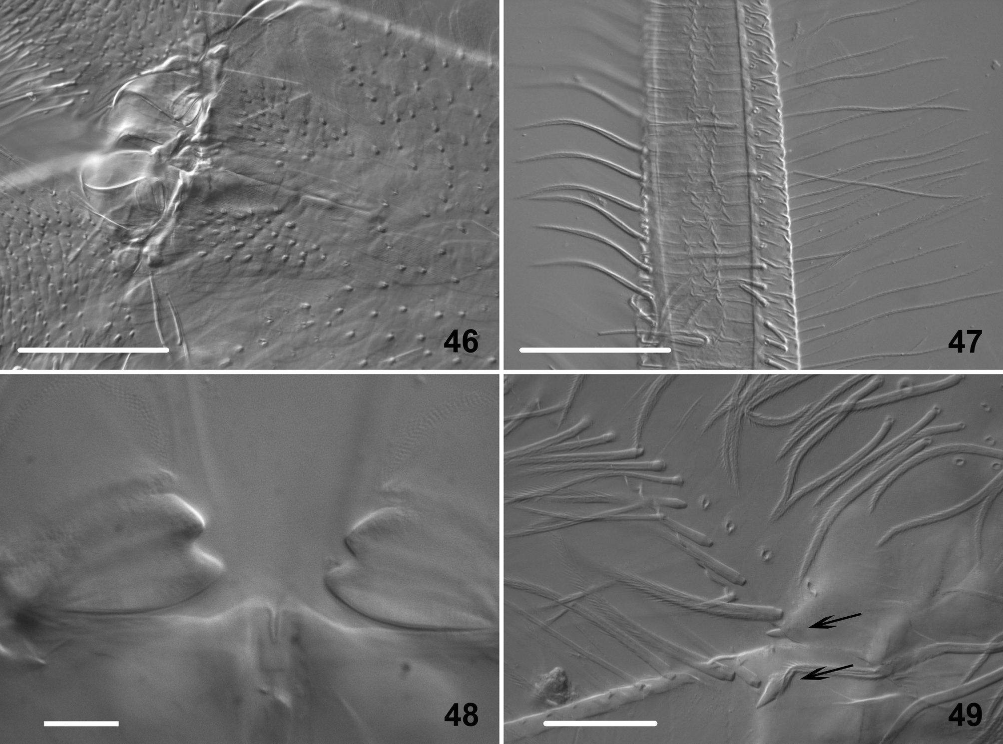
Furca ( Figs 40–49 View FIGURES 40 – 45 View FIGURES 46 – 49 ). Furca with pseudopores, smooth mac, different kinds of serrated mes and scales of various morphologies. Pseudopores as 6–8 dorsally on each side of manubrium, among distal group of chaetae. Manubrium dorsally with two longitudinal stripes of serrated mes, each one interspersed with 14 ± 2 smooth pointed mac ( Fig. 40 View FIGURES 40 – 45 ); between these stripes, loose cover of scales except in basal 1/3 where few serrated mes are present; externally, each longitudinal stripe of mes is bordered by a very narrow stripe devoid of chaetae and scales. Cross-section of manubrium as shown in Fig. 41 View FIGURES 40 – 45 . Manubrium covered ventrally by medium size scales, without chaetae, except an unpaired ventro-basal serrated and thick mac and 2+2 apical serrated mes near the axis ( Fig. 42 View FIGURES 40 – 45 ); scales evenly distributed, except on two ventro-distal areas where scale cover is more dense with larger and more elongated scales ( Fig. 46 View FIGURES 46 – 49 ). Distal manubrial thickening with 2 to 5 small teeth on each side ( Fig. 42 View FIGURES 40 – 45 ).
Dens not articulated, but constituted of a short basal part hardly annulated, a long medial part annulated dorsally and a short, thinner, non annulated distal part about 4 times as long as mucro ( Figs 43, 45 View FIGURES 40 – 45 ); covered by various chaetae and scales arranged in a complex pattern as shown in schematic cross-sections ( Fig. 44 View FIGURES 40 – 45 ). All dental chaetae serrated except one smooth mac basally on dorsal part of each dens ( Fig. 40 View FIGURES 40 – 45 ). Two types of serrated chaetae can be recognized ( Fig. 47 View FIGURES 46 – 49 ); the thickest ones arranged dorso-laterally in two longitudinal rows on basal and medial part of dens; the thinner ones, ventro-laterally, very long on basal part and shorter on medial part, arranged in two longitudinal rows, with two irregular lines of serrated chaetae internally on basal part ( Fig. 44 View FIGURES 40 – 45 ). Small serrated mes present between rows and ventral scales. Ventrally between two rows of thin serrated chaeta, dense cover of medium size elongated scales, small and rounded on basal part of dens, increasing in size laterally to large, fusiform, pointed apically ( Fig. 44 View FIGURES 40 – 45 ); scales absent in distal non-annulated part ( Fig. 45 View FIGURES 40 – 45 ). Dens thickening with two faint transversal structures ( Figs 42 View FIGURES 40 – 45 , 48 View FIGURES 46 – 49 ). One short and thick internal spine basally on each dens ( Figs 40 View FIGURES 40 – 45 , 49 View FIGURES 46 – 49 ). Ratio manubrium: dens: mucro = 10: 14: 0.5. Mucro bidentate with one basal spine, sometimes not observed (probably detached) ( Fig. 45 View FIGURES 40 – 45 ). Tenaculum with 1 serrated chaeta and 4 teeth on each ramus.
Discussion. The extensive re-description of H. (V.) absoloni given here is the first detailed description of a species of the subgenus, all existing ones being very succinct and lacking most characters used in modern taxonomy of Entomobryidae . Several morphological features, described for the first time for the subgenus, revealed interesting characters, e.g. the presence of a chaeta ms on Th. III. It was observed in two specimens only, but S-chaetae are often very difficult to detect; if it is not an aberration, it would be unique among Entomobryoidea. The detailed description of antennal chaetotaxy shows a huge diversity in chaetae morphological types. Their distribution on antennae follows recognizable but loose patterns, as chaetae-to-chaetae homologies are not possible, with a few exceptions, between adults of a same species or between whorls of Ant. III–IV. The descriptive framework set up in this paper will be used as a reference in the ongoing subgenus revision.

| DNA |
Department of Natural Resources, Environment, The Arts and Sport |
No known copyright restrictions apply. See Agosti, D., Egloff, W., 2009. Taxonomic information exchange and copyright: the Plazi approach. BMC Research Notes 2009, 2:53 for further explanation.