
Podocotyle, Dujardin, 1845
|
publication ID |
https://doi.org/10.11646/zootaxa.4638.4.3 |
|
publication LSID |
lsid:zoobank.org:pub:E8CE848C-3E8C-45DF-A093-D696D19EBD46 |
|
persistent identifier |
https://treatment.plazi.org/id/51258795-FFBF-852A-D9A1-FECDFE53D98C |
|
treatment provided by |
Plazi |
|
scientific name |
Podocotyle |
| status |
|
Podocotyle View in CoL sp. 3
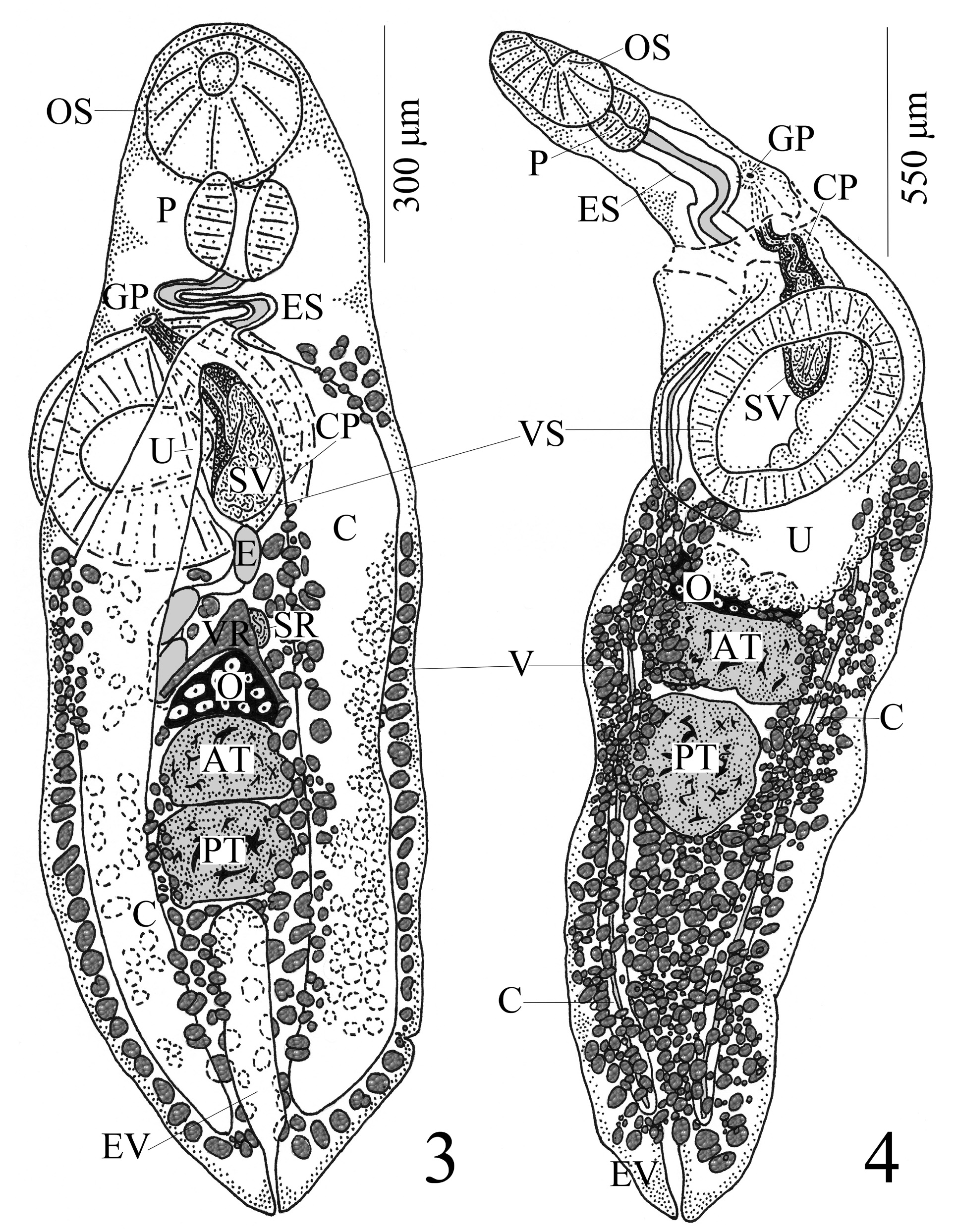
( Fig. 4 View FIGURES 3–4 )
Host: Bathygadus favosus Goode & Bean ( Gadiformes : Macrouridae : Bathygadinae ).
Locality: Off Colombia in Caribbean Sea, 11°31.8’ N, 74°24.5’ W, depth = 1,143m, 16–17/July/1970.
Site of infection: Intestine.
Prevalence: B. favosus : 1 of 10 (10.0%).
Intensity: only 1 worm.
Mean intensity: 1/1 = 1.00.
Relative density/abundance: 1/10 = 0.10.
Deposited Specimen: NHMUK 2019.4.12.24 (1 slide).
Description: [Based on 1 extended specimen with incomplete transverse tear in forebody region. Measurements and proportions given in Table 2 View TABLE 2 .] With characteristics of genus. Body elongate, extended, flattened dorsoventrally, widest in region around junction of anterior and middle 1/3 body; anterior end attenuated; posterior extremity broad, round. Forebody twisted in mount, attenuate, about 1/3 body length. Hindbody wider than forebody, gradually narrows in posterior 1/3 of body. Tegument smooth. Pre-oral lobe not observed. Oral sucker subterminal, unspecialized in lateral view. Ventral sucker conspicuous, unspecialized, muscular, transversely oval and wider than long, at junction near first and second 1/3 of body. Prepharynx very short. Pharynx muscular, sub-rectangular. Esophagus moderately long, thick-walled, slightly sinuous, passes through damaged area of worm. Intestinal bifurcation anterior to ventral sucker. Ceca long, narrow, widest at level of ventral sucker, thick-walled, terminates blindly near posterior extremity.
Testes 2, median, tandem, smooth, round or oval, almost contiguous, intercecal, post-equatorial, extends from middle 1/3 of body to near junction of middle and posterior 1/3 of body. Post-testicular region confined to posterior 1/3 of body. Cirrus pouch distinct with conspicuous wall, pyriform, posterior extent extends to mid-level of ventral sucker. Seminal vesicle internal, bi-partite; proximal portion large, saccate, occupies entire left half of cirrus pouch, loops back posterodextrally across center of pouch, and narrows to form tubular, distal portion in anterodextral area of pouch, then passes anterior. Pars prostatica near damaged area of worm, appears long and thick-walled; ejacula- tory duct and cirrus presumed present (i.e. in damaged area). Prostatic gland cells numerous, dark-stained, highest numbers in anterior portion of cirrus pouch. Genital pore submedian (sinistral), pre-acetabular, at mid- to lower esophagus level and midway between left margin and midline of worm. Genital atrium distinct.
Ovary 3-lobed, lobes anteriorly-directed, median, equatorial, post-acetabular, contiguous to and immediately anterior to anterior testis, intercaecal. Portions of proximal female system obscured by uterus and eggs and not observed; canalicular seminal receptacle and Laurer’s canal with dorsal opening expected. Vitelline reservoir subme- dian (sinistral), circular, immediately anterior to anterosinistral margin of ovary; transverse vitelline ducts run paral- lel to anterior margin of ovary with right transverse duct overlapping anteromedial margin of latter near midline of worm; main vitelline duct passes anterosinistrally from anterior margin of vitelline reservoir to join oviduct. Entire path of oviduct obscured by uterus and eggs, but observed to arise anteriorly from ovary, expected to receive both Laurer’s canal and main vitelline duct, then enter oötype which is itself directly anterior to ovary and surrounded by conspicuous Mehlis’ gland cells. Uterus conspicuous, intercaecal (a few loops of uterus pass ventrally over medial wall of left cecum between ovary and ventral sucker); loops proceed anteriorly from level of region between ovary and anterior testis, pass dorsally over left half of ventral sucker, and enters genital atrium. Metraterm thick-walled, passes ventral to cirrus pouch and occupies space between anterior border of pouch and genital atrium; transverse striations (i.e. musculature) in metraterm apparent. Vitelline follicles moderate in size, dense, round or oblong, circumcecal, extend longitudinally in 2 uninterrupted lateral bands from near posterior extremity anteriorly up to level of posterior margin of ventral sucker, encroach over lateral margins of gonads and into immediate post-acetabular region, not confluent in pre-ovarian region, in space between ovary and anterior testis and in inter-testicular region, confluent in post-testicular region. Eggs large, somewhat collapsed and/or crenulated, operculate, amber, non-embryonated, non-filamented, numerous and densely packed in uterus.
Excretory bladder I-shaped/tubular, thin-walled, extends to posterior testis. Excretory pore terminal.
Remarks: These 3 specimens were assigned to the Podocotylinae within the Opecoelidae based on their possession of the following diagnostic combination of characters: a canalicular seminal receptacle, a well-developed cirrus pouch that is relatively short (extends from mid-level to posterior margin of ventral sucker) and encloses an internal seminal vesicle that becomes long and tubular distally, blind ceca, an intercecal and pre-ovarian uterus (uterus of Podocotyle sp. 1 extends posterior to ovary), and a deep-sea piscine host ( Cribb 2005; Martin et al. 2018). The present specimens key out to the genus Podocotyle based on their possession of the same diagnostic combination of morphological characters listed earlier.
With only one individual each of what we believe are three unique species (see below), we cannot observe any intra-specific variation and any detailed comparative analyses of these individuals with accepted species of Podocotyle would be of little value. However, due to the rarity of this material (i.e. all digenean parasites originally reported from both host species [ B. favosus and N. atlantica ], including Podocotyle sp. 1, 2 & 3 herein, have been documented only by two of us – CKB and HWA – see Table 1 View TABLE 1 ), we felt that a detailed description of these three specimens would be a helpful addition to our limited knowledge of deep-sea endohelminth diversity in the Gulf of Mexico and Caribbean Sea.
A comparison among Podocotyle sp. 1 vs 2 vs 3 indicates that these individuals lack conspecificity (see Table 2 View TABLE 2 ). Armstrong (1974, p. 72) noted that Podocotyle sp. n. #1 (= Podocotyle sp. 1 this study) was characterized, in part, by a short excretory vesicle (extends to posterior testis) and vitelline fields that extended anteriorly to the posterior margin of the ventral sucker (Note: we observed the right band of vitelline follicles to extend to the posterior margin of the ventral sucker; whereas, the left band of follicles was short of the sucker by 52 μm). The anterior extent of the excretory vesicle and vitelline fields were the same in Podocotyle sp. 3 herein as well. Armstrong (1974, p. 74–75) also noted that Podocotyle sp. n. #2 (= Podocotyle sp. 2 this study) differed in the combination of the anterior extent of the vitelline follicles to the anterior margin of the ventral sucker (Note: we observed that the right band of follicles extended to the anterior margin of the sucker; whereas, the left band extended anteriorly only to the posterior margin of the sucker) and the anterior extent of the excretory vesicle (to the posterior testis). Podocotyle sp. 1 is very similar to P. sp. 2 in body size (1,592 long vs 1,512 long) and size of internal features; however, P. sp. 3 is twice the overall size (3,072 long) of P. sp. 1 & 2, and the former differs in the larger size of several internal features (e.g. hindbody, prepharynx, esophagus, and post-cecal, intra-testicular, post-testicular, pre-ovarian and post-uterine lengths) and allometric measurements (e.g. forebody length as a percentage of body length [30.2% vs 25.1% and 25.9%], sucker ratio [1:3.38 vs 1:1.83 and 1:1.59], etc.; see Table 2 View TABLE 2 ). Another difference among these three species is in egg size. Even in a collapsed and/or crenulated state, the eggs of P. sp. 2 and 3 are much larger than that of P. sp. 1 (80–100 [92.0] [n = 3] × 36–48 [44.0] [n = 3] and 80–90 [84.8] [n = 5] × 60 [60] [n = 5] vs 50–54 [51.6] [n = 5] × 20–26 [22.4] [n = 5]). Thus, we believe each of the three specimens described herein to be unique and have elected to designate them simply as Podocotyle sp. 1, 2 & 3.
Future parasitological studies of B. favosus and N. atlantica from the deeper waters off Colombia and in the NE Gulf of Mexico are recommended in hopes of obtaining additional specimens of these three species of Podocotyle to document intraspecific variability, offer a complete species identification, and/or document new species from this genus in the deep sea.
| NHMUK |
Natural History Museum, London |
| GP |
Instituto de Geociencias, Universidade de Sao Paulo |
| OS |
Oregon State University |
| HWA |
Southwest Agricultural University |
No known copyright restrictions apply. See Agosti, D., Egloff, W., 2009. Taxonomic information exchange and copyright: the Plazi approach. BMC Research Notes 2009, 2:53 for further explanation.
|
Kingdom |
|
|
Phylum |
|
|
Class |
|
|
Order |
|
|
Family |
|
|
SubFamily |
Podocotylinae |