
Stygophrynus sunda, Rahmadi & Harvey, 2008
|
publication ID |
https://doi.org/ 10.5281/zenodo.5340403 |
|
publication LSID |
lsid:zoobank.org:pub:9360EED9-AB16-4943-BB1A-30830216B9C4 |
|
persistent identifier |
https://treatment.plazi.org/id/4DD9E831-ACAD-47F3-856B-D0D2B1A4E0B0 |
|
taxon LSID |
lsid:zoobank.org:act:4DD9E831-ACAD-47F3-856B-D0D2B1A4E0B0 |
|
treatment provided by |
Diego |
|
scientific name |
Stygophrynus sunda |
| status |
sp. nov. |
Stygophrynus sunda View in CoL , new species
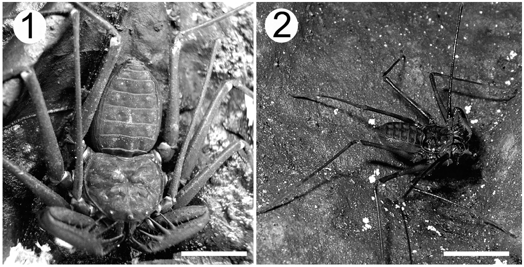
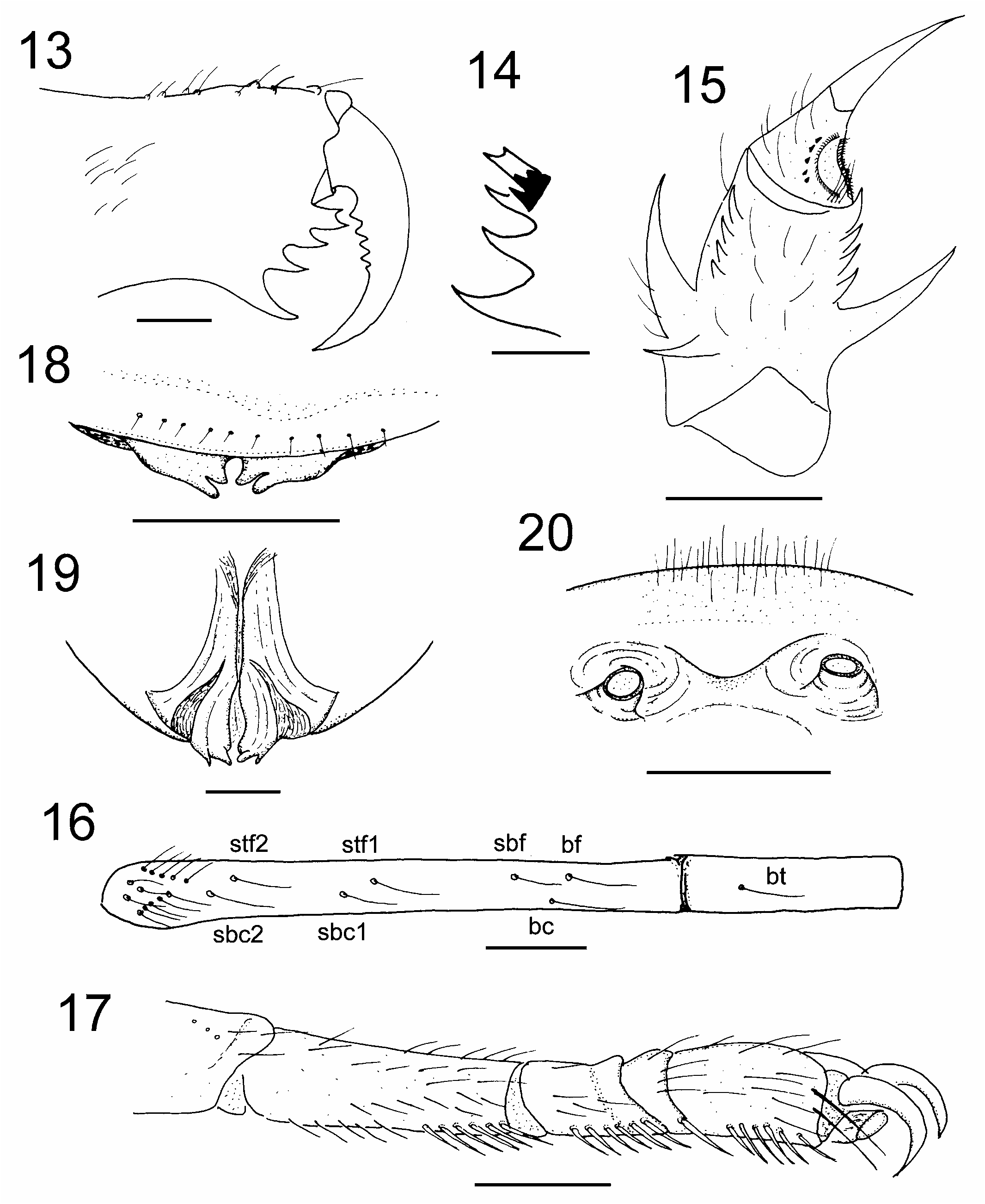
( Figs. 1 View Figs , 3–12 View Figs )
Material examined. – Holotype: male ( MZB.Ambl. 109), INDONESIA: Banten: Air Panas Cibiuk [= Hot Water spring], under stones on small limestone area (06°47'08.3"S 105°31'22.6"E, alt. 81 m a.s.l.), Desa [= Village] Taman Jaya, Kecamatan [= District] Sumur, Kabupaten [= Regency] Lebak, coll. Sidiq Harjanto and Cahyo Rahmadi, 25 Feb.2007. GoogleMaps
Paratypes. – INDONESIA: Banten: 1 female ( MZB. Ambl. 110) , 4 juveniles ( MZB. Ambl. 111–114), same locality data as holotype ; 1 male ( MZB.Ambl.021) , 2 females
( MZB. Ambl.022–023) (1 female with an egg sac on ventral abdomen) and 1 juvenile ( MZB.Ambl. 023), Ujung Kulon, Ujung Kulon National Park , coll. Team Oxford (Christopher Stewart), Aug.1993 ; 7 females ( MZB.Ambl. 025–031), Pulau Legundi (no specific location, presumably located on Lampung Bay), coll. unknown, 21 May 1955 .
Specimens for comparison. – 1 male and 1 female Stygophrynus berkeleyi ( ZRC.ARA.529: 2 ex.) MALAYSIA: Kedah, Baling, Limestone cave . coll. H. D. Collings. Apr.– May 1935. Identified by E.A.M. Speijer. All specimens of Stygophrynus dammermani .
Diagnosis. – Can be easily distinguished from other species of the genus by the following combination of characters: two teeth on the external face of the basal cheliceral segment; six teeth on the movable finger with the 2 dorsalmost teeth about equal in size, the remaining teeth decreasing in size distally; the dorsal surface of the chelicera roughened with small denticles.
Description. – Male holotype: Colour of living holotype specimens: carapace, pedipalpal femur dark black, pedipalpal tibia and tarsi reddish, pedal tibiae reddish, abdominal tergites greenish; colour in alcohol: carapace, pedipalps and legs brown; tergites yellowish-brown; femur of legs without annulations. All setae acicular.
Carapace ( Fig. 1 View Figs ): anterior margin nearly straight, with 12 setiferous tubercles, eye tubercle black, large and high with two setiferous tubercles on dorsal surface, eyes slightly directed to anterolateral margin, surface with numerous small tubercles lacking setae especially on frontal margin, several setiferous tubercles, central sulcus deep and radiating; frontal process triangular, tip visible from above.
Chelicera ( Figs. 3–4 View Figs ): dorsal surface with 7 large and 7 small setiferous tubercles; antero-dorsal surface of basal segment with 2 setiferous tubercles on outer margin and 1 setiferous tubercle on inner margin, basal segment with 4 teeth on internal margin ( Fig. 3 View Figs ), the upper-most tooth bicuspid, with lower cusp larger than upper cusp, the lower-most the largest, external margin with 2 teeth on common base ( Fig. 4 View Figs ), movable finger with 6 teeth, the 2 proximal teeth about equal in size, teeth 3 to 6 decreasing in size distally.
Sternum: anterior sternite of tritosternum elongate, median and posterior sternites rounded; anterior sternite with 2 apical setae and 2 intermediate setae; median sternite with 3 small setae and 2 large setae, posterior sternite with 2 large setae.
Pedipalp ( Figs. 5–6 View Figs ): trochanter with 8 spines and 11 setiferous tubercles on anterodorsal margin, spines with basal setae, with 7 anteroventral spines, and many setiferous tubercles; femur with 4 major spines and many small denticles on anterodorsal margin, F3 the longest, F3> F2> F4> F1, 1 small spine between F1–F2, F2–F3 and F3–F4 ( Fig. 5 View Figs ), 4 major spines and 4 small spines on anteroventral margin, FIII> FII> FI> FIV, with several small spines between FI and distal margin, 1 small spine between FI–FII; FIII and FIV situated close together on proximal margin ( Fig. 6 View Figs ); patella with 3 major spines about equal size and 2 small spines on anterodorsal margin, with 2 small spines between P1 and distal margin, with 1 small spine between P1–P2 and P4–P5, and with 3 small spines between P5 and proximal margin ( Fig. 5 View Figs ); with 5 major spines and 6 small spines on anteroventral margin, PIII> PIV> PV> PI> PII ( Fig. 6 View Figs ); tibia with 1 large submedial spine on anterodorsal margin, which has a subsidiary basal spine, with 3 contiguous spinelets on distal margin, the most distal the largest, this 3 small spinelets decreasing in size proximally, anteroventral margin with 1 large submedial spine and, with 4 spinelets distal ones (the most distal largest and three other decreasing in size proximally); tarsus with 5 denticles dorsal to cleaning organ, about three setae on proximal edge of cleaning organ; cleaning organ with row of short dorsal setae and 29 long setae ventrally; tarsus completely divided, apotele present ( Fig. 7 View Figs ).
Legs ( Figs. 8, 9 View Figs ): femora I, II, III and IV with small tubercles lacking setae. Right tibiae I with 25 segments, right tarsus I with 43 segments, left leg I is missing; tibiae II and III with 2 segments; tibiae IV with 5 segments; fourth segment with 1 trichobothrium, bt (0.48); fifth segment (distitibia) with 22 trichobothria ( Fig. 8 View Figs ), bf (0.10), sbf (0.27), stf 1 (0.35), stf2 (0.75), bc (0.21), sbc1 (0.55), sbc2 (0.75), distitibia II and III with same number and arrangement of trichobothria ( Fig. 8 View Figs ); tarsi II, III, IV with 4 segments, segment 2 with light transverse line, fourth segment without oblique slit; pulvilli present ( Fig. 9 View Figs ).
Genitalia ( Figs. 10–12 View Figs ): Male: Ventral surface with genital operculum cover the genitalia, the distal part with darker colour. Two limbs are present on ventral which is shorter than dorsal one ( Fig. 10 View Figs ), dorsal surface with two black striations
( Fig. 11 View Figs ). Female: Gonopods is soft and tube-like, with setae on margin of genital operculum ( Fig. 12 View Figs ).
Dimensions (mm), male holotype (female paratype MZB. Ambl.110): Body length (excluding chelicera) 13.00 (13.00). Carapace: median length 4.75 (4.50), width 7.00 (7.00); median eyes to anterior margin 0.15 (0.15), lateral eyes to lateral eyes 2.75 (2.40), to anterior margin 0.50 (0.50), lateral margin 0.50 (0.50). Pedipalps: trochanter length 1.75 (1.75), width 1.00 (1.00), femur length 5.00 (4.50), width 1.50 (1.50), patella length 5.50 (5.00), width 1.25 (1.25), tibia length 2.50 (2.00), width 1.25 (1.25), tarsus length 2.25 (2.40). Leg I: femur 13.75 (13.00), patella 1.00 (1.00), tibia 23.15 (22.50), tarsus 23.75 (25.00). Leg II: femur 8.25 (7.75), patella 1.25 (1.00), basitibia 7.50 (7.00), distitibia 3.50 (3.25), metatarsus and tarsus 2.75 (2.50). Leg III: femur 9.00 (8.75), patella 1.25 (1.00), basitibia 8.75 (8.25), distitibia 3.75 (3.75), metatarsus and tarsus 2.75 (2.50). Leg IV: femur 8.50 (8.00), patella 1.25 (1.00), basitibia 9.50 (9.00), distitibia 3.25 (3.00), metatarsus and tarsus 3.00 (2.75).
Etymology. – The specific epithet refers to the presence of this species on Sunda Strait. It is to be treated as a noun in apposition.
Remarks. – Stygophrynus sunda is most similar to S. dammermani but the new species is much smaller than the latter, can also be separated by the dentition of the external margin of the basal segment of the chelicera and the dentition of the movable cheliceral finger. The number of trichobothria and their arrangement is also quite different, as S. dammermani has 21 trichobothria ( Fig. 16 View Figs ) and S. sunda has 23 ( Fig. 8 View Figs ).
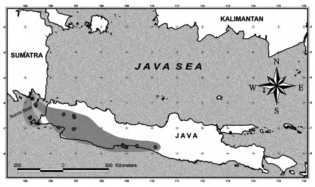
Natural history. – S. sunda is found live under stones in limestone forest in Ujung Kulon National Park. The habitat in Legundi Island is uncertain since no specific information on specimens examined.The distribution of this species is only known from Pulau Legundi located in the Sunda Strait and on the western ridge of Gunung Hondje near Cibiuk Hotspring, Ujung Kulon in western Java ( Fig. 21 View Fig ).

No known copyright restrictions apply. See Agosti, D., Egloff, W., 2009. Taxonomic information exchange and copyright: the Plazi approach. BMC Research Notes 2009, 2:53 for further explanation.
|
Kingdom |
|
|
Phylum |
|
|
Class |
|
|
Order |
|
|
Family |
|
|
Genus |