
Lepeophtheirus acutus Heegaard, 1943
|
publication ID |
https://doi.org/ 10.1080/00222933.2012.738832 |
|
DOI |
https://doi.org/10.5281/zenodo.10536628 |
|
persistent identifier |
https://treatment.plazi.org/id/52218794-812D-5D16-6684-84AA194AB3C2 |
|
treatment provided by |
Felipe |
|
scientific name |
Lepeophtheirus acutus Heegaard, 1943 |
| status |
|
Lepeophtheirus acutus Heegaard, 1943
( Figures 1–4 View Figure 1 View Figure 2 View Figure 3 View Figure 4 )
Lepeophtheirus acutus Heegaard, 1943: 4 ; Yamaguti, 1963: 71; Kik et al., 2011: 797.
Material examined
From M. alfredi : nine adult females (two immature) and one adult male, ex no. 16, 29 October 2006; 21 immature adult females and 10 adult males, ex no. 10-3-1, 20 September 2009; seven adult females, ex no. 10-3-1, 18 June 2010. From R. typus : 49 adult females (one immature), ex no. 27, 10 January 2010; one adult female, ex no. 28, 30 April 2008; seven adult females, ex no. 32, 2 October 2009; 15 adult females, ex no. 32, 28 April 2010; 23 adult females, ex no. 33, 30 October 2009; 207 adult females (one immature) and three adult males (NSMT-Cr 21848), ex no. 33, 27 January 2010. Syntypes (Type-656): three immature adult females, one mature adult male and one late chalimus female, Apamama , Gilbert Islands, Kiribati, 24 November 1917 .
Description
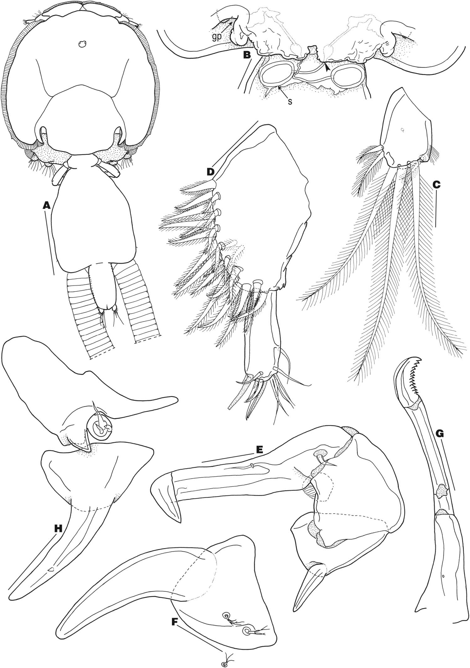
Adult female. Body ( Figure 1A View Figure 1 ) 4.22 (3.77–4.59) mm long (excluding caudal setae) (n = 10). Cephalothoracic shield subcircular, slightly longer than wide [1.99 (1.80–2.12) × 1.92 (1.70–2.17) mm], with well-developed paired frontal plates and posterior margin of thoracic zone extending beyond posterior limit of lateral zone. Free fourth pedigerous somite wider than long [208 (175–225) × 409 (350–475) µm] and indistinctly separated from genital complex. Genital complex longer than wide [1.39 (1.25–1.57) × 1.08 (0.95–1.22) mm], with oblique margins at anterior half, parallel margins at posterior half, slightly protruded posterolateral corners and genital apertures ( Figure 1B View Figure 1 ) situated ventrally near junction of abdomen. Abdomen composed of one somite, longer than wide [616 (550–675) × 350 (300–375) µm] and indistinctly separated from genital complex. Caudal ramus ( Figure 1C View Figure 1 ) longer than wide [91 (65–100) × 69 (60–80) µm], with six plumose setae (seta I absent), mid-dorsal sensillum and short row of setules along distomedial margin. Egg sacs ( Figure 1A View Figure 1 ) uniseriate.
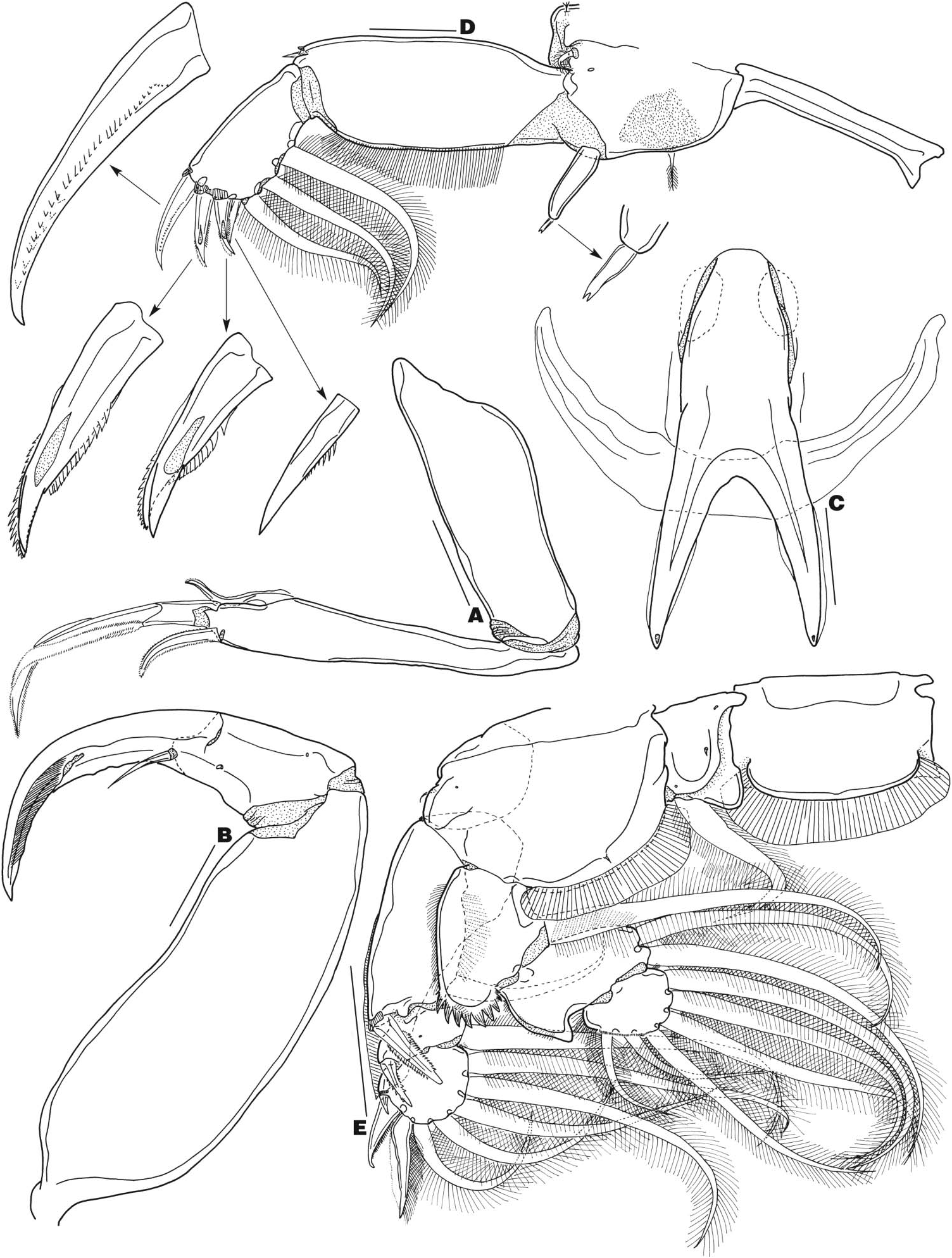
Antennule ( Figure 1D View Figure 1 ) two-segmented; proximal segment longer than distal segment, bearing two posterodistal processes and 27 setae (25 plumose, two naked); distal segment slim, bearing 12 setae (two setae near posterodistal corner share a common base) and two aesthetascs. Antenna ( Figure 1E View Figure 1 ) three-segmented, comprising coxa, basis and one-segmented endopod incorporating distal claw; coxa with long spiniform process on posterolateral corner; basis stout, with dorsolateral adhesion pad; terminal segment long, uncinate, bearing two setae. Postantennal process ( Figure 1F View Figure 1 ) slightly curved, with two inner setulose papillae at wide base and another setulose papilla adjacent to base. Mandible ( Figure 1G View Figure 1 ) modified into elongated stylet bearing distolateral hyaline membrane and 12 distomedial teeth. Maxillule ( Figure 1H View Figure 1 ) composed of trisetose papilla and long, slender dentiform process; latter with minute hyaline structure on ventral surface; sclerite anterior to papilla with triangular process that distally projects over base of dentiform process. Maxilla ( Figure 2A View Figure 2 ), brachiform, two-segmented, composed of elongate, unarmed syncoxa and slender basis; latter with long, curved flabellum and long apical calamus and short apical canna; calamus and canna each furnished with finely serrated membranes. Maxilliped ( Figure 2B View Figure 2 ) large, subchelate, three-segmented, comprising long protopod (corpus) and subchela consisting of free endopodal segment (shaft) and claw; protopod unarmed; shaft with minute pore proximally and minute hyaline element subapically; claw with long basal seta and fine surface striations distally. Tines of sternal furca ( Figure 2C View Figure 2 ) slightly shorter than box, divergent, apically pointed, ornamented with hyaline marginal membranes and overlying bow-shaped sclerite.
Armature on rami of legs 1–4 is shown in Table 2.
Leg 1 ( Figure 2D View Figure 2 ) intercoxal sclerite naked and elongate. Protopod with one outer and one inner plumose seta, one proximolateral setulose papilla, one mid-lateral pore and patch of minute spinules near insertion of inner seta. First exopodal segment with small pectinate membrane at base of small outer spine and row of setules along inner margin. Second exopodal segment with pectinate membrane at base of each apical denticulate spine; middle and inner apical spines each with accessory process; apical seta short, with row of spinules along inner margin; three inner setae plumose. Endopod cylindrical, bearing apically bifurcate element. Leg 2 ( Figure 2E View Figure 2 ) intercoxal sclerite subquadrate, with hyaline membrane along distal margin. Coxa with one inner plumose seta and one small sensillum and two minute pores on anterior surface. Basis with one outer short, naked seta, one minute pore near outer margin, one small inner sensillum and hyaline membrane along dorsolateral and posterior margins. Exopod three-segmented, with large hyaline membrane covering dorsal surface of ramus. First segment with one inner plumose seta, row of setules along inner margin and pectinate membrane at base of outer serrate spine. Second segment with one inner plumose seta, short row of setules along proximomedial margin, one outer serrate spine and one minute pore on anterior surface. Third segment with five inner plumose setae, few setules along proximomedial margin, pectinate membrane at base of outermost naked spine, row of spinules along inner margin of outer apical spine and finely serrate membrane along outer margin and row of setules along inner margin of apical spine. Endopod three-segmented. First segment with one inner plumose seta, row of setules along most of outer margin and row of large denticles on distolateral corner. Second segment with two inner plumose setae, spiniform projection on distolateral corner and row of setules along inner and outer margins. Third segment with spinules at base of three outermost plumose setae and row of setules along proximolateral and proximomedial margins.
Leg 3 ( Figure 3A View Figure 3 ) protopod with one outer naked and one inner plumose setae, velum between rami, corrugated patch on dorsolateral surface, three marginal membranes, minute pores scattered on ventral surface, two short sensilla along posterior margin and patch of fine spinules between pair of short sensilla on ventromedian surface. Exopod two-segmented. First segment with one inner plumose seta and one outer spinulate spine; latter straight, with hyaline membrane at base. Second segment with five plumose setae, four spinulate spines, one minute pore and setules along outer margin. Endopod two-segmented. First segment with one inner plumose seta. Second segment with six plumose setae and setules along outer margin. Leg 4 ( Figure 3B View Figure 3 ) uniramous, composed of protopod and two-segmented exopod. Protopod with one distolateral pinnate seta. First exopodal segment with pectinate membrane at base of outer spinulate spine. Second exopodal segment with three apical spinulate spines, one distal pore and pectinate membrane at base of innermost spine; outer and middle spines subequal, about one-third length of inner spine; middle spine typically reflexed over inner spine. Leg 5 ( Figure 3C View Figure 3 ) vestigial, situated near posterolateral corners of genital complex, composed of two setiferous lobes (anterior lobe with one plumose seta; posterior lobe with one pore and three plumose setae). Leg 6 (not figured) rudimentary, represented by unarmed genital operculum at gonopore opening.
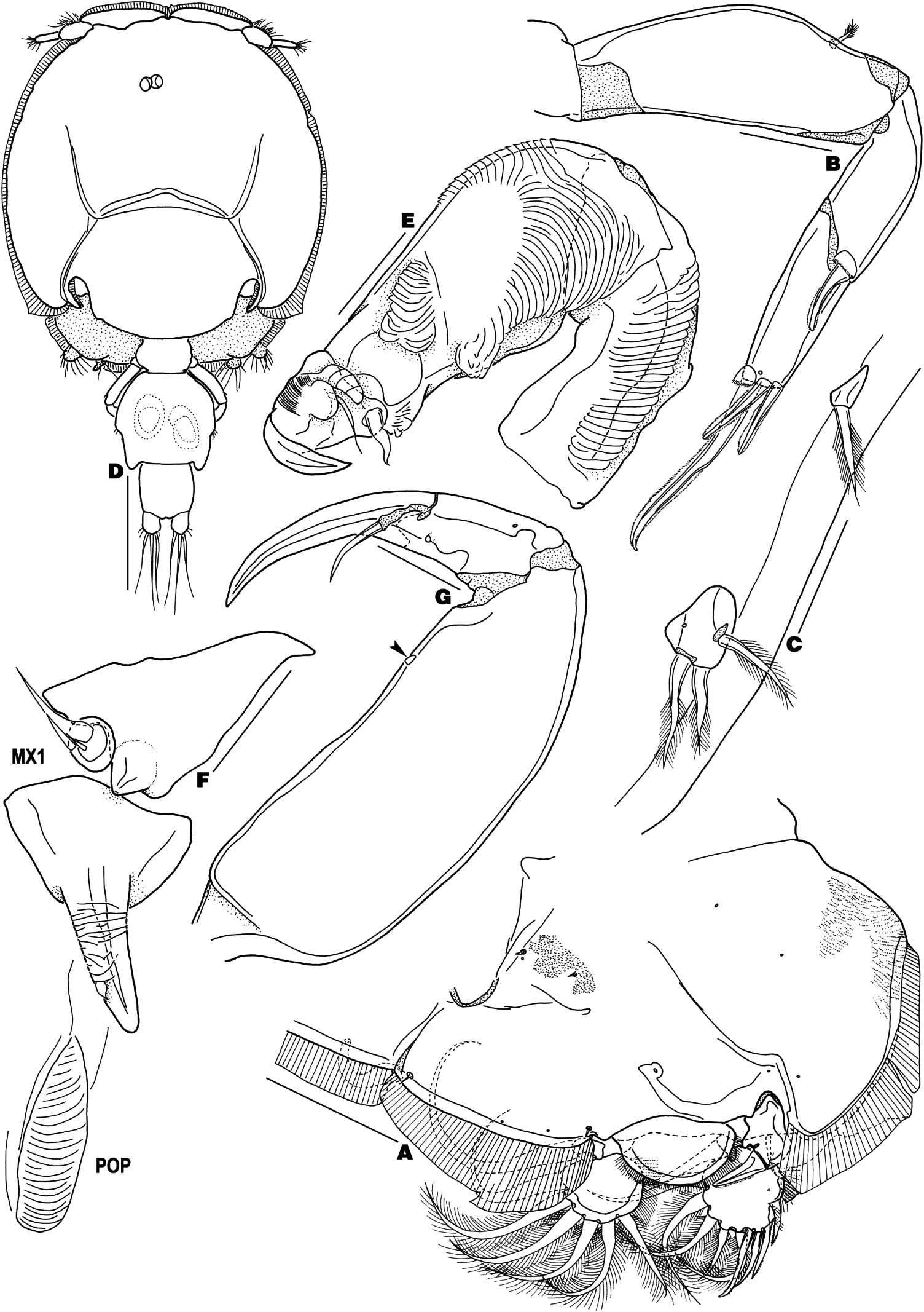
Adult male. Body ( Figure 3D View Figure 3 ) 1.92 (1.76–2.32) mm long (excluding caudal setae) (n = 7). Cephalothoracic shield slightly longer than wide [1.21 (1.10–1.45) × 1.11 (1.04–1.27) mm]. Free fourth pedigerous somite wider than long [113 (100–140) × 209 (190–260) µm]. Genital complex slightly wider than long [344 (310–390) × 357 (330–400) µm]. Abdomen composed of one somite, longer than wide [210 (190–275) × 192 (180–210 µm]. Caudal ramus longer than wide [95 (90–110) × 65 (60–75) µm], armed as in female.
All limbs as in female, except for the following. Antenna ( Figure 3E View Figure 3 ) threesegmented, comprising coxa, basis, and one-segmented endopod incorporating distal claw; coxa with corrugate structure along inner margin; basis with one large and two small corrugate structures; terminal segment forming short claw and bearing two setae, outer surface striations, one corrugate, linguiform structure and round process. Maxillule ( Figure 3F View Figure 3 ) with corrugate surface and one stout hyaline structure on dentiform process. Postoral process ( Figure 3F View Figure 3 ) elongate, with corrugate surface. Maxilliped ( Figure 3G View Figure 3 ) similar to that of female, except protopod with small hyaline structure on myxal area and claw without surface ornamentation. Tines of sternal furca ( Figure 4A View Figure 4 ) noticeably shorter than box and unornamented.
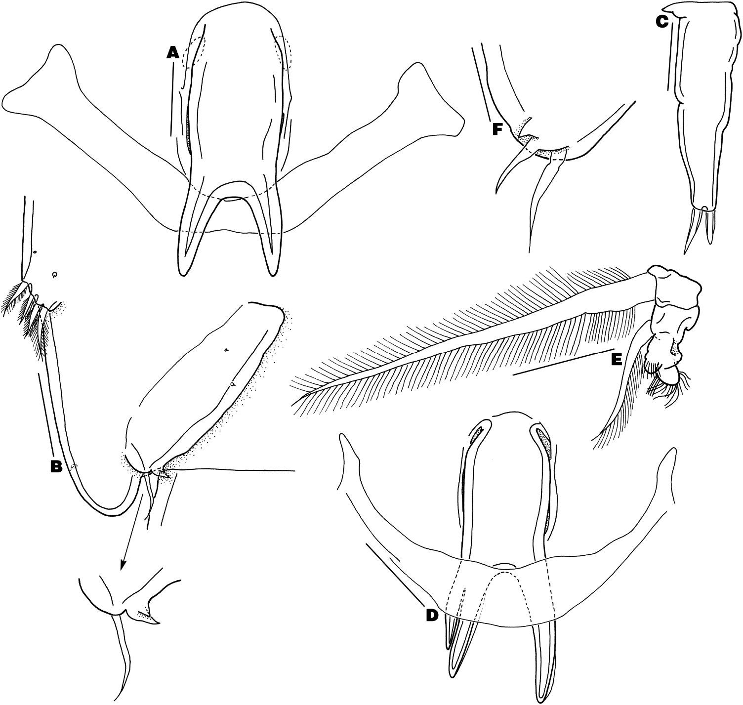
Leg 5 ( Figure 4B View Figure 4 ) vestigial, situated mid-laterally on genital complex and bearing four plumose setae. Leg 6 ( Figure 4B View Figure 4 ) forming genital operculum, with one seta and two subequal, spiniform processes on distolateral corner.
Variability
Some females with two apical elements on leg 1 endopod ( Figure 4C View Figure 4 ). One immature adult female syntype with one bifurcate tine on sternal furca ( Figure 4D View Figure 4 ). One male with an abnormal distal endopodal segment of left leg 3 ( Figure 4E View Figure 4 ). One male with very long spiniform process on leg 6 ( Figure 4F View Figure 4 ).
Attachment sites
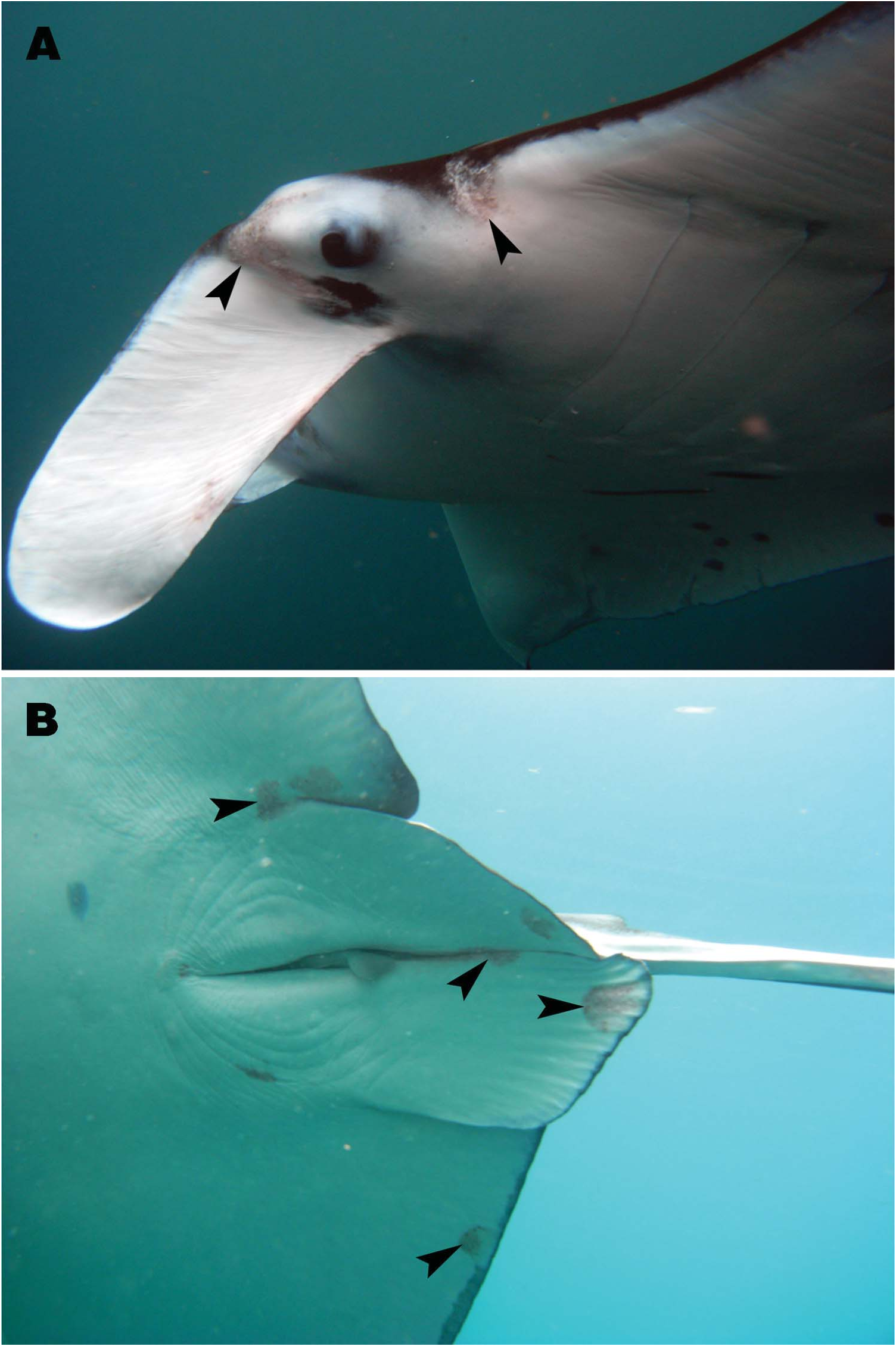
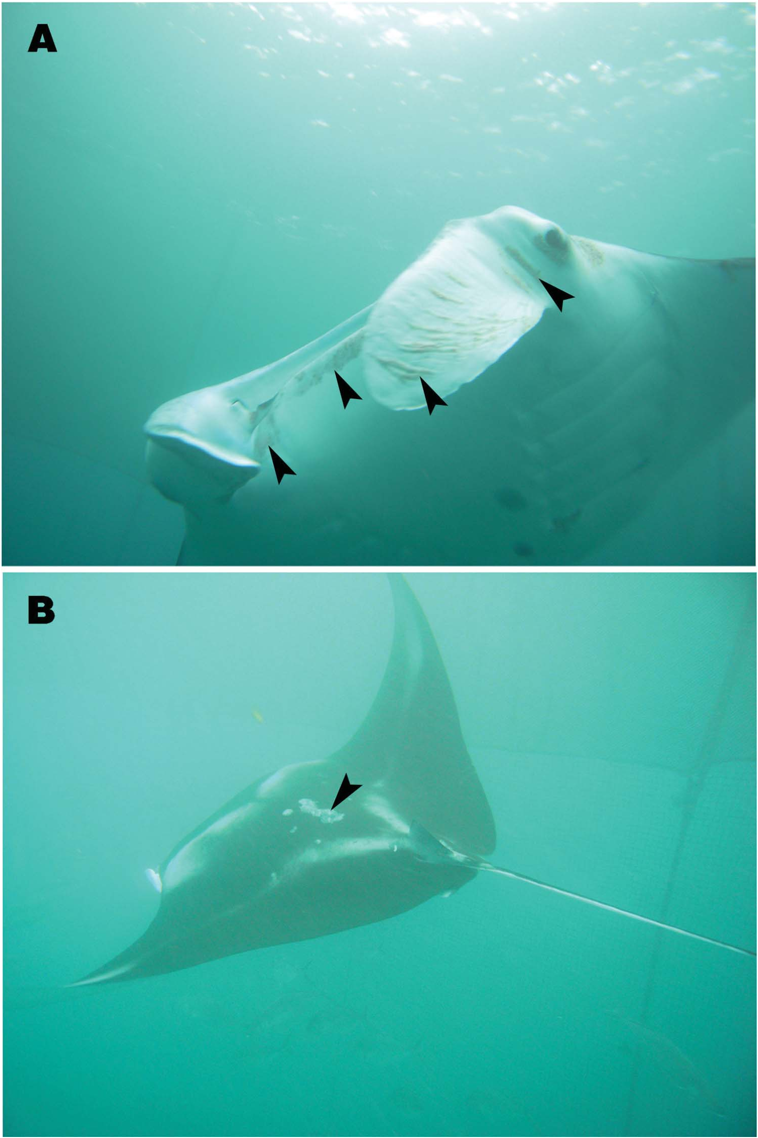
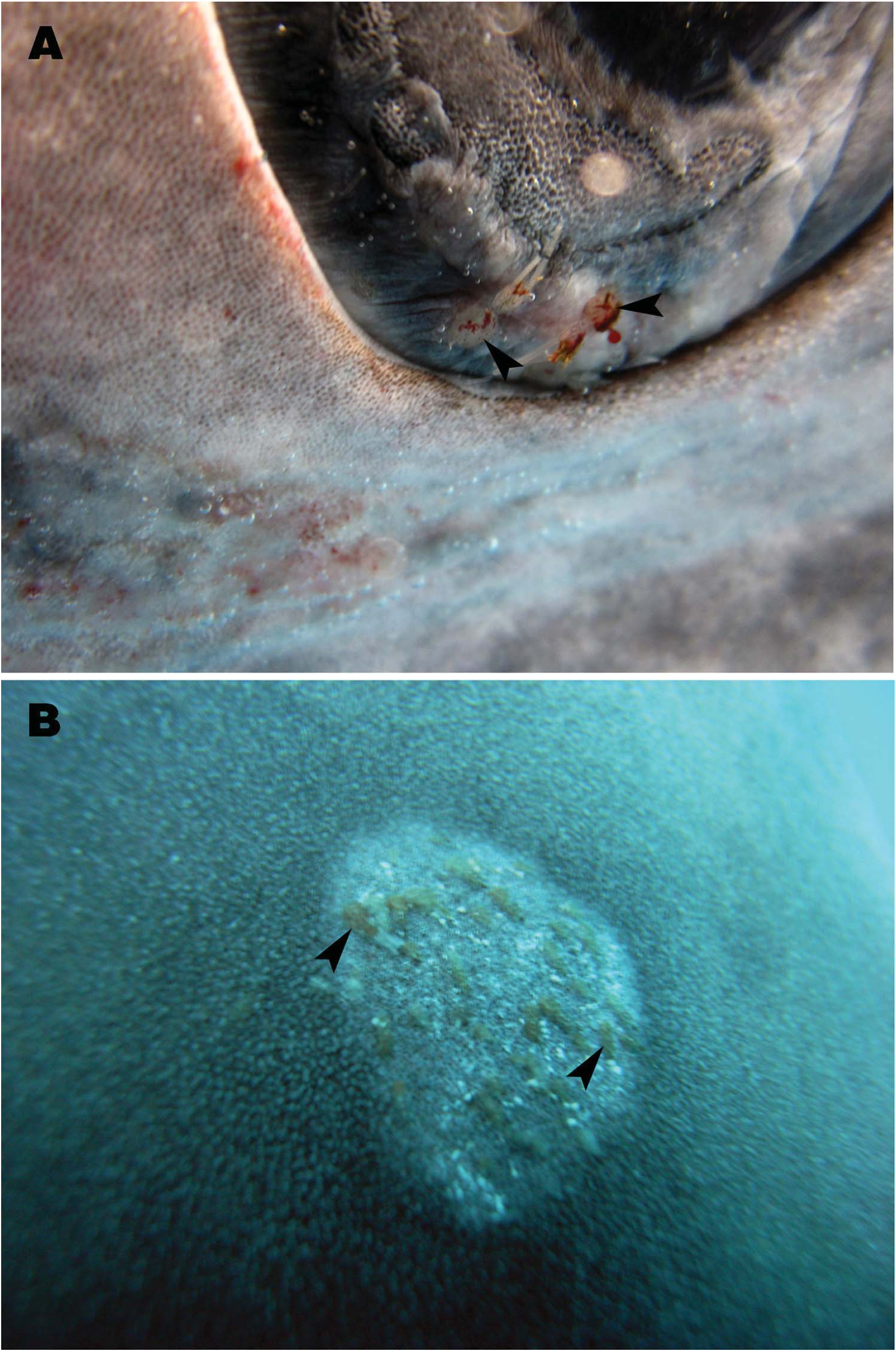
Lepeophtheirus acutus individuals were found attached around the eyes and mouth, on the cephalic fins, on and around the pelvic fins and on the dorsal body surface of M. alfredi ( Figures 5 View Figure 5 and 6 View Figure 6 ) and on the eyes and dorsal body surface of R. typus ( Figure 7 View Figure 7 ).
No known copyright restrictions apply. See Agosti, D., Egloff, W., 2009. Taxonomic information exchange and copyright: the Plazi approach. BMC Research Notes 2009, 2:53 for further explanation.
|
Kingdom |
|
|
Phylum |
|
|
Class |
|
|
Order |
|
|
Family |
|
|
Genus |
Lepeophtheirus acutus Heegaard, 1943
| Tang, Danny, Maran, B. A. Venmathi, Matsumoto, Yousuke & Nagasawa, Kazuya 2013 |
Lepeophtheirus acutus
| Kik MJL & Janse M & Benz GW 2011: 797 |
| Yamaguti S 1963: 71 |
| Heegaard P 1943: 4 |