
Portelmis kinonatilis, Fernandes, André S., Passos, Maria Inês S. & Hamada, Neusa, 2010
|
publication ID |
https://doi.org/ 10.5281/zenodo.196163 |
|
DOI |
https://doi.org/10.5281/zenodo.3505177 |
|
persistent identifier |
https://treatment.plazi.org/id/523FAE58-FFC2-3B15-51D1-F99CFC993BBC |
|
treatment provided by |
Plazi |
|
scientific name |
Portelmis kinonatilis |
| status |
sp. nov. |
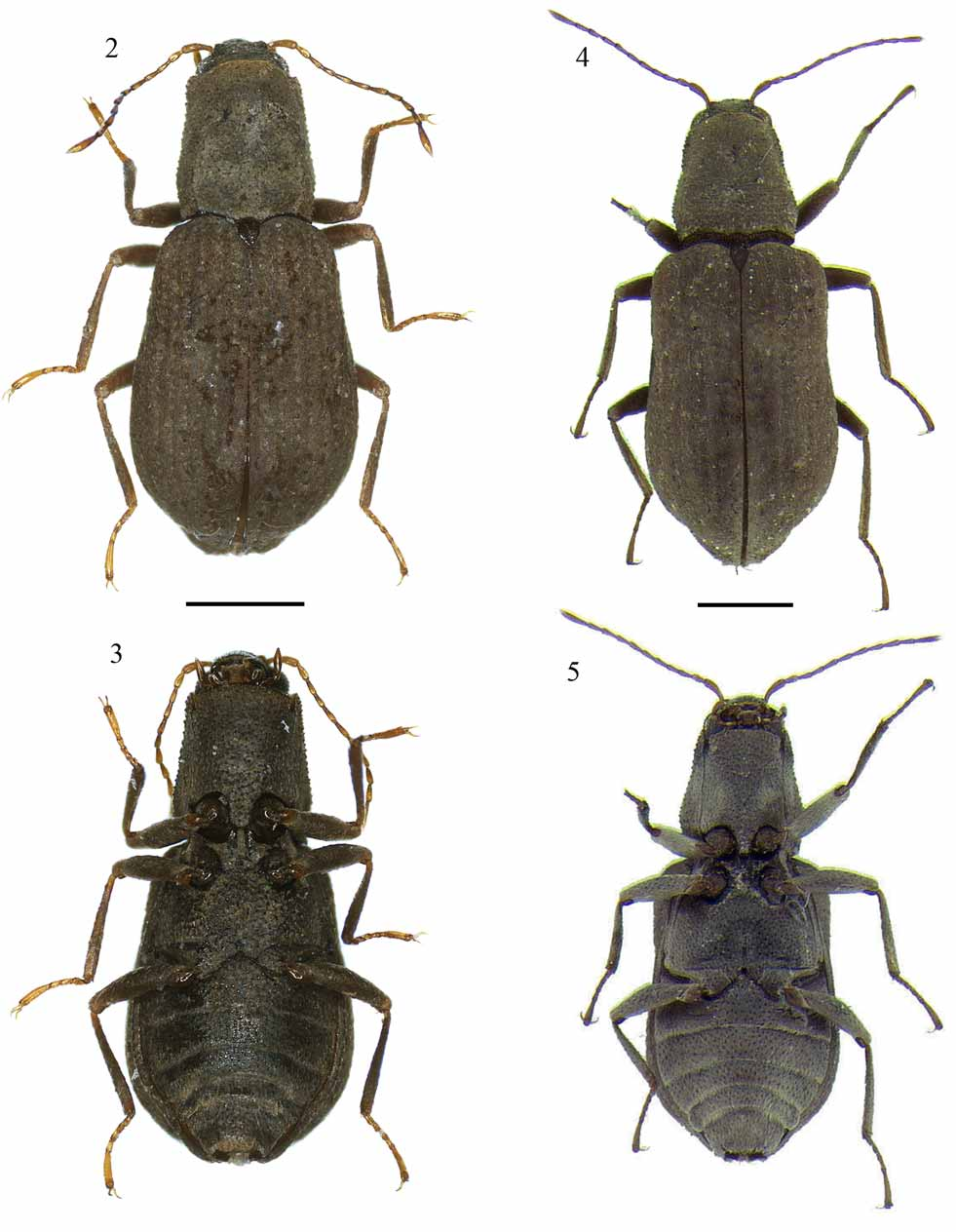
Portelmis kinonatilis View in CoL sp. nov.
( Figs. 2 View FIGURES 2 – 5 ; 3; 10; 11; 14)
Diagnosis. Pronotum with sublateral carinae extending to the basal 1/4; pronotal surface densely granulate. Prosternal process gradually and feebly narrowed to apex; lateral margin nearly straight; apex rounded. Disc of ventrite I densely granulate, without punctures. Disc of metasternum with median longitudinal impression restricted to posterior 1/2. Male genitalia, in ventral view, with parameres slightly elongated, with 3/4 the length of median lobe, apex rounded with spongy structures; median lobe short and abruptly narrowed in the apical 1/4, with apex rounded.
Description. Holotype: male ( Figs. 2 View FIGURES 2 – 5 ; 3; 10; 11). Length 2.07 mm, greatest width 0.87 mm. Body ( Figs. 2 View FIGURES 2 – 5 ; 3) elongate, subovate; surface of head, pronotum and ventral surface of body with granules 3/4 the diameter of eye facets and usually separated by about three times their diameter (prosternum, disc of metasternum and disc of ventrite I with coarser granules); dorsum sparsely covered with fine, short, recumbent and pale setae, with sparse long setae near elytral apex, scutellum glabrous; surface of the venter generally as the dorsum, with plastron present.
Head ( Figs. 2 View FIGURES 2 – 5 ; 3): Without distinct impressions; frontal margin truncate. Eyes moderately protuberant; laterally rounded; separated by a distance two times wider than eye. Antenna with 11 segments; long and slender; first and last segments slightly swollen and twice as long as the remaining segments. Frontoclypeal suture present between bases of antennae. Clypeus slightly wider and longer than labrum; anterior margin truncate; lateral angles rounded. Labrum rectangular; anterior margin slightly convex; anterolateral angles rounded, with some long and pale setae. Maxillary palpus with four segments; last segment slightly swollen, as long as second and third segments combined. Labial palpus with three segments; last segment swollen, two times wider than second segment, as long as the remaining segments combined. Gula 2.5 times narrower than submentum.
Color ( Figs. 2 View FIGURES 2 – 5 ; 3): Cuticle yellowish-brown and opaque, except base of head, mouth parts, antennae, scutellum, trochanters and tarsus which are shiny.
Thorax ( Figs. 2 View FIGURES 2 – 5 ; 3): Pronotum ( Fig. 2 View FIGURES 2 – 5 ) longer (0.60 mm) than wide at base (0.53 mm); wider at base than at apex (0.44 mm); one sublateral carina, on each side, present on basal 1/4; impressions on disc (one longitudinal, median, oval, on apical 3/4; one oblique on each side, on basal 1/3); anterolateral angles slightly produced, subacute; anterior margin broadly convex, sinuate behind eyes, extending over base of head; lateral margin nearly straight, crenate; posterior angles slightly produced, acute; posterior margin with three arches, two broad, one on each side in front of the elytron, and one narrow in front of scutellum. Elytra ( Fig. 2 View FIGURES 2 – 5 ) subovate; longer (1.4 mm) than wide, (maximum width, at apical 1/6, 0.87 mm); intervals flat; humeral angle broadly rounded, slightly sinuate at middle; without sublateral carinae; lateral margins crenate; disc with punctures separated by twice their diameters, half as wide as intervals between striae. Scutellum ( Fig. 2 View FIGURES 2 – 5 ) flat; subovate; longer than wide; wider at base; angles rounded. Prosternum ( Fig. 3 View FIGURES 2 – 5 ) with anterior margin convex; without impression or carinae. Prosternal process nearly as long (0.14 mm) as wide at base (0.135 mm), wider at base than at apex (0.08 mm); not extending beyond anterior coxae; gradually and gently narrowed to apex; lateral margin nearly straight, apex rounded. Mesosternum ( Fig. 3 View FIGURES 2 – 5 ) longer (0.17 mm) than wide between coxae (0.14 mm); shorter than prosternum; posterior margin between mesocoxae concave, wider than anterior margin between procoxae. Metasternum ( Fig. 3 View FIGURES 2 – 5 ) with median, longitudinal impression extending until basal 1/ 2; disc flat; anterior margin between mesocoxae convex; posterior margin between metacoxae concave; posterior portion in front of metacoxae with transverse arched impression. Legs ( Figs. 2 View FIGURES 2 – 5 ; 3) long; pro- and mesocoxae globular; tibiae with indistinct fringes of tomentum, two fringes on apical 1/2 (anterior and posterior margin) on the mesotibiae and only one fringe (inner margin) on pro- (apical 1/4) and metatibiae (apical 1/2); tarsal claws without basal teeth.
Abdomen ( Fig. 3 View FIGURES 2 – 5 ): Nearly as long (0.8 mm) as wide (maximum width, ventrite I, 0.78 mm). Ventrite I ( Fig. 3 View FIGURES 2 – 5 ) with anterior margin between metacoxae strongly convex; without carinae or impression. Ventrite V ( Fig. 3 View FIGURES 2 – 5 ) with postero-lateral angle with strong toothlike projection; posterior margin between projections truncate, with long setae extending beyond posterior margin.
Male Genitalia ( Figs. 10 View FIGURES 10 – 19 ; 11): Parameres ( Figs. 10 View FIGURES 10 – 19 ; 11) slightly elongate, 3/4 the length of median lobe. Paramere in dorsal view ( Fig. 10 View FIGURES 10 – 19 ) gradually narrowed to apex; apex rounded, with nearly indistinct spongy structures. Paramere in lateral view ( Fig. 11 View FIGURES 10 – 19 ) with lateral margins nearly parallel; apex rounded, with nearly indistinct spongy structures. Median lobe ( Figs. 10 View FIGURES 10 – 19 ; 11) short, about 1/2 the length basal lobe. Median lobe in dorsal view ( Fig. 10 View FIGURES 10 – 19 ) about the same width of parameres; abruptly narrowed in the apical 1/3, with apex rounded. Median lobe in lateral view ( Fig. 11 View FIGURES 10 – 19 ) about 1/2 the width of parameres; moderately curved to venter; gonopore extending beyond the apex of parameres; apex rounded.
Plastron: Present on the genae, and ventral surface of thorax and abdomen except pro- and mesocoxae, tarsus and trochanters.
Female. Externally similar to male.
Female Genitalia ( Fig. 14 View FIGURES 10 – 19 ): Coxites less than 1/3 the length of the styli; in dorsal view wider at base than long. Styli elongate; in dorsal view basal segment narrowed from base to middle, and then slightly widened until apical portion; with numerous pores distributed along the apical 1/2; external lateral margin with some pairs of very short and slender setae, apex of each basal segment rounded and with some short and stout setae; apical segment narrow, cylindrical and 1/6 the length of the basal segment.
Intraspecific variation. Size range (n= 7): length 2.01 – 2.16 mm, greatest width 0.82 – 0.88 mm. Color: little variation in cuticle tonality. The specimens examined did not have significant morphological variation.
Type-locality. Igarapé Marta Stream, Coari Municipality, Amazonas State, Brazil (04°49’38”S, 65°01’55”W).
Type-series. Holotype (Male): BRAZIL: Amazonas: Coari, Igarapé Marta, 04°49’38”S, 65°01’55”W, Dnet, Kinon, R. L. Ferreira-Keppler leg. 21/IV/2007 (INPA). Paratypes: 1 male, same data as holotype (INPA); 1 female, same data as holotype (INPA); 1 male, same data as holotype (DZRJ); 1 female, same data as holotype (DZRJ); 1 male, same data as holotype (NMNH); 1 female, same data as holotype (NMNH).
Habitat notes. The type series was collected with a D-net in the kinon. The kinon was first defined by Fittkau (1977) and consists of organic material (especially plant fragments) floating and retained by a barrier (such as a log) in Amazonian running waters. Specimens of P. g u r n e y i and a probable larva of Portelmis were also found at the same sampling of Kinon. The larva does not appear to belong to any of the South American genera for which larvae have been described. However, the larval stage of several Elmidae genera are still unknown, therefore it is uncertain if the larva in question belongs to Portelmis . The occurrence of specimens of P. kinonatilis and P. g u r n e y i in kinon represents the first report of the natural habitat of Portelmis .
Etymology. The specific epithet is a reference to the habitat where the type-series was found, the kinon ( kinonatilis , from latin “found in kinon).
Comparative notes. Portelmis kinonatilis sp. nov. is closely related to P. nevermanni . Both species have the pronotal surface and the disc of ventrite I densely granulate; disc of ventrite I without punctures; and disc of metasternum with median longitudinal impression restricted to the posterior 1/2 ( Figs. 2 View FIGURES 2 – 5 ; 3). Portelmis kinonatilis sp. nov. can be distinguished from P. nevermanni by the presence of short sublateral carinae on pronotum (pronotum without carinae in P. nevermanni ); elytra subovate (elytra subparallel in P. nevermanni ); prosternal process with lateral margin nearly straight (prosternal process subcapitate in P. nevermanni ) ( Figs. 2 View FIGURES 2 – 5 ; 3). Portelmis kinonatilis sp. nov. can be distinguished from all known species of Portelmis by the spongy structures on the apices of its parameres ( Figs. 10 View FIGURES 10 – 19 ; 11).

No known copyright restrictions apply. See Agosti, D., Egloff, W., 2009. Taxonomic information exchange and copyright: the Plazi approach. BMC Research Notes 2009, 2:53 for further explanation.
|
Kingdom |
|
|
Phylum |
|
|
Class |
|
|
Order |
|
|
Family |
|
|
SubFamily |
Elminae |
|
Genus |