
Charinus tocantinensis, Souza & Reis-Venâncio & Torres & Ferreira, 2024
|
publication ID |
https://doi.org/ 10.11646/zootaxa.5399.4.7 |
|
publication LSID |
lsid:zoobank.org:pub:FBC2E993-1342-4B65-B322-A15C7EFD41AC |
|
DOI |
https://doi.org/10.5281/zenodo.10517216 |
|
persistent identifier |
https://treatment.plazi.org/id/527987E0-FF8C-FFD9-E5F0-1DBAFCF1FCA9 |
|
treatment provided by |
Plazi |
|
scientific name |
Charinus tocantinensis |
| status |
sp. nov. |
Charinus tocantinensis sp. nov.
urn:lsid:zoobank.org:act:BD98ED75-8DB4-49E8-BADC-1ADC84C9D3DE
( Figs. 1–4 View FIGURES 1–4 , 5–11 View FIGURES 5–6 View FIGURES 7–11 , 12–14 View FIGURES 12–14 , 18 View FIGURES 15–18. 15 )
Type material. BRAZIL. Tocantins. Natividade, Natividade Cave (11°36’39.82’’S 47°37’33.11’’W), R. L. Ferreira leg., 05.V.2013 (ISLA 103964), ♀ holotype. GoogleMaps
Etymology. The species name is an adjective derived from Tocantins, the Brazilian state in which the type locality is located.
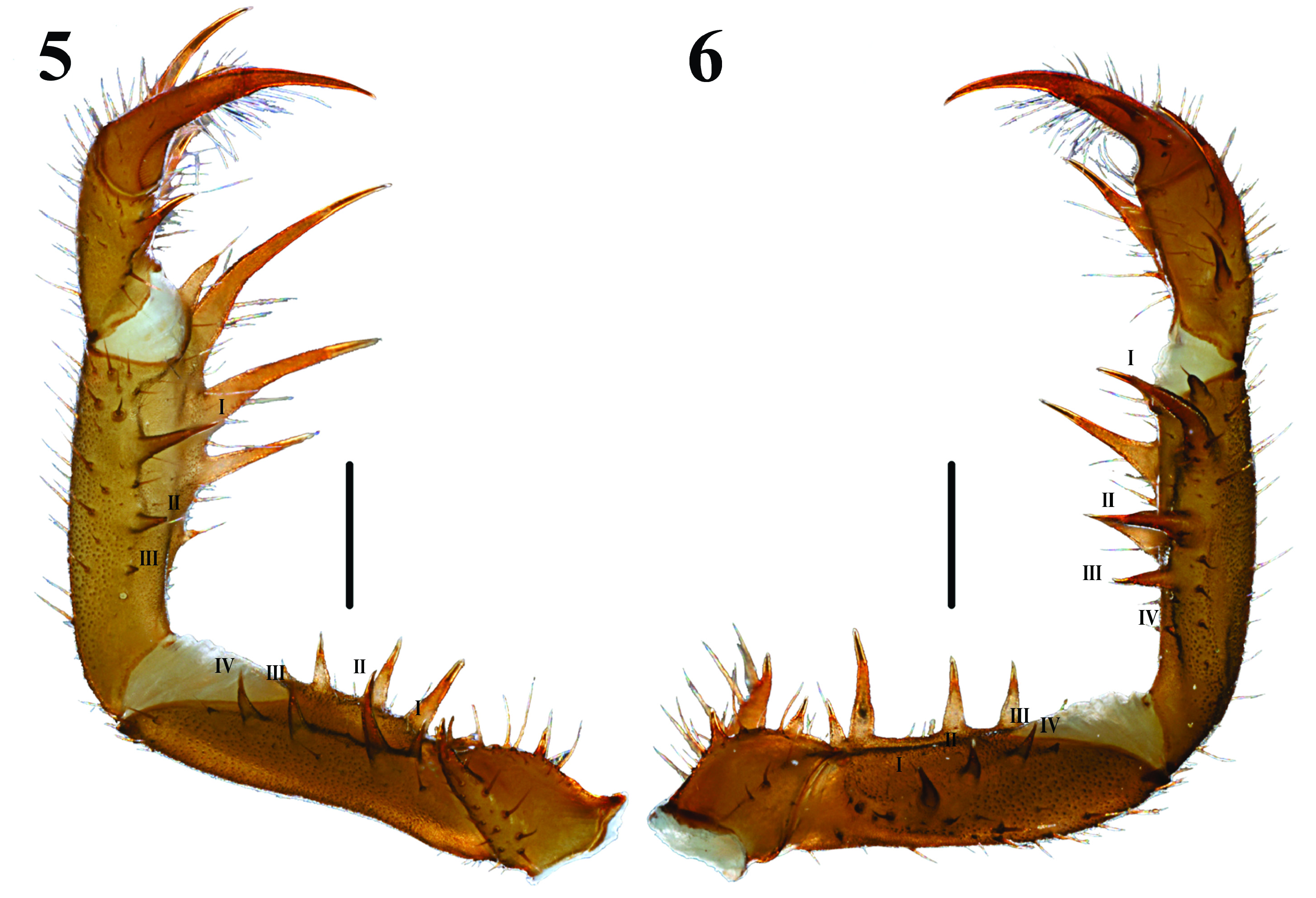
Diagnosis. This species may be separated from other Charinus in eastern South America by means of the following combination of characters: carapace frontal process large and triangular, clearly visible in dorsal view; pedipalp femur with four dorsal and four ventral spines; pedipalp patella with four dorsal and three ventral spines; pedipalp tibia with two dorsal and one ventral spine; female gonopods sucker-like, sclerotized basally and opening rounded, margins with disc-shaped folds; median and lateral eyes and median ocular tubercle well developed; chelicerae with a small distinguishable tooth on retrolateral surface of the basal segment, claw with ten teeth; tibia of leg I with 23 articles, tarsus I with 41 articles.
Charinus tocantinensis sp. nov. resembles C. renneri and C. iuiu , as these possess i) median and lateral eyes and median ocular tubercle developed; ii) anterior margin of carapace with six setae; iii) female gonopods sucker-like, whit opening rounded and edges with a fold and a posterior slit; iv) leg IV distitibia with 18 trichobothria, trichobothrium bc situated closer to sbf than to bf, sc and sf series each with six trichobothria; v) tibia of leg I with 23 articles, tarsus I with 41 articles. However, Charinus tocantinensis sp. nov. can be readily distinguished by the presence of four dorsal spines on the pedipalp patella ( Figure 6 View FIGURES 5–6 ), being five dorsal spines in C. renneri ( Miranda et al. 2021: Figure 73E) and five ou six dorsal spines in C. iuiu (Vasconcelos et al., 2015: Figure 18 View FIGURES 15–18. 15 ). Furthermore, the new species C. tocantinensis is also differs from C. renneri by the presence of cheliceral claw with ten teeth (compare ( Figure 7 View FIGURES 7–11 ; Miranda et al. 2021: Figure 10I View FIGURES 7–11 : in C. renneri cheliceral claw with eight teeth). Charinus tocantinensis sp. nov. is also differs from C. iuiu by having basally sclerotized sucker-shaped gonopods (compare Figure 7 View FIGURES 7–11 ; Vasconcelos et al., 2015: Figure 21: in C. iuiu sucker-like gonopods unsclerotized basally).
Description. Female (holotype).
Measurements. Total length: 10.27 mm. Carapace: length: 3.24 mm; width: 4.72 mm; sulcus-posterior margin distance: 1.03 mm; anterior margin-ocular tubercle: 0.12 mm; length of the ocular tubercle: 0.22 mm; median eye distance: 0.13 mm; lateral eyes distance: 1.74 mm; lateral eyes-lateral margin distance: 0.35 mm. Pedipalp (length): femur 2.80 mm; patella: 2.84 mm; tibia: 1.49 mm; tarsus: 0.96 mm; claw: 0.84 mm. Legs: femur length (I>III>IV>II), I: 10.25 mm; II: 6.45 mm; III: 7.59 mm; IV: 6.54 mm.
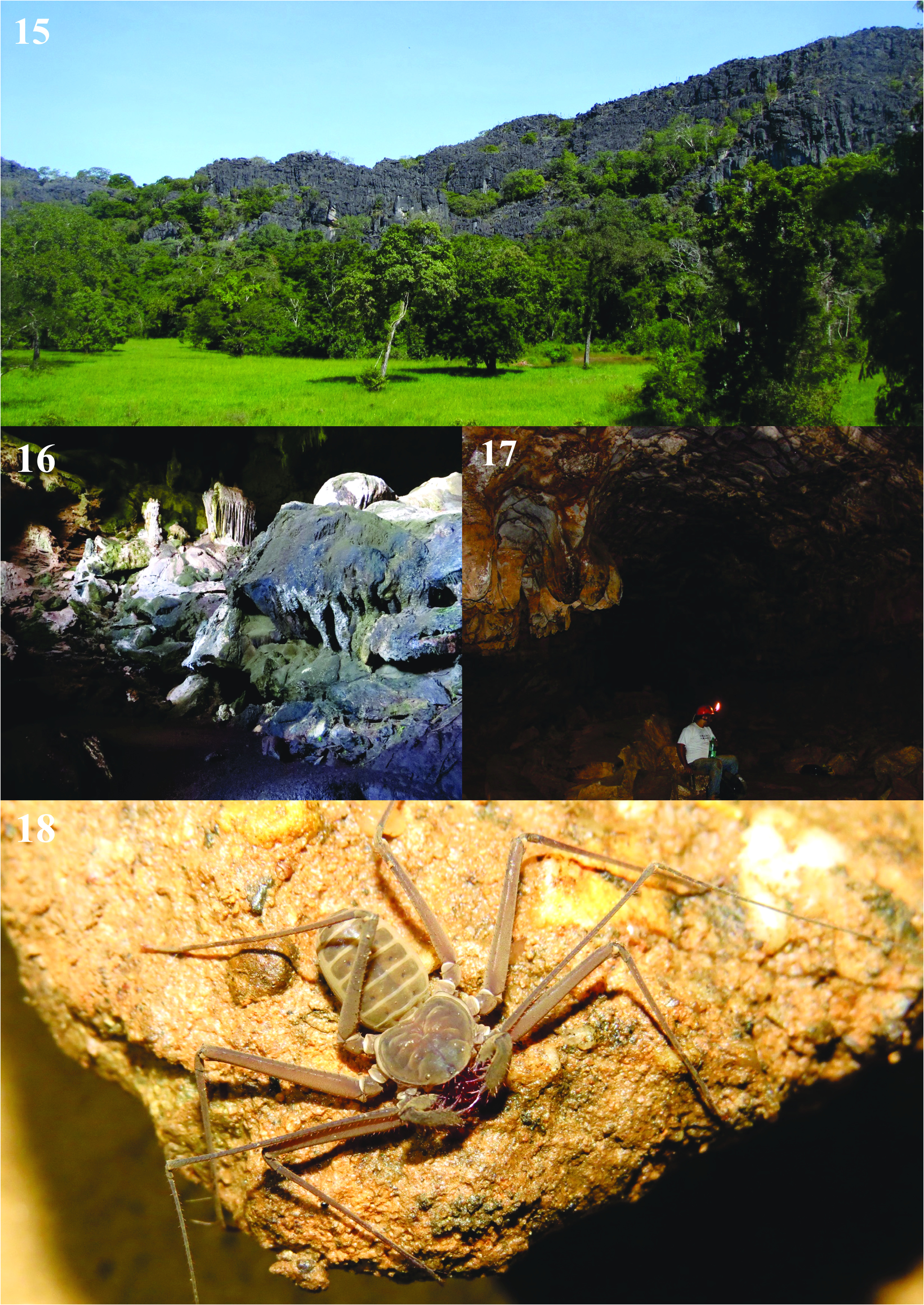
Color. Live specimen ( Fig. 18 View FIGURES 15–18. 15 ): carapace grayish olive green, abdomen moderate yellow and chelicerae deep red. In alcohol ( Figs. 1–7 View FIGURES 1–4 View FIGURES 5–6 View FIGURES 7–11 ): appendices and carapace strong yellow brown to dark or pale orange yellow, abdomen moderate orange yellow.
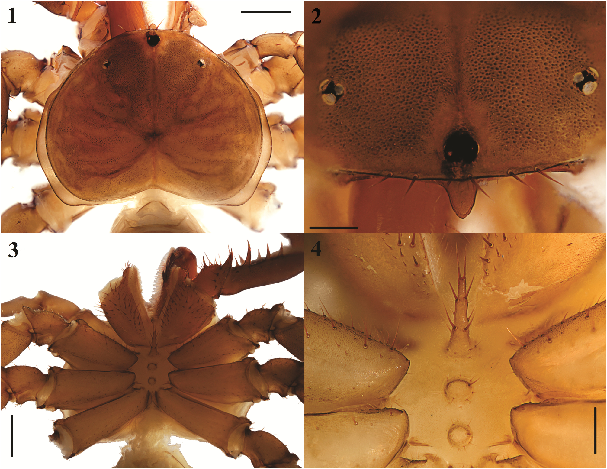
Carapace ( Fig. 1, 2 View FIGURES 1–4 ). Cordiform and slightly convex. Ratio length/width approximately 3/4. Surface with multiple small setae and scattered granules, more concentrated in the central region. The carina starts at the sides of the anterior margin and expands from the coxa of leg II to the corners of the posterior margin. Anterior margin slightly rounded, with six strong and long setae, posterior and lateral margin with thinner and smaller setae. Frontal process triangular in shape with an oval apex, clearly visible when observed from a dorsal view ( Figs 1, 2 View FIGURES 1–4 ). Median eyes and median ocular tubercle well developed; lateral eyes well developed, with dark internal pigmentation and posterior setae; triad with an anterior and posterior setae before the carina. Fovea deep and slightly triangular posteriorly. Median sulci extend from the ocular tubercle to the posterior margin, two pairs of sulci directed anteriorly and posteriorly in x-shape.
Sternum ( Figs 3, 4 View FIGURES 1–4 ). Tetra-segmented, all platelets markedly sclerotized. Tritosternum elongated projected anteriorly, truncated vertex, surpassing base of pedipalp coxae, with pair of large anterior, two median, and two posterior setae, which are surrounded by numerous tiny setae. Tetrasternum convex, with pair of large setae anteriorly, and several small setae posteriorly. Pentasternum convex and slightly smaller than the previous one, with pair of large setae anteriorly, and several small setae posteriorly.
Opisthosoma. Oblong, longer than wide; ventral sacs and ventral sac cover absent.
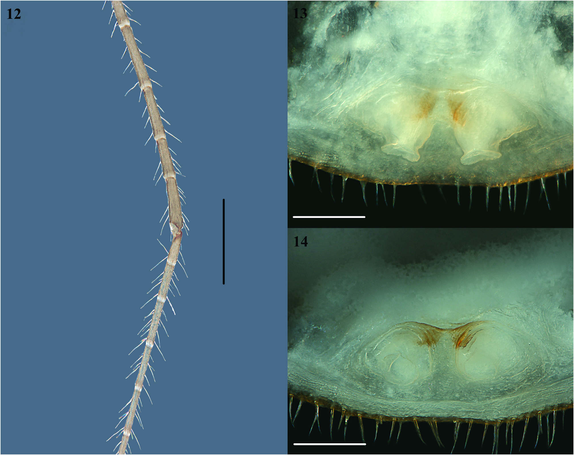
Genitalia. Female genital operculum with prominent setae posteromedially and some smaller on the ventral surface. Gonopods are sucker-like, basally sclerotized, longer than wide ( Fig. 13 View FIGURES 12–14 ), opening rounded and edges with a fold and a posterior small slit ( Fig. 14 View FIGURES 12–14 ). Gonopods separated from each other by a distance smaller than the diameter of each one and from the margin of the operculum by a distance slightly greater than its length.
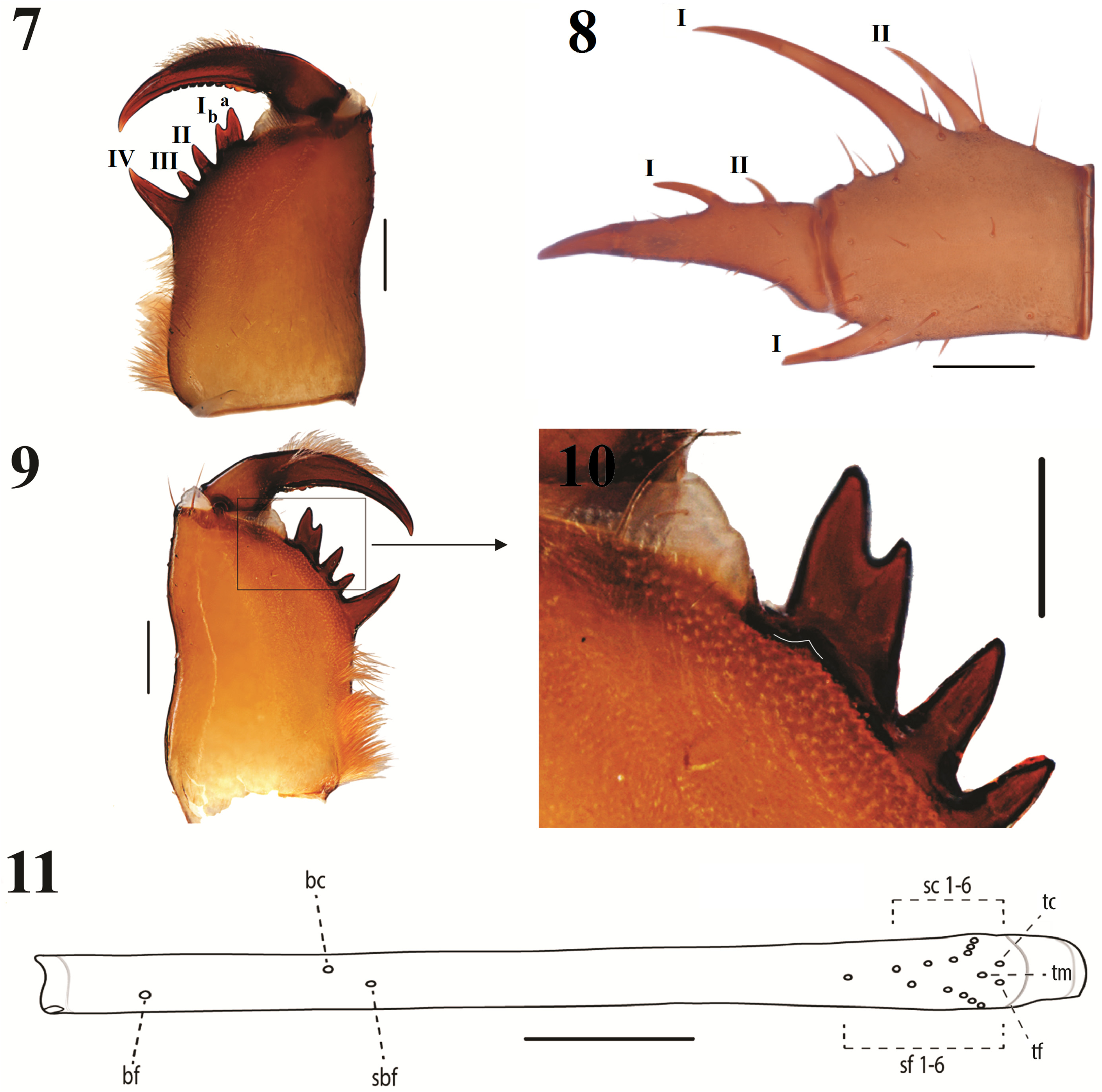
Chelicera ( Figs 7, 9 View FIGURES 7–11 ). Basal segment with four inner teeth. The distal tooth is bifid, with the distal cusp being larger than the proximal. Teeth length: IV>Ia>Ib>II>III. Small tooth in retrolateral row, opposite to bifid tooth ( Figs. 9, 10 View FIGURES 7–11 ). Claw with ten teeth. Setae on the prolateral and dorsal surface of the basal segment.
Pedipalp ( Figs 5, 6 View FIGURES 5–6 , 8 View FIGURES 7–11 ). With numerous setae. Coxal dorsal carina with one small setae on the rounded carina and three setae on the anterior margin. Trochanter with ventral spiniform apophysis pointed forwards with a series of strong setiferous tubercles. Two spines of similar size on the prolateral face, the first being close to the medial region and the second above the projection of the apophysis and close to the femur. Three setiferous tubercles aligned between the two spines, and two setae others are located proximally. Femur with two distinct setiferous tubercles proximal to spine I. Primary series with four dorsal spines (I>II>III>IV) and four ventral spines (II>III>IV>I); first two ventral spines closest to trochanter, setiferous tubercle between ventral spine 1 and proximal margin, another setiferous tubercle aligned with the fourth spine distally. Patella with four dorsal spines in primary series (I>II>III>VI), prominent setiferous tubercle between spine I and distal margin, one-quarter length of spine I; three ventral spines (I>II>III), three setiferous tubercles and three setae between spine I and distal margin, another setiferous tubercle aligned between spine I and spine II. Tibia with two dorsal spines, distal spine two times larger than the proximal spine; three setiferous tubercles, two at the base of distal spine and one at the base of proximal spine. One ventral spine approximately equal to the dorsal proximal spine; two setiferous tubercles close to anterior margin. Tarsus with three dorsal spines, on the cleaning organ -well developed- in ascending order of size; most distal spine approximately one-third length of tarsus, two smaller proximal spines close together, the most proximal spine about one-third length of its neighboring spine; Holotype with only two dorsal spines on the left tarsus, the most proximal small spine absent. Ventral row of cleaning brush with 26 setae. Apotele (claw) long and curved distally.
Legs. All setose. Subdistal part of the second segment of tarsus II–IV with a thin white ring. Leg I with 23 tibial articles and 41 tarsal articles; tarsal segment 1 is 0.8 smaller than tarsal segment 2 ( Fig. 12 View FIGURES 12–14 ). Leg IV: basitibia with four pseudo-articles, first pseudo-article with a trichobothrium medially and last pseudo-article with a trichobothrium proximally; distitibia with 3 trichobothria positioned proximally and 15 trichobothria positioned distally (bc situated closer to sbf than to bf, sc and sf series each with six trichobothria), trichobothriotaxy as in Figure 11 View FIGURES 7–11 . Basitibia-distitibia length: BT1>DT>BT4>BT3>BT2. The tarsus/metatarsus ratio is approximately 5/6, with the tarsus composed of three pseudo-articles.
Natural history. The newly discovered species was found in Natividade Cave, which is situated within an extensive limestone formation approximately 14 km to the northeast of the city of Natividade. While this cave is associated with the limestones of the Bambuí group, it is geographically isolated from the primary and continuous Bambuí group areas in the state. The cave has two entrances, with the larger one leading to a spacious chamber ( Fig. 16 View FIGURES 15–18. 15 ). Characterized by its labyrinthine structure, the cave spans a minimum horizontal projection of 500 meters, with most of its passages being completely dry ( Fig. 17 View FIGURES 15–18. 15 ). The innermost section of the cave connects with the water table, resulting in moister substrates. In the deepest and most secluded chamber, there is a pond supplied by the water table, creating a highly humid environment. Significantly, the cave provides a habitat for numerous cave-restricted species, including millipedes, pseudoscorpions, spiders, and bugs ( Polhemus & Ferreira 2018).
The cave displays noticeable human-induced alterations, primarily concentrated near its main entrance. Historical records reveal that saltpeter extraction from the cave, used in gunpowder production, was a practice in the past. Local residents report this activity continuing until the 1940s ( Polhemus & Ferreira 2018). Consequently, excavated areas and remnants of burned materials are visible in proximity to the entrance and within the initial chamber. However, in the innermost regions of the cave, no evidence of prior human visitation is apparent. It seems that access was restricted to the entrance area, leaving the deeper sections undisturbed and fortuitously preserved in their natural state. The surrounding landscape has undergone significant changes, with native forests converted to pastureland and used for agricultural purposes ( Fig. 15 View FIGURES 15–18. 15 ). The individual of C. tocantinensis sp. nov. herein described was found within the deeper, light-deprived zone ( Fig. 18 View FIGURES 15–18. 15 ).
| R |
Departamento de Geologia, Universidad de Chile |
No known copyright restrictions apply. See Agosti, D., Egloff, W., 2009. Taxonomic information exchange and copyright: the Plazi approach. BMC Research Notes 2009, 2:53 for further explanation.
|
Kingdom |
|
|
Phylum |
|
|
Class |
|
|
Order |
|
|
Family |
|
|
Genus |