
Triconia pacifica, Cho & Kim & Böttger-Schnack & Lee, 2013
|
publication ID |
https://doi.org/ 10.1080/00222933.2013.771757 |
|
DOI |
https://doi.org/10.5281/zenodo.10527241 |
|
persistent identifier |
https://treatment.plazi.org/id/551B87A1-B87F-3152-B88D-FB5E3D1CFE73 |
|
treatment provided by |
Carolina |
|
scientific name |
Triconia pacifica |
| status |
sp. nov. |
Triconia pacifica sp. nov.
( Figures 2–5 View Figure 2 View Figure 3 View Figure 4 View Figure 5 )
Type locality
Northeast equatorial Pacific (10 ◦ 30 ′ N, 131 ◦ 20 ′ W, 0–100 m).
Material examined
Holotype. One female ( NIBRIV0000244998 ) dissected and mounted on 10 slides, collected from the type locality on 21 August 2009 by D.J. Ham.
Paratypes. Three females ( NIBRIV0000244999 , NIBRIV0000245000-1 ), each dissected and mounted on nine slides, respectively. Four males ( NIBRIV0000245002-5 ), each dissected and mounted on eight or nine slides. All from the type locality .
Other material examined
Two females ( NIBRIV0000245019 , NIBRIV0000245022 ) each dissected and mounted on nine slides; collected from the Korea Strait , 33 ◦ 44 ′ 50.50 ′′ N, 128 ◦ 15 ′ 39.02 ′′ E on 7 October 2008 by K.H. Cho .
Description of female
Body length: 613 µm [traditional method: 582 µm] [traditional method, range: 553–582 µm, n = 4 individuals, based on specimens from the type locality].
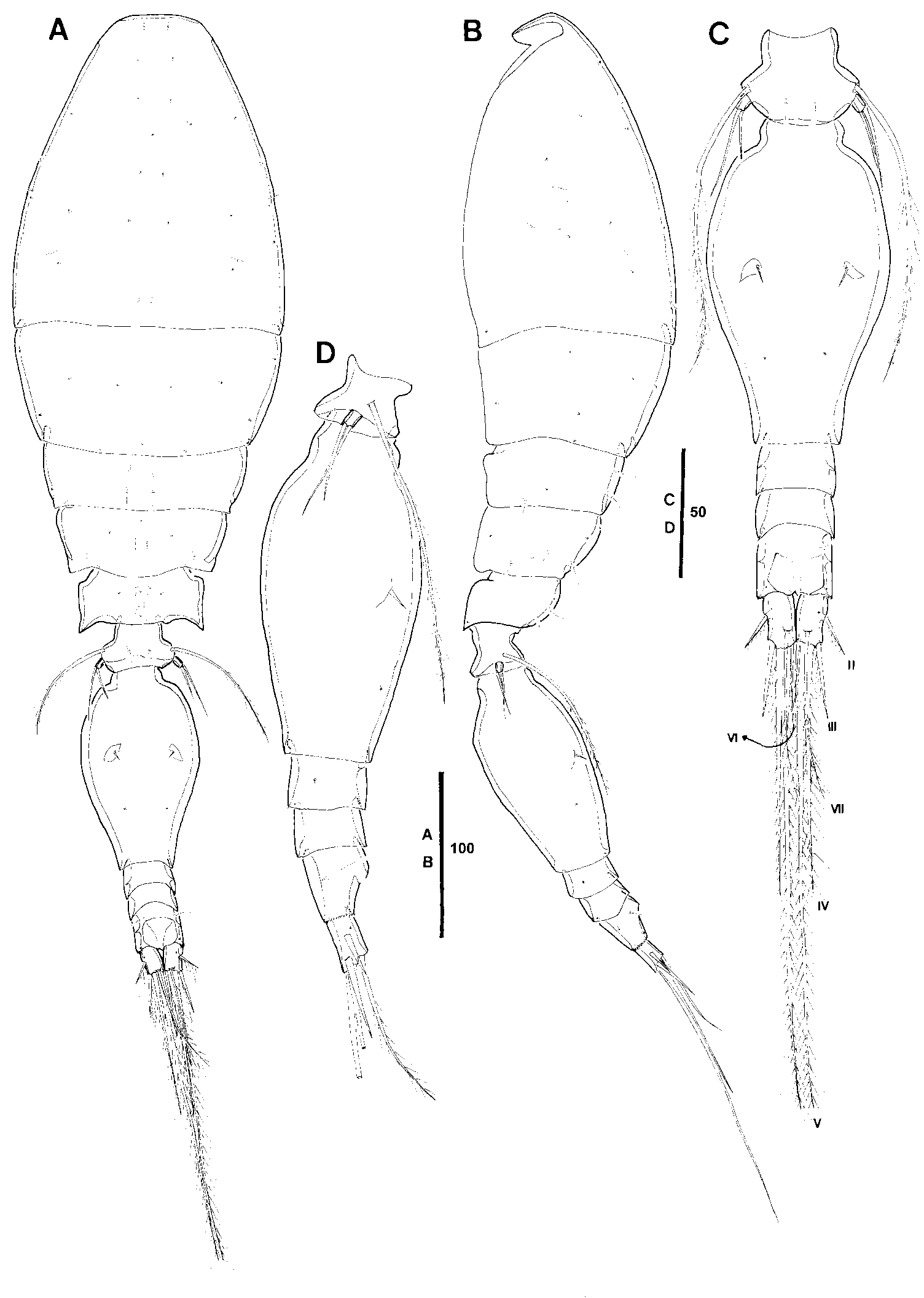
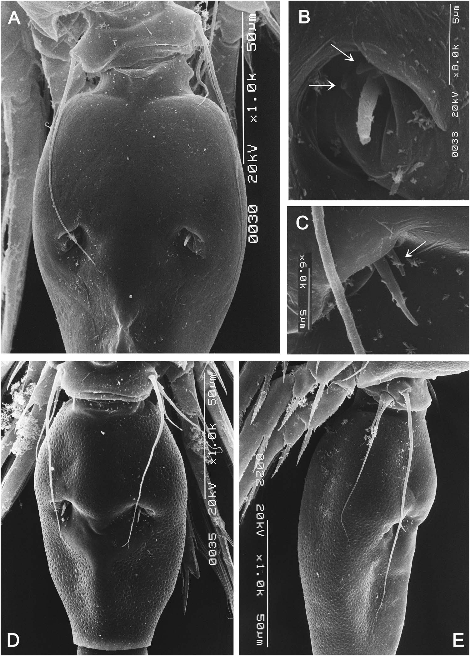
Exoskeleton well chitinized, surface covered with numerous small pits, similar to the skin of an orange (not figured). Prosome 1.7 times length of urosome excluding caudal rami, about 1.5 times urosome length including caudal rami. P2-bearing somite without conspicuous dorso-posterior projection in lateral aspect ( Figure 2B View Figure 2 ). Integumental pores on prosome as indicated in Figure 2A, B View Figure 2 . Pleural areas of P4-bearing somite with posterolateral corners elongated and pointed ( Figure 2A, B View Figure 2 ).
Urosome five-segmented, comprising P5-bearing somite, genital double-somite and three free abdominal (= postgenital) somites. P5-bearing somite with pair of middorsal secretory pores ( Figure 2A, C View Figure 2 ), other integumental pores on urosomites and CR as figured ( Figure 2C, D View Figure 2 ).
Genital double-somite 1.8 times as long as maximum width at anterior half (measured in dorsal aspect) and 1.9 times as long as postgenital somites combined, flask-like shape; lateral margins distinctly rounded, largest width at anterior half, posterior third tapering distinctly. Paired genital apertures located at about half distance from dorsal anterior margin; armed with one spine and two minute spinous processes (arrowed in Figure 13B View Figure 13 ). Surface with paired secretory pores at one quarter distance from posterior margin.
Anal somite about as wide as long; slightly longer than caudal rami ( Figure 2C, D View Figure 2 ).
Caudal ramus ( Figure 2C View Figure 2 ) about 1.6 times as long as wide, inner margin unornamented; with six setae, which are numbered using Roman numerals in Figure 2C View Figure 2 : seta I and II spiniform, unipinnate along medial margin, others setiform and plumose; length data of setae II–VII of holotype and three paratype females as shown in Table 1; range of variation of setal lengths relative to longest seta V as follows: II: 10–11%, III: 16–17%, IV: 51–61%, VI: 16–22%, VII: 33–40%.
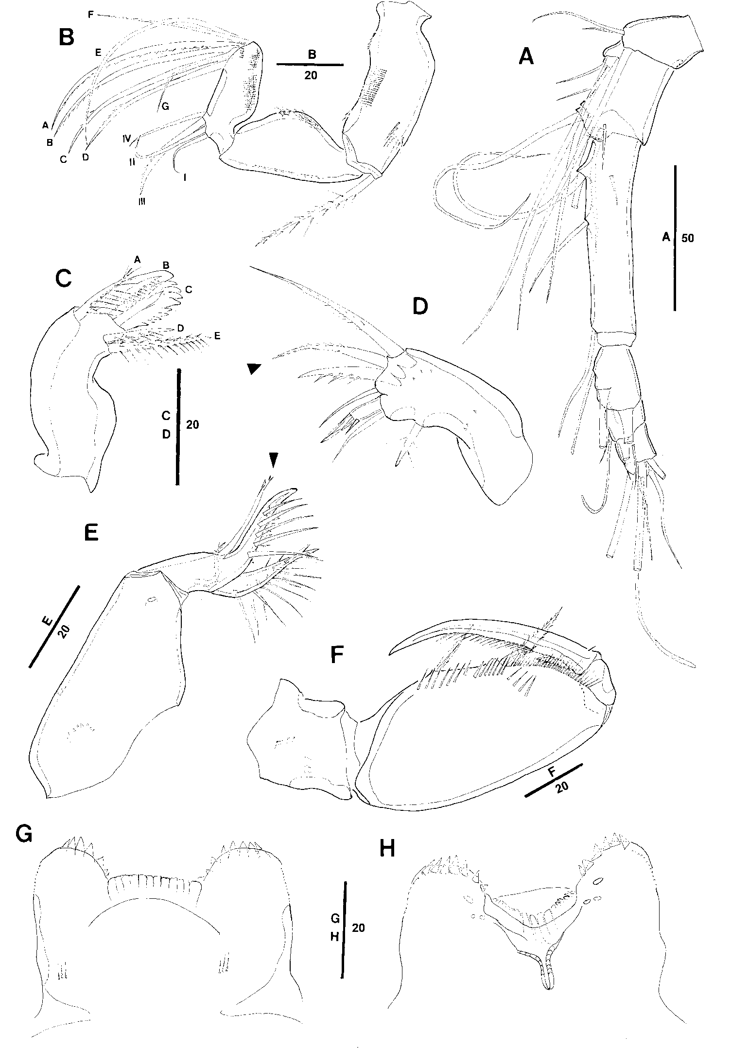
Antennule six-segmented ( Figure 3A View Figure 3 ). Armature formula: 1-[3], 2-[8], 3-[5], 4-[3+ae], 5-[2+ae], 6-[6+(1+ae)].
Antenna three-segmented, distinctly reflexed ( Figure 3B View Figure 3 ). Relative lengths (%) of segments approx. 39: 33: 28. Coxobasis with row of long, fine spinules along outer and inner margins and with few additional denticles on proximal and distal part of outer margin. Proximal endopod segment subtriangular forming outer lobate outgrowth bearing patch of spinules and single row of denticles along inner posterior margin. Distal endopod segment with two rows of short spinules along outer margin of posterior surface and additional row of minute spinules at distal part of anterior surface. Lateral armature consisting of one spiniform seta (III), pectinate at distal half, and three naked setae, seta I shortest ( Figure 3B View Figure 3 ); distal armature consisting of five curved setae (A–E) and two slender naked setae (F, G): setae A–D unipinnate, setae A–C similar in length, seta D slightly shorter; seta E longest and naked; seta G shorter than seta F, both setae shorter than seta D.
Labrum ( Figure 3G, H View Figure 3 ) distinctly bilobed, with six to seven large dentiform processes of graduated length medially on distal margin of each lobe and row of minute spinules along outer margins. Medial concavity covered anteriorly with single hyaline lamella ( Figure 3G View Figure 3 ), anterior surface with short spinular row either side of median swelling, integumental pockets absent. Posterior wall of medial concavity with four strong dentiform processes (“teeth”) and paired sclerotized indented elements either side of teeth ( Figure 3H View Figure 3 ); posterior surface with group of three secretory pores on proximal part of each lobe.
Mandible ( Figure 3C View Figure 3 ) without surface ornamentation; gnathobase with five elements numbered using capital letters in Figure 3C View Figure 3 : element A at subdistal ventral corner about as long as ventral blade B, with long, fine setules along dorsal margin, tip of element not reaching as far as tip of blade B; ventral blade B strong and broad, with row of setules on posterior surface; dorsal blade C with dentiform processes along entire dorsal and distal margins; dorsal element D setiform and bipinnate, shorter than multipinnate element E.
Maxillule ( Figure 3D View Figure 3 ) weakly bilobed, surface ornamented with few spinules as figured. Inner lobe with three elements: innermost one pinnate, located at some distance from others, middle element naked, about same length as outermost element, which is strong and spiniform, fringed with few strong spinules at midregion and spinulose at distal part. Outer lobe with four elements: two outer elements setiform and bipinnate, outermost element longest, element next to innermost spiniform and strong, with double row of short spinules, similar in length to innermost element, which is setiform and naked.
Maxilla ( Figure 3E View Figure 3 ) two-segmented, comprising syncoxa and allobasis. Syncoxa unarmed, surface ornamented with few curved spinular rows and one large secretory pore; allobasis produced distally into slightly curved spinous process (claw) bearing two rows of strong spinules along inner (= medial) margin; stout seta on outer margin reaching as far as tip of allobasal claw, ornamented with few spinules distally (arrowed in Figure 3E View Figure 3 ); medial margin with slender spinulose seta and strong spinous seta ornamented with two large spinular rows along medial margin and few spinules at outer (= lateral) margin.
Maxilliped ( Figure 3F View Figure 3 ) four-segmented; syncoxa unarmed, posterior surface ornamented with two short rows of spinules. Basis robust, with two spiniform spinulose elements on inner (= palmar) margin, distal element stronger and 1.4 times longer than proximal one; fringe of long pinnules between distal seta and articulation with endopod, row of spinules between proximal and distal seta and short transverse row on anterior surface ( Figure 3F View Figure 3 ); other ornamentation on outer margin of anterior surface absent or not discernable. Proximal endopodal segment unarmed. Distal endopodal segment (claw) with row of fine pinnules along proximal three-quarters of concave margin; with minute naked seta on outer proximal margin and unipectinate spine fused basally to inner proximal corner of claw.
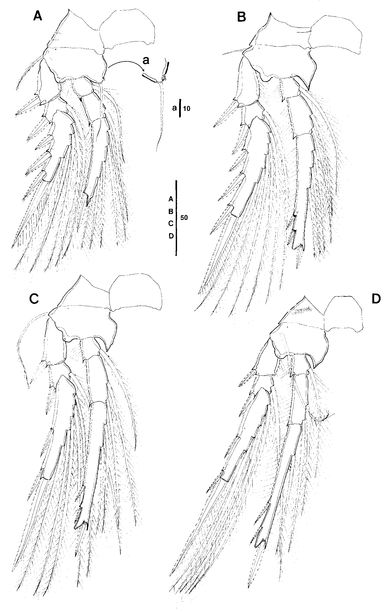
Swimming legs 1–4 biramous ( Figure 4 View Figure 4 A–D), with three-segmented rami. Intercoxal sclerites well developed, with paired row of few minute spinules or denticles on posterior surfaces (not figured). Coxae and bases of P1–P4 with sparse surface ornamentation as shown in Figure 4 View Figure 4 A–D, coxa of P4 with tuft of very long setules on posterior surface ( Figure 4D View Figure 4 ). Basis with relatively short (P1, P2) or longer (P3, P4) outer seta. Insertion of spiniform inner basal seta on P1 ornamented with minute denticles anteriorly ( Figure 4A, a View Figure 4 ). Endopod of P1 about same length as exopod, those of P2–P4 longer than exopod (particularly in P4). Surface of all segments unornamented, except for few secretory pores on posterior surface of distal exopodal and endopodal segments as figured.
Swimming leg armature formula (Roman numerals indicate spines, Arabic numeral indicate setae):
Exopods. Outer margin of segments with small serrated hyaline lamellae. Hyaline lamellae on outer spines moderately developed (P1–P3) or narrow (P4). Outer and distal spines of P1 exopod with subapical tubular extension, lacking on proximalmost spine of distal segment, outer spines on distal segment increasing in length distally ( Figure 4A View Figure 4 ). Distal spine, similar in length to (P1, P3), slightly shorter than (P2) or longer than (P4) distal segment.
Endopods. Distal margin of P1 with small pointed protrusion close to distalmost outer seta ( Figure 4A View Figure 4 ); distal margin of P2–P4 produced into long, unornamented conical process with apical pore ( Figure 4 View Figure 4 B–D). Length data of endopodal spines of holotype and three female paratypes as shown in Table 1; length ranges of outer subdistal spine ( OSDS) and outer distal spine ( ODS) relative to distal spine are as follows: P2 enp-3, OSDS: 86–111%, ODS: 67–79%; P3 enp-3, OSDS: 44–59%, ODS: 42–56%, P4 enp-3, OSDS: 41–56%, ODS: 34–41%. Distal endopodal spine reaching far beyond tip of distal conical process in P2–P4; outer distal spine on enp-3 not reaching (P2, P3) or almost reaching (P4) as far as tip of conical process; outer subdistal spine reaching as far as (P2) or almost as far as (P4) insertion point of outer distal spine, or not reaching (P3) that point. Hyaline lamellae on outer endopodal spines narrow (P2–P3) or almost lacking (P4) .
P5 ( Figure 2C, D View Figure 2 ) with small free exopodal segment and very long plumose outer basal (= protopodal) seta, more than three times longer than outer exopodal seta and extending as far as four-fifths from anterior margin of genital double-somite, reaching beyond genital apertures as far as paired pores on dorsal surface ( Figure 2D View Figure 2 , seta probably not shown in full length in Figure 13A View Figure 13 ). Exopod a little longer than wide, with two setae, which are spiniform and unornamented, the outer one slightly longer than the inner one.
P6 ( Figures 2C View Figure 2 , 13B View Figure 13 ) represented by operculum closing off each genital aperture; armed with one long spine and two minute blunt processes (arrowed in Figure 13B View Figure 13 ).
Description of male
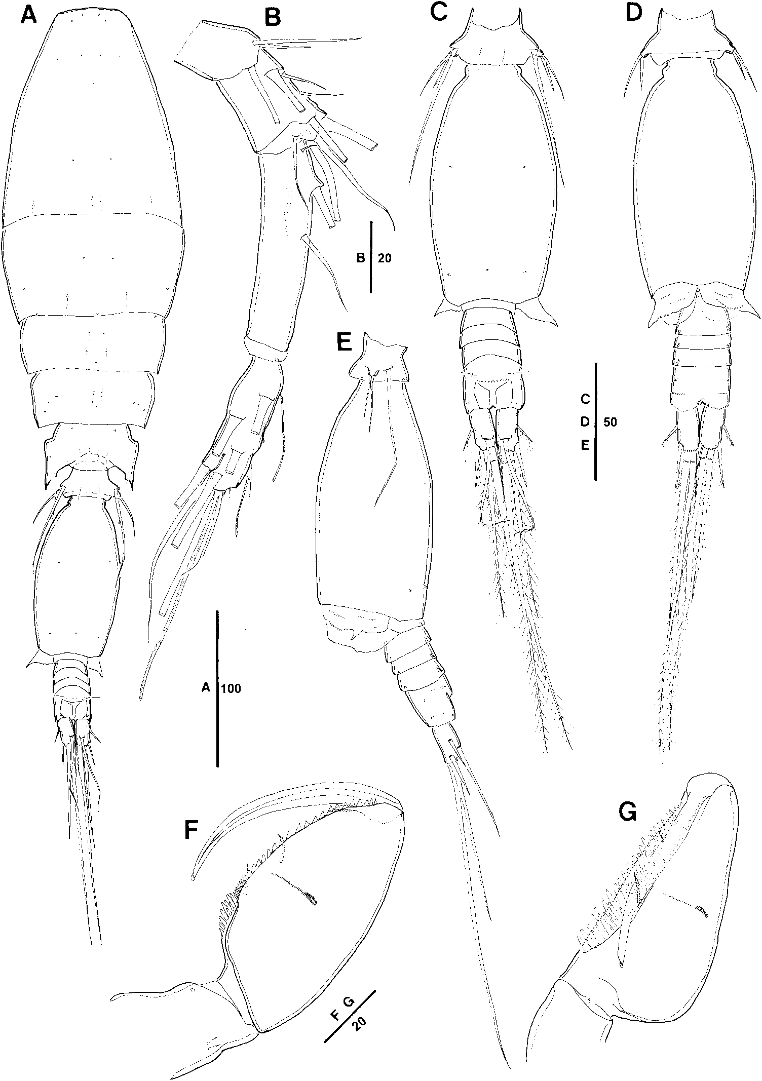
Body length (traditional method): 472 µm. Sexual dimorphism in antennule, maxilliped, endopodal spine lengths on P2, P5, P6, genital segmentation and CR.
Urosome six-segmented, comprising P5-bearing somite, genital somite and four free abdominal somites. Genital somite about twice as long as postgenital somites combined. Dorsal surface of genital somite with five secretory pores ( Figure 5C View Figure 5 ). Surface ornamentation on genital flaps and on ventral surface of anal segment as indicated in Figure 5D View Figure 5 .
Caudal rami with length to width ratio about 2: 1, larger than in female; length data of setae II–VII of three paratype males as shown in Table 1, range of variation of setal lengths relative to longest seta V as follows: II: 8–11%, III: 12–16%, IV: 49–54%, VI: 14–17%, VII: 37–40%, slightly different from female in proportional lengths of setae III and IV.
Antennule ( Figure 5B View Figure 5 ) four-segmented, distal segment corresponding to fused segments 4–6 of female; armature formula: 1-[3], 2-[8], 3-[4], 4-[11+2ae+(1+ae)].
Maxilliped ( Figure 5F, G View Figure 5 ) three-segmented, comprising syncoxa, basis and onesegmented endopod. Syncoxa unarmed, ornamented with single secretory pore at inner distal margin and few spinules on surface. Basis robust and expanded, armed with two small naked setae of equal length within longitudinal cleft; anterior surface with one to three transverse spinular rows and row of short flat spinules along inner margin ( Figure 5G View Figure 5 ), without expanded flap; posterior margin with two to three longitudinal rows of spatulated setules of graduated length ( Figure 5G View Figure 5 ).
Swimming legs with armature and ornamentation as in female, length data of endopodal spines of three male paratypes as shown in Table 1; length ranges of outer subdistal spine ( OSDS) and outer distal spine ( ODS) relative to distal spine as follows: P2 enp-3, OSDS: 106–145%, ODS: 88–92%; P3 enp-3, OSDS: 52–65%, ODS: 39–57%, P4 enp-3, OSDS: 40–47%, ODS: 34–38%, slight sexual dimorphism in proportional spine lengths on P2, with OSDS and ODS being relatively longer than in female .
P5 ( Figure 5C, E View Figure 5 ) exopod fused to somite, slightly shorter than in female, armature as in female; outer basal seta about 2.7 times as long as outer exopodal seta, relatively shorter than in female.
P6 ( Figure 5D View Figure 5 ) represented by posterolateral flap closing off genital aperture on either side; surface ornamentation as indicated in Figure 5D View Figure 5 . Posterolateral corners protruding laterally, clearly visible in dorsal aspect ( Figure 5A, C View Figure 5 ).
Etymology
The specific name refers to the type locality, the Pacific Ocean.
Remarks
Triconia pacifica sp. nov. is closely related to T. dentipes ( Giesbrecht, 1891) with a striking similarity to its sibling in general habitus and in the form of the female genital double-somite. Triconia dentipes was originally described by Giesbrecht (1891) based on females collected at a single station in the equatorial Pacific (03 ◦ S, 99 ◦ W), not very far from the type locality of the new species (10 ◦ 30 ′ N, 131 ◦ 20 ′ W), but he gave only a short Latin diagnosis. Subsequently, he found a few females of the same species also in the Gulf of Naples, Mediterranean Sea, and described it in some more detail in his comprehensive monograph, mentioning also its occurrence in the Pacific ( Giesbrecht 1893 [“1892”], p. 744). It remains uncertain, however, whether or not he compared the morphology of specimens from both regions in detail, as he did not specify the source of specimens examined, but gave only a short footnote stating that one to three females had been available for the study (Footnote on p. 593: “Material:... von den 3 kleineren Arten lagen mir 1-3 weibliche Tiere vor,...”). Böttger-Schnack (1999) redescribed both sexes of T. dentipes in great detail based on copepod material from the Red Sea in comparison with specimens from the Adriatic Sea in the Mediterranean, and summarized the taxonomic history of the species since Giesbrecht’s original account. For comparison with the new Pacific sibling species the taxonomic data provided by Böttger-Schnack (1999) were used as long as not otherwise noted, because Giesbrecht’s original description is insufficient for a detailed comparison and the type material deposited in the Zoological Statione Napoli is no longer available for distribution (cf. Böttger-Schnack 1999).
The morphometric differences between T. pacifica and T. dentipes are expressed for both sexes in (1) the proportional spine lengths on the endopod of P4, with (1a) the outer distal spine only reaching as far as the tip of the distal conical process in T. pacifica , whereas this spine is much longer, reaching far beyond the tip of the cone in T. dentipes [the legs of male T. dentipes had not been figured by Böttger-Schnack (1999), but re-examination of Red Sea material from RBS collection during the present study verified that proportional endopodal spine lengths are similar to that of the female]; and (1b) the outer subdistal spine on P4 enp-3 reaching as far as the insertion of the outer distal spine in T. pacifica , while the spine does not reach (Red Sea) or almost reaches (Adriatic Sea) that point in T. dentipes (cf. Böttger-Schnack 1999, figs. 11D and 13E, respectively); (2) the relative length of the outer basal seta on P5, reaching as far as four-fifths of the distance from the anterior margin of the female genital double-somite in T. pacifica , but less than half this distance in T. dentipes [cf. Böttger-Schnack, 1999, fig. 9C (Red Sea), fig. 13A (Adriatic); see also Di Capua and Boxshall 2008, scanning electron micrograph, fig. 2D], and about half the length of the genital somite in male T. pacifica , but less than one-third the distance from the anterior margin in male T. dentipes (cf. Böttger-Schnack, 1999, fig. 12D–F). Further slight morphometric differences between the two species may be found in the proportional spine lengths on P2 enp-3, with the outer subdistal spine always being longer than the outer distal spine in T. pacifica (cf. Table 1) and reaching as far as the insertion of the outer distal spine ( Figure 4B View Figure 4 ), but this is not, or is only just, the case in T. dentipes ; however, due to the length variation displayed by this spine in the latter species, being similar in length to the outer spine in some cases (cf. Böttger-Schnack 1999, fig. 11a,b), the difference is of restricted use for separating the two sibling species. Another slight morphometric difference is apparent in the proportional length of antennary setae D, being relatively shorter in T. pacifica compared with T. dentipes , but the actual length of this seta is difficult to discern and not directly comparable, because it may be curved to varying degrees.
A number of differences in micro-structures have been found separating female T. pacifica from T. dentipes , which are summarized in Table 2: (1) the additional ornamentation of the antennary coxobasis and setae C+D, (2) the spinular patch either side of the median swelling on the anterior surface of the labrum, (3) the bipinnate ornamentation of the element next to the outermost one on the outer lobe of the maxillule (arrowed in Figure 3D View Figure 3 ), (4) the ornamentation of the seta on the outer allobasal margin of the maxilla (arrowed in Figure 3E View Figure 3 ), and (5) the plumose ornamentation of the outer basal seta on P5.
Mx1, outer Orn. of element Sparse spinules Sparse spinules Sparse spinules Naked Sparse Naked Naked
lobe next to spinules
outermost one
Mx2, Orn. of seta on Few spinules Few spinules Few spinules Hyaline lamella Naked Few spinules Naked
allobasis outer margin distally distally distally distally
P2 enp-3 OSDS reaching Yes, beyond No / Almost No / Almost No / almost Yes, beyond Yes, beyond No
insertion of (Red Sea);
ODS yes
(Med.Sea)
P4 enp-3 L of ODS: CP About equal Shorter Shorter Longer About equal Shorter Shorter
OSDS reaching Yes No No No Yes No Yes
insertion of
ODS
P5, outer L, reaching Far beyond Reaching Far beyond Not reaching Reaching Beyond Far beyond
basal seta genital
apertures or
(far) beyond
ornamentation Plumose Plumose Plumose Spinulose Plumose Naked Naked
distally
References Present study Present study Present study Böttger- Böttger- Böttger- Wi et al. (2012) Schnack Schnack Schnack
(1999) (1999) (1999)
Notes: Med.Sea: Mediterranean Sea, Orn.: ornamentation, L: Length, W: Width, Mx1: Maxillule, Mx2: Maxilla, OSDS: outer subdistal spine, ODS:
outer distal spine, CP: distal conical process, A2: Antenna.
∗ Pacific Ocean near Galapagos Islands (3 ◦ S, 99 ◦ W).
The body length of female T. pacifica is similar to that of T. dentipes from the Mediterranean Sea, which measure 510–520 µm according to Giesbrecht (1893 [“1892”]), and 500–580 µm according to unpublished data by RBS based on copepods from the western Mediterranean Sea (cf. Table 2), but they are distinctly larger than specimens from the Red Sea, which measure only between 440 and 490 µm (Böttger- Schnack 1999). A reduced body length of oncaeid copepod species is a common phenomenon in the Red Sea and has been discussed as being a result of the extreme environmental conditions (temperature, food shortage) in that area ( Böttger-Schnack et al. 1989).
The identity of T. dentipes sensu H. Itoh , which was described from the adjacent waters of Japan ( Itoh 1997), is discussed below under T. elongata .
| V |
Royal British Columbia Museum - Herbarium |
No known copyright restrictions apply. See Agosti, D., Egloff, W., 2009. Taxonomic information exchange and copyright: the Plazi approach. BMC Research Notes 2009, 2:53 for further explanation.
|
Kingdom |
|
|
Phylum |
|
|
Class |
|
|
Order |
|
|
Family |
|
|
Genus |