
Paraphysifer Sinaiko & Dietrich, 2020
|
publication ID |
https://doi.org/ 10.11646/zootaxa.5254.2.9 |
|
publication LSID |
lsid:zoobank.org:pub:A86C7AF5-C6C0-40E4-AEF9-BA2F2A711EAD |
|
DOI |
https://doi.org/10.5281/zenodo.7727501 |
|
persistent identifier |
https://treatment.plazi.org/id/55408791-FF50-E657-FF0F-008013FDF9E7 |
|
treatment provided by |
Plazi |
|
scientific name |
Paraphysifer Sinaiko & Dietrich, 2020 |
| status |
|
Genus Paraphysifer Sinaiko & Dietrich, 2020 View in CoL
Type species: Paraphysifer wilsoni Sinaiko & Dietrich, 2020 , by original designation.
Diagnosis. Sinaiko & Dietrich (2020) provided the detailed description and diagnosis of Paraphysifer . Here we broaden the scope of the genus to include species which bear symmetrical paraphysis-like structures that are articulated to the connective but fused with the aedeagus.
Key to species
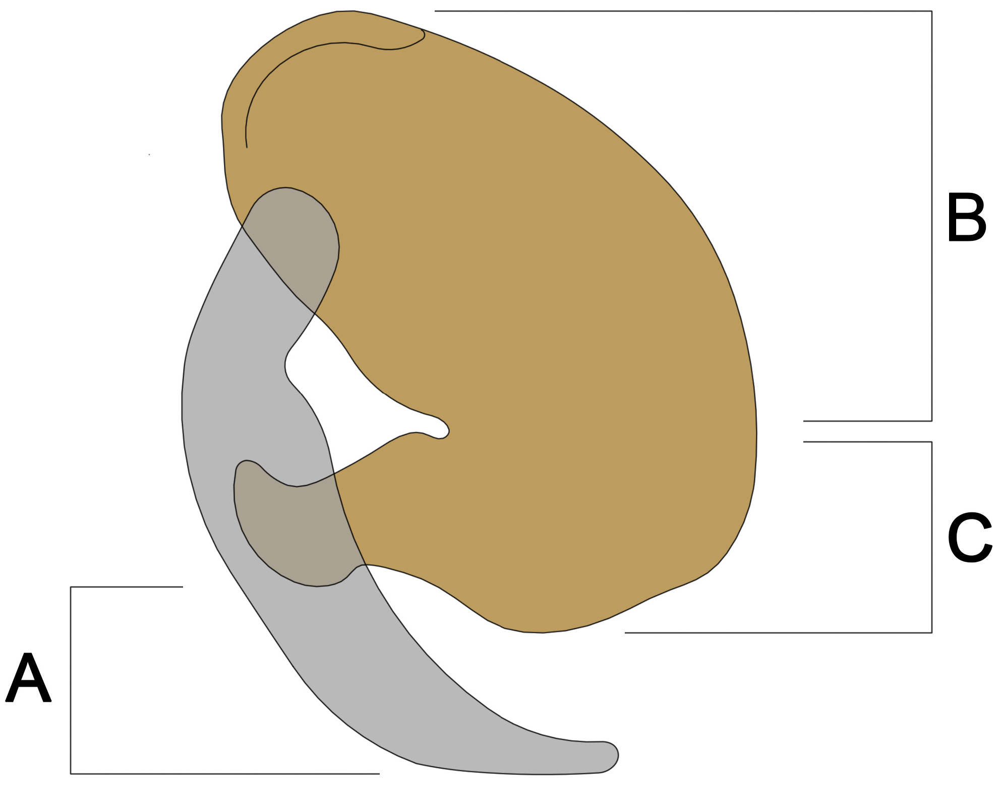
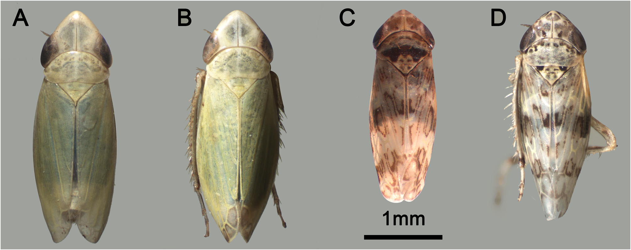
The terms “paraphysis ventral extension” and “aedeagus dorsal \ ventral lobe” are as shown in Figure 1 View FIGURE 1 . Full habitus photos are shown in Figure 2 View FIGURE 2 , genital structures are shown in Figure 3. View FIGURE 3
1. General color of specimen cream with extensive, symmetrical fuscous marks ( Figs. 2C, 2D View FIGURE 2 ); anterior tip of vertex in dorsal view less produced; aedeagus articulated to a paraphysis that has a ventral extension ( Figs. 1 View FIGURE 1 , 3 View FIGURE 3 : C1, C3)....... Group A (2)
- General color of specimen uniform green, unmarked ( Figs. 2A, 2B View FIGURE 2 ); anterior tip of vertex in dorsal view well produced; aedeagus fused to a paraphysis-like ventral process that lacks a ventral extension ( Fig. 3 View FIGURE 3 : A1, A3, B1, B3)...... Group B (3)
2. Dorsal lobe of sac-like aedeagus in lateral view as broad as its ventral lobe; ventral extension of paraphysis strongly produced and elongated posteriorly, ending in a sharp tip; subgenital plates short, rounded apically. P. pictipennis ( Kirschbaum, 1868) View in CoL
- Dorsal lobe of sac-like aedeagus in lateral view about half as broad as its ventral lobe; ventral extension of paraphysis slightly produced and elongated ventrally, ending in a blunt tip ( Fig. 3 View FIGURE 3 : C1, C3); subgenital plates long, sharp apically ( Fig. 3 View FIGURE 3 : C2)............................................................................. P. perpictus ( Logvinenko, 1978) View in CoL
3. Dorsal edge of sac-like aedeagus in lateral view oriented postero-dorsally, parallel to paraphysis-like structure ( Fig. 3 View FIGURE 3 : B1, B3). Margins of dorsal lobe of sac-like aedeagus not spread ( Fig. 3 View FIGURE 3 : B3, B4). Subgenital plates long ( Fig. 3 View FIGURE 3 : B2)...................................................................................... P. salsuginosus ( Logvinenko, 1961) View in CoL
- Dorsal edge of sac-like aedeagus in lateral view oriented dorsally, not parallel to paraphysis-like structure ( Fig. 3 View FIGURE 3 : A1, A3). Margins of dorsal lobe of sac-like aedeagus well spread ( Fig.3 View FIGURE 3 : A3, A4). Subgenital plates short ( Fig. 3 View FIGURE 3 : A2)......................................................................................... P. narsikulovi ( Dlabola, 1960) View in CoL

No known copyright restrictions apply. See Agosti, D., Egloff, W., 2009. Taxonomic information exchange and copyright: the Plazi approach. BMC Research Notes 2009, 2:53 for further explanation.