
Diskeria gigantea, Schockaert, Ernest R., Curini-Galletti, Marco, Ridder, Wouter De & Artois, Tom, 2011
|
publication ID |
https://doi.org/10.5281/zenodo.203197 |
|
DOI |
https://doi.org/10.5281/zenodo.6182153 |
|
persistent identifier |
https://treatment.plazi.org/id/55718796-FFC5-FFEE-DB96-67EEFAEDFD3D |
|
treatment provided by |
Plazi |
|
scientific name |
Diskeria gigantea |
| status |
sp. nov. |
Diskeria gigantea n.sp.
Figs 1–4 View FIGURE 1 View FIGURE 2 View FIGURE 3 View FIGURE 4 , 9 View FIGURE 9 A
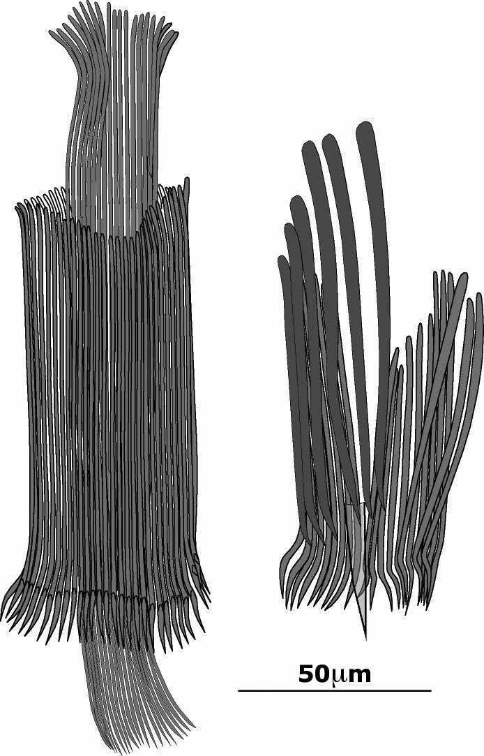
Diagnosis. Species of Diskeria with a copulatory organ with an inner ring of 40–50 needles of ±225µm (190– 320µm) forming a stylet-like structure and an outer ring of ±60 needles ranging from ±100 to ±125µm. The accessory organ opens into the male atrium and has a stylet of 20–35µm surrounded by needles in two groups, ±12 thin needles (37–72µm) and ±15 stouter needles (70–125µm).
Occurrence. Port aux Français, Kerguelen (Territoires Australes Françaises), between the pebbles beneath the little building at the port ( 10 December 1992). Occurs in high numbers, along with many amphipods and isopods on which it feeds.
Material studied. Many animals studied alive, six whole mounts, two animals squashed for karyology and mounted and five sectioned specimens (16 slides). One whole mount is designated the holotype ( SMNH nr. 7354); all other material designated paratypes (UH 419-439).
Derivation of the name. The genus name is derived from the local expression “DisKer”, designating the District Commissioner of Kerguelen; the species name refers to the size of the species, which is gigantic for a proseriate.
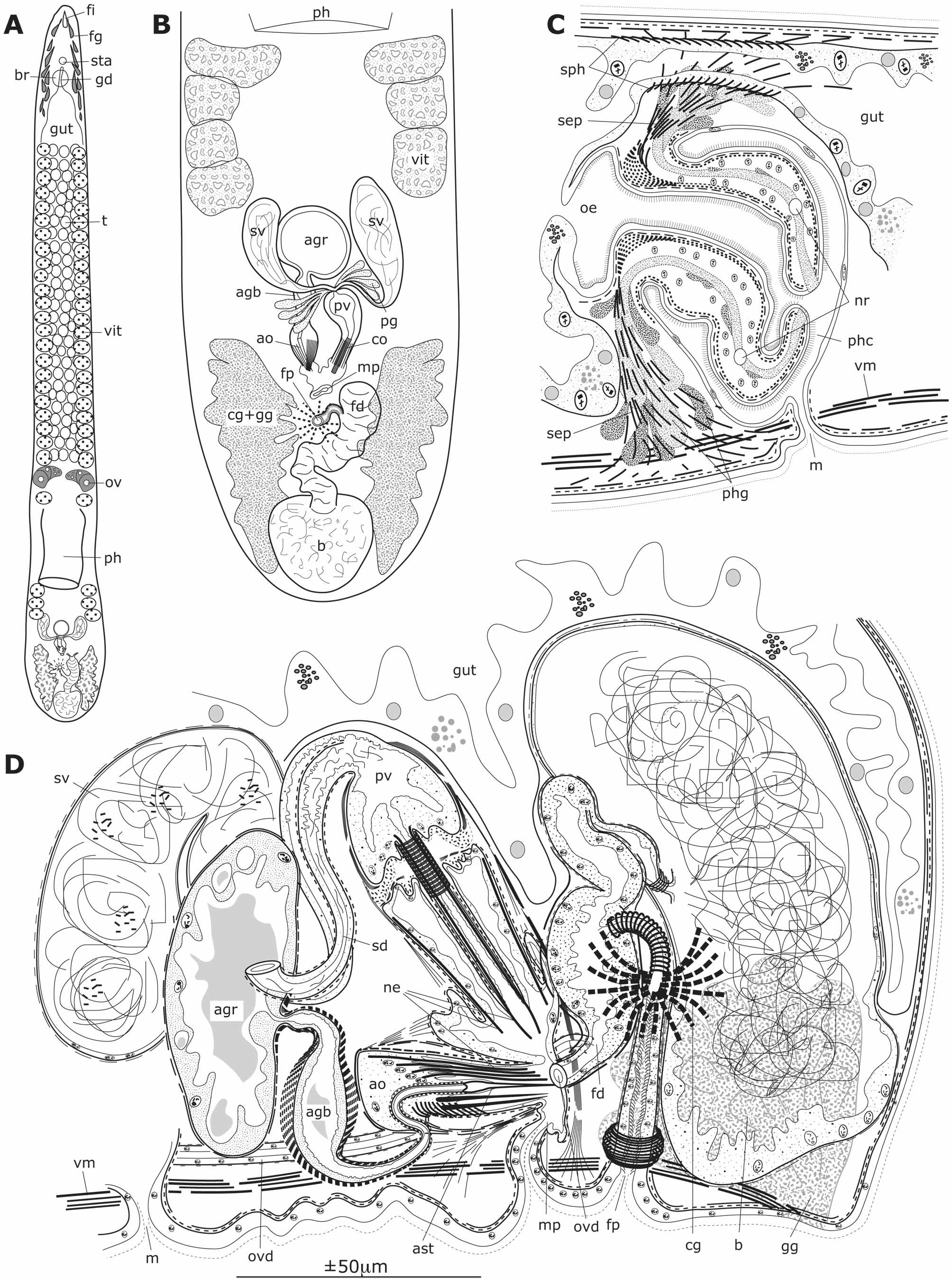
Description. The adult animals are around 2cm, rather broad, flattened and very actively crawling between the pebbles. They are whitish, the head region somewhat paler and have no eyes. Under the microscope the animals are rather opaque, making the internal anatomy difficult to study and many individuals had to be sacrificed before the internal structures became clear.
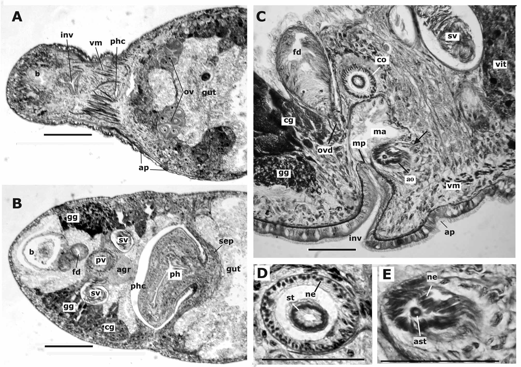
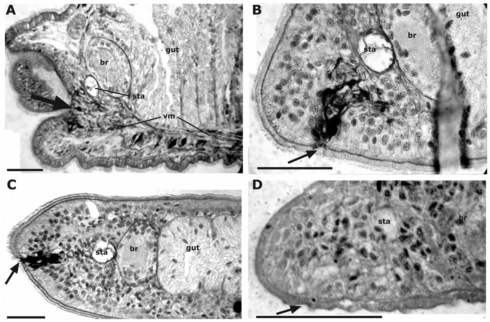
The pre-cerebral gut diverticulum reaches the statocyst (but does not extend beyond it) and the intestine continues until the very caudal end of the animal. The epidermis, moderately vacuolated, about 10µm high, with intra-epithelial nuclei, is slightly thicker at the ventral side. The cilia are shorter (3µm) on the dorsal side than on the ventral side (5µm). There are no rhabdite glands. In the sections, small retracted adhesive patches were detected, surprisingly only on the latero-dorsal sides (see Fig. 2 View FIGURE 2 ). The large eosinophilic and basophilic frontal glands converge towards a small subterminal pit ( Fig. 9 View FIGURE 9 A). In the caudal part of the body two large gland masses were seen in the living animal ( Fig. 1 View FIGURE 1 A, B). In the sections these masses appear to consist of large glands, mixed with the large cement glands of the female system and open at either side of the very caudal end ( Figs 1 View FIGURE 1 D and 2B). Apart from the longitudinal muscle layer of the body wall, two latero-ventral bundles of strong longitudinal muscles are present, inserting on the body wall behind the female pore and in the brain region ( Figs 1 View FIGURE 1 D, 2A and 9A).
The pharynx is 350–400µm long, 3–4 times as long as wide and in the anterior half of the last body quarter. The epithelium that covers the pharynx at its inner and outer side is low, with insunk nuclei, and ciliated ( Fig. 1 View FIGURE 1 C). At the most distal end of the pharynx, the epithelium is devoid of cilia. Here open the glands that lie outside the pharyngeal parenchyma. Under the epithelium there is a weak outer longitudinal and an inner circular muscle layer, the latter forming an obvious sphincter at the base of the pharynx. The longitudinal muscles under the inner epithelium continue up to the body wall and form a septum between the pre- and post-pharyngeal parts of the body ( Figs 1 View FIGURE 1 C and 2B). Dorsally, the intestine pierces the septum as a narrow canal surrounded by circular muscles ( Fig. 1 View FIGURE 1 C). There is no obvious oesophagus: the epithelium of the pharynx lumen continues proximally from the sphincter for about 100µm. The epithelium covering the pharyngeal pocket is very low with some intra-epithelial nuclei, without cilia or underlying muscles, except near the mouth.
The vitellaria run from some distance behind the brain to the level of the copulatory organ with only three to four follicles behind the pharynx. The ovaries, in front of the pharynx, are very large, C-shaped with the developing oocytes anteriorly and the ripe oocytes at the caudo-ventral side ( Fig. 1 View FIGURE 1 A). There are at least 70 testes in front of the pharynx, three to four next to each other, between and ventral to the vitellaria.
The ovo-vitelloducts run backwards and join each other behind the copulatory organ, where they form a wide and rather muscular female duct with a high epithelium devoid of cilia ( Figs 1 View FIGURE 1 B and 1D). This duct continues towards the bursa in the very caudal end of the animal. This bursa has a nucleated epithelium of a variable thickness, related to the degree of its fullness. In some individuals, the bursa is filled with sperm and its epithelium is stretched thin (as in Fig. 2 View FIGURE 2 B); in other individuals the bursa is empty and the epithelium high. No obvious signs of a resorbing function were seen. The bursa can be closed from the female duct by a sphincter. The distal-most portion of the female duct is a narrow canal running to the female pore. Cement glands discharge in this canal at some distance from the point where it leaves the wider part of the female duct. It is surrounded by very weak circular and longitudinal muscle fibres, but the circular muscles form a strong sheath around the atrium between the female duct and the point where the cement glands enter. There is also a strong sphincter near the female pore.
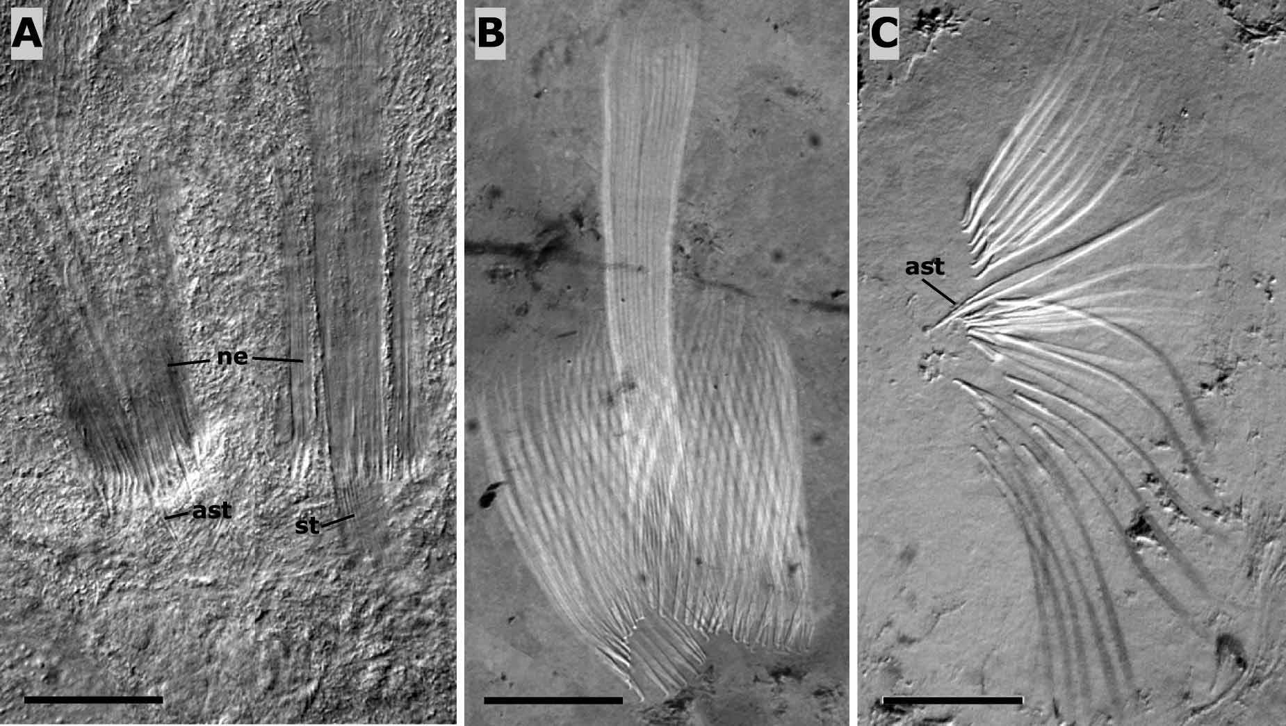
The body wall around the pores can be retracted, forming a triangular invagination ( Fig. 2 View FIGURE 2 A). The male pore is a transverse slit and at a very short distance in front of the female pore. The copulatory organ is in the dorsal part of the male atrium, somewhat at the right, while the accessory organ is at the left and ventrally in an almost horizontal position ( Figs 1 View FIGURE 1 D and 2C). This accessory apparatus consists of a glandular reservoir ( Fig. 1 View FIGURE 1 D and 2B) from where departs a muscular duct that is swollen into a highly muscular bulb. This continues as a narrow duct towards a short conical stylet that is surrounded dorsally by 10–12 thin needles and ventrally by ±15 thicker and slightly longer needles ( Figs 3 View FIGURE 3 and 4 View FIGURE 4 C). All these needles are clearly a product of the high atrial epithelium that fills the proximal part of the accessory apparatus. Stylet and needles are surrounded by circular muscles and strong protractor muscles ( Fig. 2 View FIGURE 2 E). Measurements of stylet and needles in eight individuals in whole mounts are given in Table 1.
The copulatory organ has two seminal vesicles, on either side of the accessory glandular reservoir ( Figs 1 View FIGURE 1 D and 2B) which taper into two narrow canals and join at the entrance of the prostate vesicle. That vesicle has a high, nucleated epithelium, obviously glandular. External prostate glands are depicted in Fig. 1 View FIGURE 1 B, but no signs of such glands were found in the sectioned material and may be an error of observation made while studying these opaque animals. From the prostate vesicle depart some 40–50 needles ( Figs 1 View FIGURE 1 D, 3 and 4A–B), about 191µm long (in the holotype; mean length in all eight individuals: 227µm), arranged in a ring and so forming a tube that is easily confused with the stylet found in many other proseriates. This ring of needles is surrounded by a second ring of some 60 needles, with a length ranging from 109µm to 114µm in the holotype (mean lengths in the eight individuals: 100 to 123µm) ( Table 1). All needles are embedded in the epithelium of the male atrium, those of the outer ring in the outer epithelium, those of the inner ring in a central continuation of the epithelium ( Figs 1 View FIGURE 1 D and 2D). The needles protrude from this epithelium only over a short distance where the atrium enlarges to continue to the part where the needles and the stylet of the accessory organ enter.
Copulatory organ Accessory organ
shortest longest "stylet" shortest longest stylet 1 107 120 204 37 70 37 2 107 126 241 72 91 19 3 89 130 213 62 71
4 101 123 194 55 109 22 5 96 116 226
6 104 128 318 67 105 31 7 87 129 228 92
Holotype 109 114 191 72 126 34 Mean 100 123 227 61 95 29
| SMNH |
Saskatchewan Museum of Natural History |
No known copyright restrictions apply. See Agosti, D., Egloff, W., 2009. Taxonomic information exchange and copyright: the Plazi approach. BMC Research Notes 2009, 2:53 for further explanation.
|
Kingdom |
|
|
Phylum |
|
|
Class |
|
|
Order |
|
|
Family |
|
|
Genus |