
Paracalviria islandica, Schockaert, Ernest R., Curini-Galletti, Marco, Ridder, Wouter De & Artois, Tom, 2011
|
publication ID |
https://doi.org/10.5281/zenodo.203197 |
|
DOI |
https://doi.org/10.5281/zenodo.6182157 |
|
persistent identifier |
https://treatment.plazi.org/id/55718796-FFCF-FFE2-DB96-668CFE6CFC13 |
|
treatment provided by |
Plazi |
|
scientific name |
Paracalviria islandica |
| status |
sp. nov. |
Paracalviria islandica n.sp.
Figs 7 View FIGURE 7 , 8 View FIGURE 8 , 9 View FIGURE 9 C
Synonym: Calviriidae gen. nov sp. nov. in Curini-Galletti et al. (2010).
Diagnosis. Diagnosis of the genus; with about 60 atrial needles of 130–180µm.
Occurrence. Iceland, Keflavik, close to the Sandgerdi Marine Centre, in the intertidal zone of a beach with medium sand (July, 1999).
Material studied. Many animals studied alive, nine animals in eight whole mounts (only one female), one specimen squashed for karyology and mounted, and 10 sectioned specimens (two “adult males” and one young male; three fully developed females and two young females, two juveniles). One whole mount is designated the holotype ( SMNH nr. 8098); all other material designated paratypes (UH 440-457).
Derivation of the name. The beautifully arranged needles of the copulatory organ resemble those of the species of Calviria ; the species name refers to the location where the species was found.
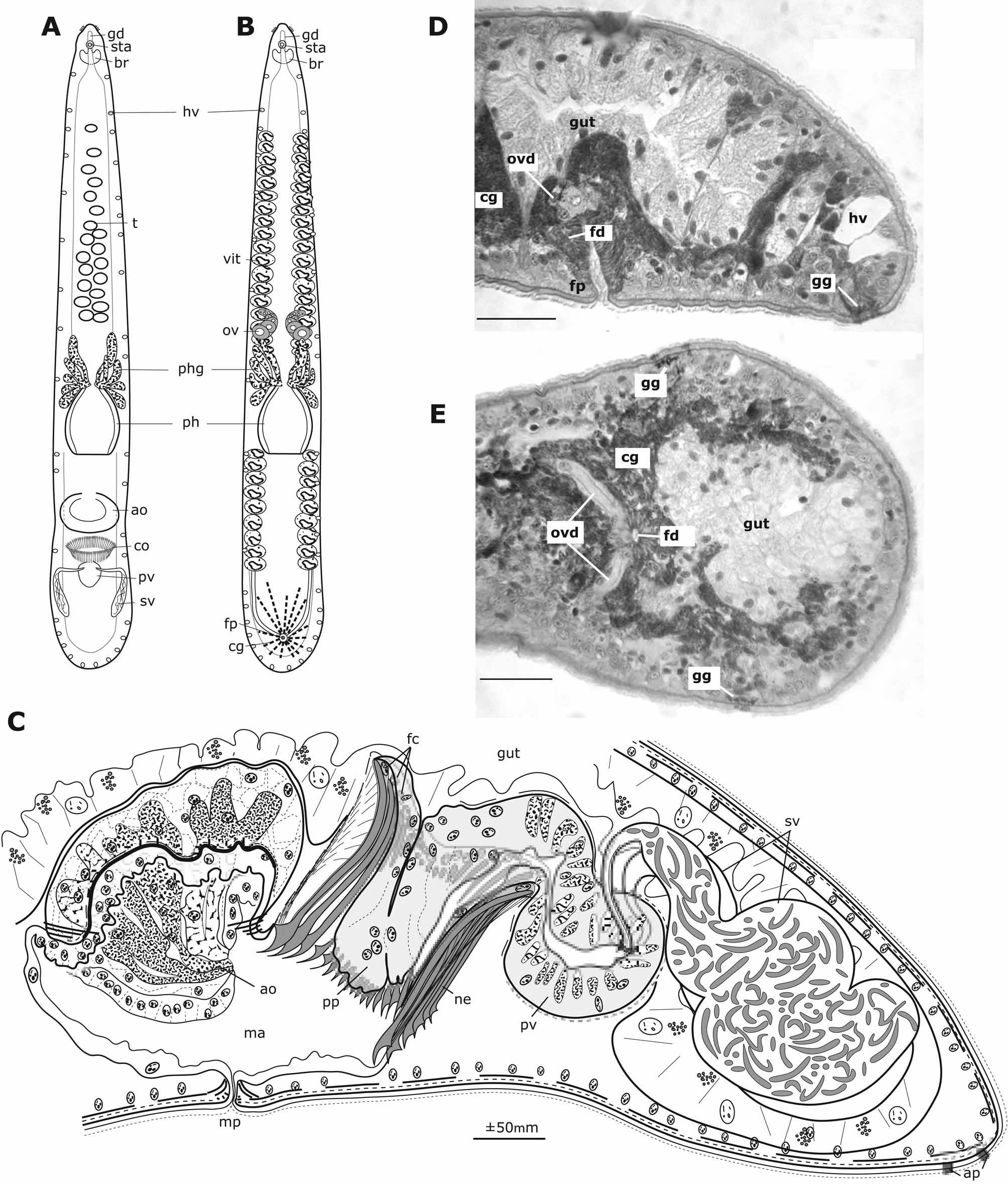
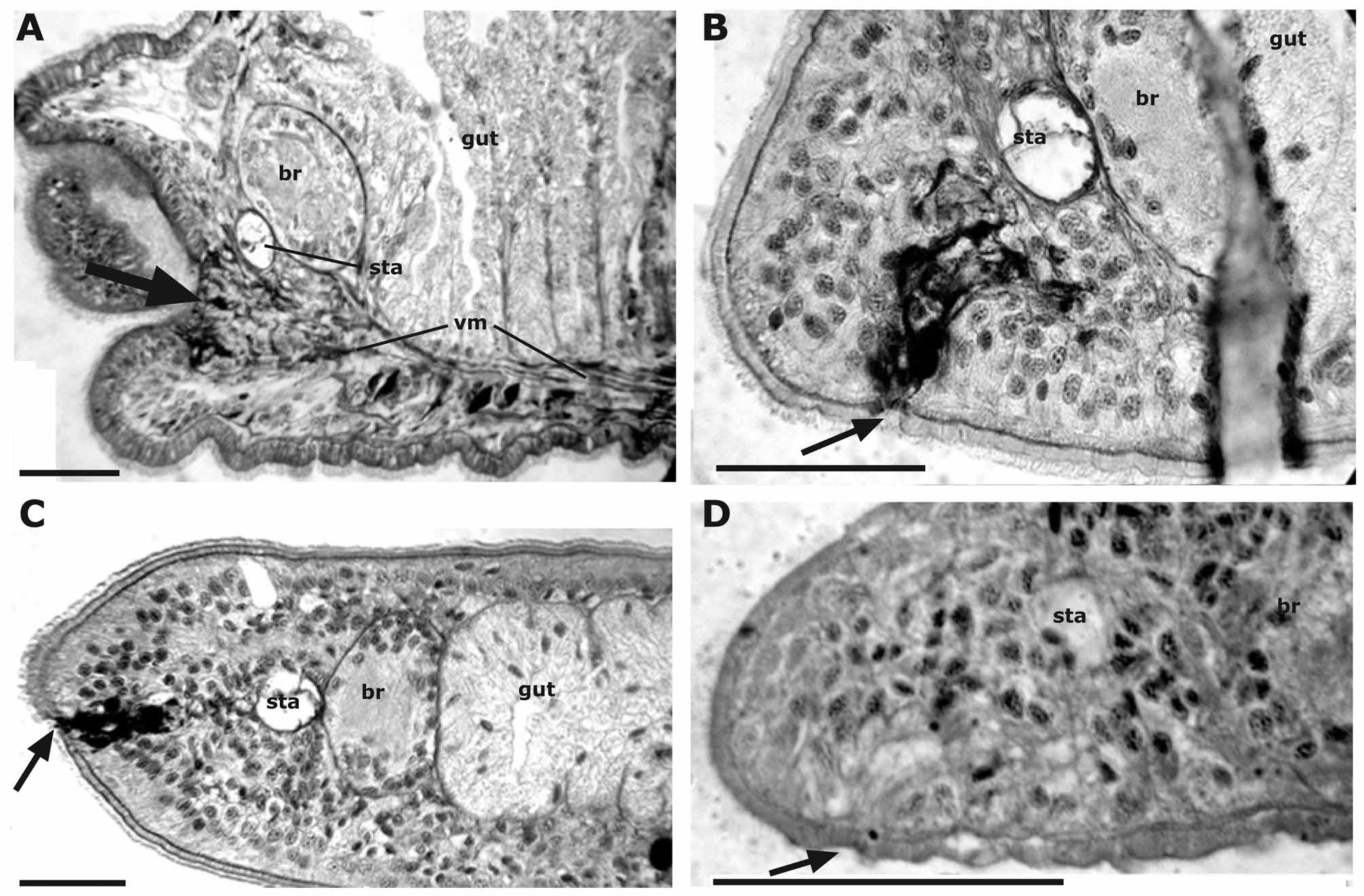
Description. The animals are whitish, 5–7mm long (measured in the whole mounts); there is seemingly no difference in size between the individuals in the male and in the female stage. There is a slight narrowing at the level of the brain and at the genital pore in the male stage. There are some sensory bristles at either side of the head ( Fig. 7 View FIGURE 7 A and B). The epidermis and cilia are around 3µm (on the dorsal as well as at the ventral side) with the nuclei sunk under the muscle layer, which consists of fine circular and slightly thicker longitudinal muscles. Large hyaline vacuoles, about 20µm high and 17µm broad occur all over the body, but mainly at the lateral sides and in the caudal region. In the male stage there are a few adhesive papillae at the tail, while in the female stage there are large glands opening in a row from the lateral sides over the ventral side ( Fig. 7 View FIGURE 7 D, E). The frontal glands converge to a slightly depressed sub-terminal area ( Fig. 9 View FIGURE 9 B). The anterior diverticulum of the gut runs clearly over the statocyst, and the gut itself runs to the very caudal end. The pharynx is at about 2/3 of the body length and has a similar construction to that in the other Calviriidae , albeit the internal circular muscle layer is highly developed. The glands that open at the tip of the pharynx are extremely developed and are even found in front of the ovaries.
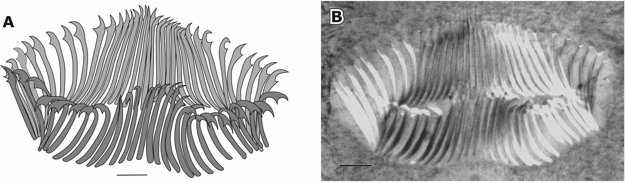
In the male stage ( Fig. 7 View FIGURE 7 A and C) there are 20–30 testes from some distance behind the brain to the level of the pharynx glands; the most anterior (and smaller) testes are in a single row, while those more towards the pharynx are in two alternating rows. Two large seminal vesicles, filled with unusually thick sperm, are in the very caudal end of the animal and enter the prostate vesicle laterally through S-shaped seminal ducts. The prostate vesicle, with coarse basophilic glands proximally and fine eosinophilic glands distally, continues with a duct, the epithelium of which is much thicker dorsally than ventrally. This duct protrudes as a penis papilla in a ring of ±60 atrial needles. There are weak spiral muscles around the prostate vesicle, but no muscles were detected around the duct that forms the penis papilla. The atrial needles ( Fig. 8 View FIGURE 8 ) are arranged in an oval ring, about 400–500µm in lateral direction and 100µm in antero-caudal direction. The number of needles is constant around 60 in the five “adult” males in the whole mounts. The length of the needles varies from 130 to 180µm and they are about 45µm in the two developing males. They end in a strong hook; at the base of each hook is inserted a bundle of muscles that originates from the proximal end of the needle. In front of the copulatory organ there is a large oval accessory glandular organ in the male atrium, about 300x460 to 360x730µm (measured in the whole mounts). Towards the atrium it is covered by a cuboidal hyaline epithelium. The central area of the organ is depressed, the whole organ forming a kind of sucker. The central area of the basement membrane under the epithelium is thick with some weak muscles under it. Large basophilic and eosinophilic glands open at the right and left sides of the sucker-like depression. The rest of the atrium is covered with a low epithelium and the male pore is guarded by a small sphincter.
In the fully developed female stage ( Fig. 7 View FIGURE 7 B, D and E) the ovaries, lying in front of the pharynx glands, are very large, C-shaped. The vitellaria start at some distance behind the brain and continue until about midway between the pharynx and the caudal end, with seven to eight post-pharyngeal follicles. The ovovitelloducts enter the short female duct (±65µm long) from both sides. The cement glands are highly developed and enter the female duct over its entire length. Anteriorly they almost reach the last vitellarian follicle; posteriorly they reach the caudal end, where they are mixed with the row of adhesive glands.
In one of the “males” in whole mount there was a faint indication of developing ovaries, but in none of the sectioned male stages was any indication of ovaries seen. On the other hand, in two of the developing females remnants of the male system were seen in the gut. In one of these individuals, the copulatory organ could clearly be recognised; in the other individuals there were some spines in the post-pharyngeal part of the gut that can be interpreted as partly resorbed needles of the copulatory organ. Probably the male organs are digested during the development of the female organs.
| SMNH |
Saskatchewan Museum of Natural History |
No known copyright restrictions apply. See Agosti, D., Egloff, W., 2009. Taxonomic information exchange and copyright: the Plazi approach. BMC Research Notes 2009, 2:53 for further explanation.
|
Kingdom |
|
|
Phylum |
|
|
Class |
|
|
Order |
|
|
Family |
|
|
Genus |