
Hydroglyphus balkei Hendrich
|
publication ID |
https://doi.org/ 10.5281/zenodo.198996 |
|
DOI |
https://doi.org/10.5281/zenodo.5621558 |
|
persistent identifier |
https://treatment.plazi.org/id/56298791-CC13-282A-08BA-8964FE55F929 |
|
treatment provided by |
Plazi |
|
scientific name |
Hydroglyphus balkei Hendrich |
| status |
|
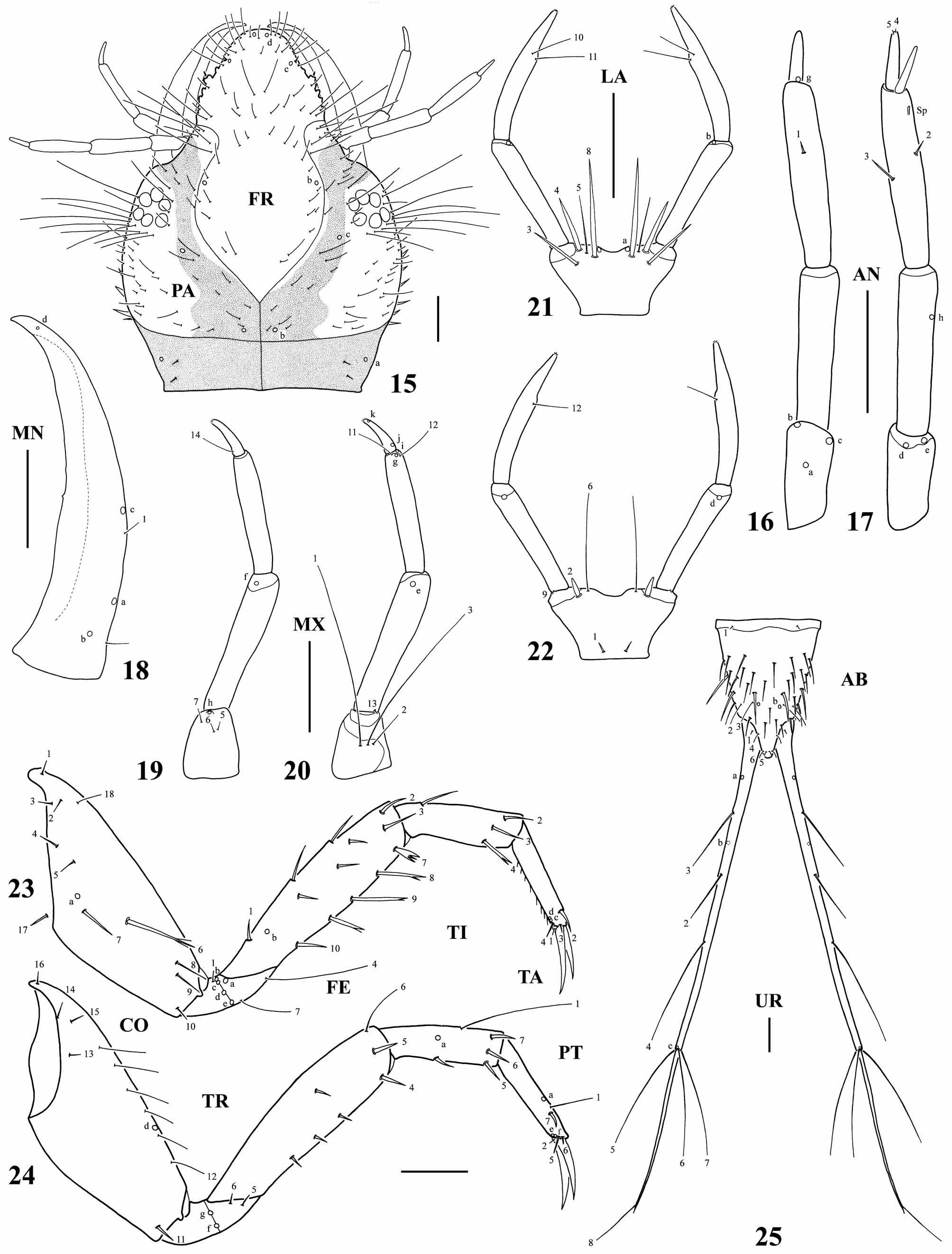
Hydroglyphus balkei Hendrich View in CoL , third-instar larva
Source of material. Australia; Watson, 14 km W Herberton Qld., 31/iii/1996; C. H. S. Watts.
Diagnosis. Antennomere 3 with a ventroapical spinula ( Fig. 17 View FIGURES 15 – 25 ); pore PAj absent; U without secondary setae ( Fig. 25 View FIGURES 15 – 25 ); absence of anterior secondary setae on CO and presence of 8–9 secondary setae on mesoFE ( Table 2); ratios A4/A3, LP2/LP1, L3/L1, L3/HW and LAS/HW ( Table 1 View TABLE 1 ).
Description. Color. Head capsule pale yellow, area posterior to occipital suture light brown, V-shaped band contiguous to ecdysial line light brown; head appendages pale yellow, mandible light brown; thoracic tergites pale yellow with light brown laterals, meso- and metatergite with a light brown anterior macula at each side of mid line; abdominal tergites I–VI predominantly pale yellow, with light brown laterals, sclerites VII–VIII predominantly light brown; membranous parts pale; legs pale yellow; urogomphus light brown.
Body. Subcylindrical, narrowing towards abdominal apex. Measurements and ratios that characterize the body shape are shown in Table 1 View TABLE 1 .
Head. Head capsule ( Fig. 15 View FIGURES 15 – 25 ). Longer than broad; posterolateral surface covered with minute spinulae; maximum width posterior to stemmata, without neck constriction; occipital suture present; ecdysial line well marked, coronal line short; occipital foramen broadly emarginate ventrally; posterior tentorial pits visible ventrally; FR elongate, lateral margins sinuate; nasale moderately elongate, subtriangular, rounded apically, slightly sinuate laterally, with one small branch at each side; ventrodistal surface with spinulae of different shapes, ventrolateral margin with several robust spinulae; anteroventral margin of nasale with a half circle of 33–35 spatulate setae of different lengths, directed downward; six dorsolateral stemmata at each side forming a circle. Antenna ( Figs 16–17 View FIGURES 15 – 25 ). Elongate, composed of four antennomeres, shorter than HW; A4 the shortest, A2 and A3 the longest, subequal; A3 with a ventroapical spinula; A3’ relatively elongate. Mandible ( Fig. 18 View FIGURES 15 – 25 ). Prominent, broad basally, distal half projected inwards and upwards, apex sharp; mandibular channel present. Maxilla ( Figs 19–20 View FIGURES 15 – 25 ). Cardo fused to stipes; stipes short, broad; galea and lacinia absent; MP elongate, shorter than antenna, composed of three palpomeres, MP3 the shortest, MP1 and MP2 the longest, subequal. Labium ( Figs 21–22 View FIGURES 15 – 25 ). Prementum small, subtrapezoidal, somewhat broader than long, without lateral spinulae, anterior margin slightly indented medially; LP elongate, composed of two palpomeres; LP2 slightly longer than LP1.
Thorax. Terga convex, pronotum slightly shorter than meso- and metanotum combined, meso- and metanotum subequal; protergite subrectangular, more developed than meso- and metatergite; meso- and metatergite transverse, with anterotransverse carina; sagittal line well visible; sterna membranous; spiracles present on mesothorax. Legs ( Figs 23–24 View FIGURES 15 – 25 ). Long, composed of six articles sensu Lawrence 1991), L1 the shortest, L3 the longest; CO robust, elongate, TR divided into two parts, FE, TI and TA slender, subcylindrical, PT with two long, slender, slightly curved claws; posterior claw shorter than anterior claw on L1 and L2, posterior claw longer than anterior claw on L3; most surface of legs covered with minute slender spinulae in transverse rows; ventral surface of pro- and mesoTA and to a lesser extent pro- and mesoTI with elongate spinulae.
Abdomen. Eight-segmented; segments I–VI sclerotized dorsally, membranous ventrally; tergites I–VI narrow, transverse; segments VII–VIII completely sclerotized, ring-like; all sclerites without sagittal line, with anterotransverse carina, covered with minute spinulae in transverse rows; spiracles present on laterals of segments I–VII; LAS ( Fig. 25 View FIGURES 15 – 25 ) the longest, siphon short, subconical. Urogomphus ( Fig. 25 View FIGURES 15 – 25 ). Very long, composed of two urogomphomeres; U1 long, much longer than LAS, basal portion covered with minute spinulae; U2 narrow, setiform, shorter than U1.
Chaetotaxy. Head capsule with numerous secondary setae; parietal with 4–5 short, spine-like, secondary setae on each lateroventral margin; MN with one hair-like, secondary seta on basoexternal margin; thoracic and abdominal sclerites I–VII with several secondary setae, mainly on posterior half; secondary leg setation detailed in Table 2 and Figs 23–24 View FIGURES 15 – 25 ; LAS with numerous spine-like secondary setae ( Fig. 25 View FIGURES 15 – 25 ). Primary chaetotaxy similar to that of generalized Hydroporinae larva (Alarie & Harper 1990; Alarie et al. 1990; Alarie 1991; Alarie & Michat 2007) except for the following features: pore FRc submarginal, contiguous to seta FR7; pores PAd, PAe and PAj absent; pore PAg present; pore ANf absent; setae MX 4, MX 8, MX 9 and MX 10 absent; seta MX 1 inserted distally on the stipes; seta LA7 absent; seta TR 2 absent; pore FEa absent; seta TI7 short, spine-like; pores ABa and ABc absent; seta AB10 spine-like; we were unable to find pore ABd and setae AB7 and AB8; however, we could not establish if they are really absent due to the presence of spinulae on the siphon; setae UR2, UR3 and UR4 inserted far from each other; setae UR5, UR6 and UR7 elongate; seta UR8 inserted distally.
No known copyright restrictions apply. See Agosti, D., Egloff, W., 2009. Taxonomic information exchange and copyright: the Plazi approach. BMC Research Notes 2009, 2:53 for further explanation.