
Daptonema hyalocella, Aryuthaka, Chittima & Kito, Kenji, 2012
|
publication ID |
https://doi.org/ 10.5281/zenodo.211983 |
|
DOI |
https://doi.org/10.5281/zenodo.5670632 |
|
persistent identifier |
https://treatment.plazi.org/id/563DAB49-FFDC-B454-37A7-B880FDA9F9F2 |
|
treatment provided by |
Plazi |
|
scientific name |
Daptonema hyalocella |
| status |
sp. nov. |
Daptonema hyalocella View in CoL sp. n.
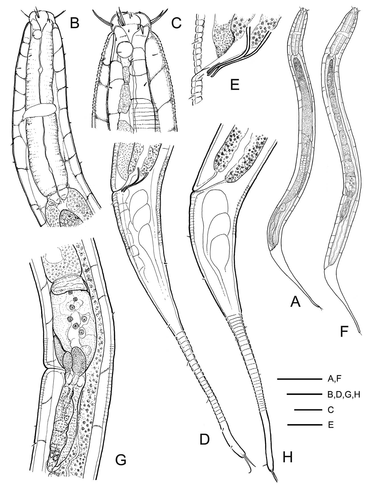
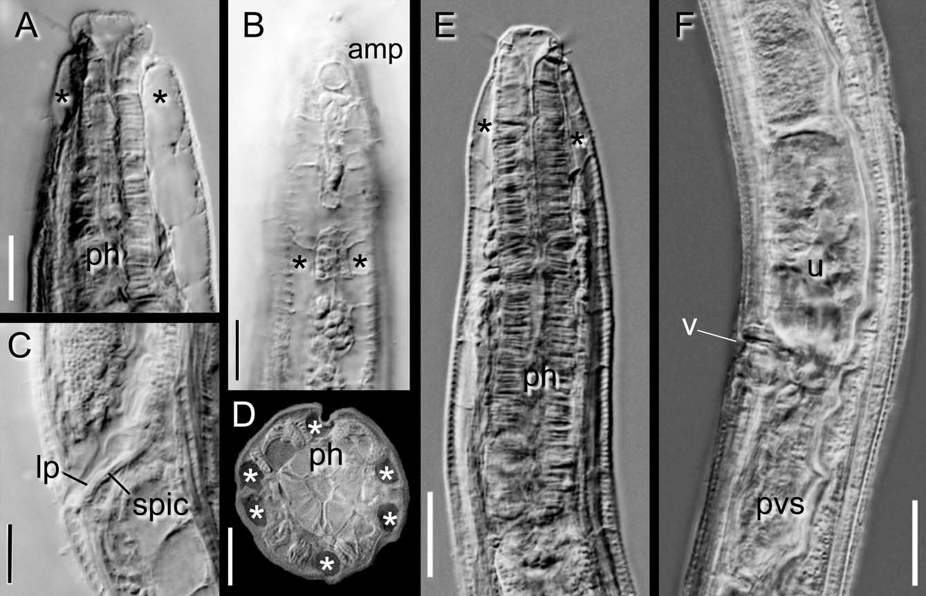
( Fig. 2 View FIGURE 2 A–H, 3A–F)
Type specimens. Five males and 5 females. Holotype: male ( ZIHU 3811). Allotype: female ( ZIHU 3817). Paratypes: 4 males ( ZIHU 3812-3814; KUMF, T0009) and 4 females ( ZIHU 3815,3816 & 3818; KUMF, T0010). Collected on 4th February 2003 by C. Aryuthaka.
Type locality and habitat: in Halophila ovalis patches at Ban Pa Khlok, Phuket Province, Thailand; 8º 01' 20'' N, 98º 24' 40'' E.
Measurements. Table 1.
Holotype: L = 741 µm, a = 20.6, b = 7.0, c = 4.6.
Males (n = 5, including holotype): L = 676–890 (773 ± 79.6) µm, a = 17.3–21.7 (19.4 ± 1.7), b = 6.0–7.0 (6.6 ± 0.4), c = 4.4–5.2 (4.8 ± 0.3).
Females (n = 5): L = 694–859 (783 ± 60.7) µm, a = 15.8–20.5 (18.7 ± 1.9), b = 6.1–6.5 (6.4 ± 0.2), c = 4.5–5.4 (5.0 ± 0.3), V (%) = 55.9–58.3 (57.2 ± 1.0).
Description. MALE (Holotype ZIHU 3811: range and/or avg ± sd in parentheses). Body short, about 0.7 (0.6–0.9) mm long, almost uniform diameter except pharyngeal and tail regions; posterior portion bent dorsally ( Fig. 2 View FIGURE 2 A). Cuticle weakly striated, annules about 1.5 (1.3–2.0) µm apart but somewhat obscure on lateral chords. Epidermal cells of dorsal, ventral and lateral chords highly transparent, well thickened, extending inwards into body cavity, especially in pharyngeal region ( Fig. 2 View FIGURE 2 B, 3A, D); a central row of cells in lateral chords somewhat reticulated, not as thickened as adjacent rows of transparent cells ( Figs. 2 View FIGURE 2 B, 3B). In lateral view, transverse boundaries of each 2 adjoining transparent epidermal cells appear like bridges that subdivide the body cavity into compartments. Short somatic setae sparsely distributed throughout body, usually less than 6 µm long; peculiar cervical setae not observed.
Head ( Fig. 2 View FIGURE 2 C, 3A) blunt, clearly set off; labial region low, about 7 (6–7) µm in height. Head equipped with 6 tiny papillae and 12 short cephalic setae, 0.7 (0.6–0.7; 0.6 ± 0.04) hd long at maximum. Amphids circular, small, about 6 (5.6–6.6; 6.1 ± 0.4) µm in diameter, occupying 27/27 (25–33; 28.9 ± 2.9) % cbd; anterior margin of amphids 0.8/0.8 (0.5–0.9; 0.8 ± 0.1) hd from anterior body end. Buccal cavity cup-shaped, about 8 (7–8) µm wide, posterior half surrounded by pharynx. Pharynx ( Fig. 2 View FIGURE 2 C, 3E) muscular, almost cylindrical but gradually enlarging toward posterior portion, 0.15 (0.14–0.17) body length. Cardia short, 7 (7–12) µm long. Nerve ring indistinct, anterior margin 49 (42–49; 45.5 ± 2.6) % of pharyngeal length from anterior body end. Ventral gland and excretory pore not observed.
Reproductive system diorchic. Anterior testis outstretched, to the left of the intestine, extending to near pharyngeal end, 464 (396–578; 475 ± 75.5) µm or 63 (59–65; 61.8 ± 2.6) % of body length from cloaca; posterior testis, right of intestine, reflexed at 294 (245–355; 296 ± 39.5) µm or 40 (36–40; 38.2 ± 1.5) % of body length from cloaca and extending backward to level 156 (109–175; 140.0 ± 25.7) µm or 21 (14–22; 18.2 ± 3.2) % of body length from cloaca. Spicules ( Fig. 2 View FIGURE 2 E, 3C) weakly cuticularized, remarkably thin (about 1 µm in diameter at middle portion) and short, 0.8/0.7 (0.6–0.8; 0.73 ± 0.05) abd long on arc or 0.8/0.7 (0.6–0.8; 0.69 ± 0.05) on chord; proximal end not cephalated, proximal and distal portions loosely curved in counter directions (loose S-shaped). Gubernaculum indistinct, small lateral pieces (crura) located near distal end of spicules. Ejaculatory glands between distal end of posterior testis and cloaca on each side of the intestine but exact number not clearly observed.
Tail ( Fig. 2 View FIGURE 2 D) conico-cylindroid, 5.8 (5.5–5.8; 5.7 ± 0.11) abd long; posterior cylindroid portion about twofifths (0.3–0.4) of tail length; a long terminal seta located on each side, 12 (8–14) µm long. Spinneret short, not protruded. Three caudal glands in line and partially overlapping.
FEMALE (Allotype ZIHU 3817; range and/or avg ± sd in parentheses). Similar to males in most features. Body short about 0.8 (0.6–0.9) mm long ( Fig. 2 View FIGURE 2 F). Epidermal chords consisting of transparent cells, especially in pharyngeal region ( Fig. 3 View FIGURE 3 E). Amphids slightly smaller than in male, 5.5/5.4 (5.1–6.1; 5.6 ± 0.3) µm in diameter, occupying 26/24 (23–32; 25.7 ± 2.6) % of corresponding body diameter. Nerve ring located at 45 (39–46; 43.7 ± 2.5) % of pharyngeal length from anterior.
Reproductive system monodelphic, prodelphic. Ovary outstretched, left of intestine, extending near to pharyngeal end, 321 (276–332; 305 ± 22.0) µm or 41 (37–41; 39.0 ± 1.4) % of body length from vulva. Vulva transverse, located slightly posterior to midbody, 5.9 (4.8–6.8; 6.0 ± 0.8) abd long anterior to anus; postvulval uterine sac present, 1.6 (1.1–1.6; 1.4 ± 0.2) vd long ( Figs. 2 View FIGURE 2 G, 3F). Small vaginal glands, probably 3, located on each side of vulva.
Tail ( Fig. 2 View FIGURE 2 H) conico-cylindroid, 5.4 (4.9–6.1; 5.3 ± 0.5) abd long; posterior one-third almost cylindroid (24–35% of tail length). Two terminal setae located at tail end as in male, 8 (8–15) µm long.
Etymology. The specific name hyalocella refers to the peculiar transparent cells of the epidermal chords which appear to divide the body into compartments.
Differential diagnosis. Daptonema hyalocella sp. n. is characterized in the genus by the following features: short body length (0.6–0.9 mm), small de Man’s c ratio (4.4–5.4), peculiar epidermal chords consisting of large cells with transparent appearance (bridge-like structures observed in lateral view as if they are in body cavity), two testes and one ovary, male copulatory apparatus consisting of thin (about 1 µm in diameter at middle portion), short (0.6–0.8 abd long) and loosely S-shaped spicules with no distal cephalation, indistinct gubernaculum with small lateral piece (crura), and conico-cylindroid tail with 2 long terminal setae (8–14 µm).
Daptonema hyalocella sp. n. is distinguished from most other species of Daptonema by the epidermal chords consisting of large cells with a transparent appearance together with the male copulatory apparatus consisting of conspicuously thin, S-shaped spicules with no proximal cephalation and an indistinct gubernaculum with a lateral piece. Similar epidermal chords have been reported in Daptonema trabeculosum ( G. Schneider, 1906) by Schneider (1906), Riemann (1966) and Lorenzen (1977) and in D. conicum ( Filipjev, 1922) by Lorenzen (1977), though they did not refer to the epidermal cells as such but described the transverse boundaries of each 2 adjoining epidermal cells as “Trabekel” ( Schneider, 1906) or “Trabekeln” ( Riemann, 1966); terms which mean a cytoplasmic bridge between the cuticle and alimental canal. According to Riemann (1966), this feature of the epidermal chords has no taxonomic significance because “Trabekeln” did not always exist in his specimens. In the seagrass beds that have been studied, besides D. hyalocella sp. n. and the new species described below, there are several species of Daptonema and Paramonohystera Steiner, 1916 that also have similar epidermal chords (Aryuthaka & Kito, unpublished data), though their bridge-like structures were usually not clearly evident. However, in D. hyalocella sp. n., these bridge-like structures were distinct in all of the specimens examined, rendering individuals easily distinguishable from other nematodes viewed with transmitted light; they are a valuable taxonomic feature.
In having the distinctive epidermal chords, Daptonema hyalocella sp. n. is similar to D. conicum , D. trabeculosum and the new species described below. However, D. hyalocella sp. n. is easily distinguished from the former three species by the features of male copulatory apparatus; the thin, loose S-shaped spicules with no proximal cephalation and an indistinct gubernaculum with a lateral piece, compare with curved spicules with proximal cephalation and a gubernaculum lacking a lateral piece in D. conicum (cf. Filipjev, 1922; Lorenzen, 1977), thick, almost L-shaped spicules with proximal cephalation in D. trabeculosum (cf. G. Schneider, 1906; Lorenzen, 1977), and thick, L-shaped spicules with proximal cephalation and a gubernaculum with a dorsal apophysis and a lateral piece in the following new species. Daptonema hyalocella sp. n. somewhat resembles D. trabeculosum redescribed by Riemann (1966) in having the thinner spicules (about 2 µm in diameter, calculated from his drawings, Abb. 22), that bend weakly and are poorly cephalated proximally, and in having a gubernaculum with a lateral piece. However, D. hyalocella sp. n. differs from his D. trabeculosum by the short body length and the small value of de Man’s c ratio; 0.6–0.9 mm and c = 4.4–5.4 vs. 0.8–1.1 mm and c = 6.7–7.3 for D. trabeculosum . Daptonema trabeculosum sensu Riemann was reported from the mouth of the Elbe on the North Sea. Considering both the male copulatory apparatus and geographic distribution, it might be that D. trabeculosum sensu Riemann is not the same species as D. trabeculosum sensu Schneider (1906) and Lorenzen (1977) reported from Tvarminne, on the Baltic Sea coast of Finland (type locality).
Except for the feature of epidermal chords, D. hyalocella sp. n. rather resembles D. microspiculum ( Gerlach, 1953) in having the short, S-shaped spicules with no proximal cephalation. However, D. hyalocella sp. n. is distinguished from D. microspiculum by the following features (cf. Gerlach, 1953): larger de Man’s b ratio (6.0–7.0 vs. 3.5–4.2 in D. microspiculum ), 12 shorter cephalic setae 8–10 µm in length (vs. 10 setae 14–16 µm length); smaller and more anterior amphids (6–7 µm diameter, located 0.5–0.9 hd from anterior body end in both sexes vs. about 16 µm diameter in male, located at about 1.5 hd length from anterior body end in female; calculated from original drawings); the presence of an indistinct gubernaculum with a lateral piece (vs. no gubernaculum); and shorter tail terminal setae (8 – 14 µm vs. 25–30µm).
No known copyright restrictions apply. See Agosti, D., Egloff, W., 2009. Taxonomic information exchange and copyright: the Plazi approach. BMC Research Notes 2009, 2:53 for further explanation.
|
Kingdom |
|
|
Phylum |
|
|
Class |
|
|
Order |
|
|
Family |
|
|
Genus |
Daptonema hyalocella
| Aryuthaka, Chittima & Kito, Kenji 2012 |
D. microspiculum (
| Gerlach 1953 |
D. conicum (
| Filipjev 1922 |
Paramonohystera
| Steiner 1916 |
Daptonema trabeculosum (
| G. Schneider 1906 |
D. trabeculosum sensu
| Schneider 1906 |