
Psilomyzon sarcotragusicola, Zagami & Costanzo & Brugnano, 2014
|
publication ID |
https://doi.org/ 10.1080/00222933.2014.917209 |
|
persistent identifier |
https://treatment.plazi.org/id/567F878C-FFBB-FF8E-18AB-78701D73AEDC |
|
treatment provided by |
Felipe |
|
scientific name |
Psilomyzon sarcotragusicola |
| status |
sp. nov. |
Psilomyzon sarcotragusicola sp. nov.
( Figures 7–9 View Figure 7 View Figure 8 View Figure 9 )
Material examined
Holotype female plus 3 female and 2 male paratypes are stored in the collections of the Zoological Museum Cambria ( ZMC), Department of Biological and Environmental Sciences , University of Messina , Italy, Reg. Nos 2013.36 (holotype female), 2013.3741 (paratypes). Dissected female and male paratypes were mounted on 10 and 5 slides, respectively, Reg. Nos 2013.42 (female) and 2013.43 (male). In addition, in the collections of the Natural History Museum , London are stored 1 female and 1 male paratypes Reg. Nos 2013.88 and 2013.89, respectively. One further paratype female and 1 male (intact) are stored in the authors’ collection.
Description
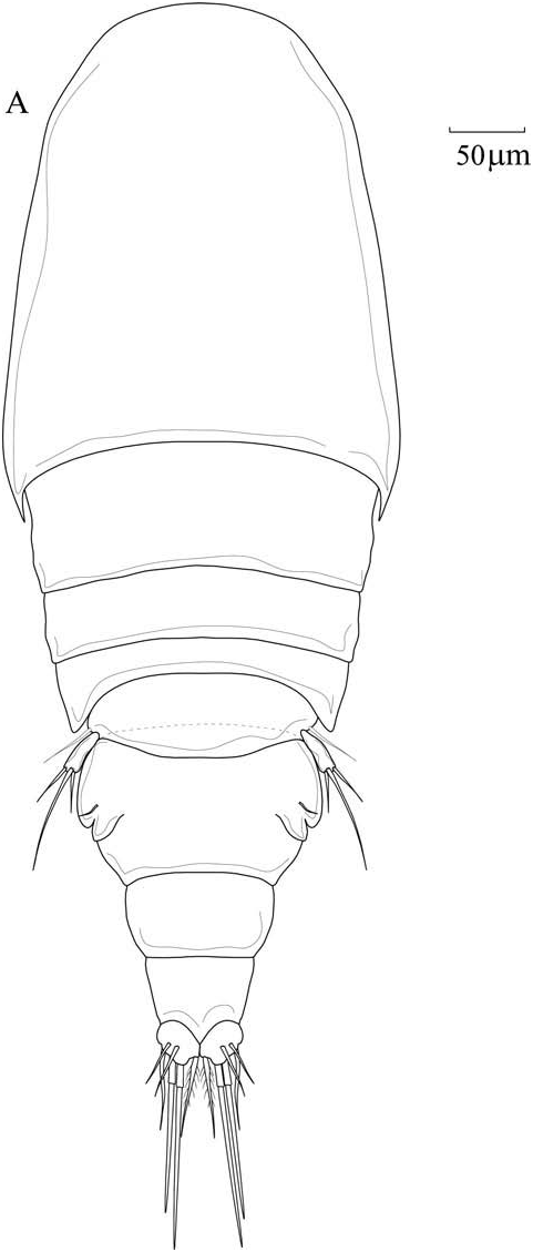
Adult female. Holotype total length 0.70 mm. Mean body length 0.65 mm, with range of 0.62–0.70 mm (based on four specimens).
Body ( Figure 7A View Figure 7 ) with weakly modified cyclopiform shape, narrow in dorsal view. Prosome elongate, about 1.8 times longer than wide, 4-segmented, first pedigerous somite fully incorporated into cephalothorax, with acute lateral margins. Free pedigerous somites with rounded lateral margins. Urosome 4-segmented, comprising fifth pedigerous somite, genital double-somite, formed by fusion of genital and first abdominal somites, free postgenital and anal somites. Genital double-somite about 1.6 times wider than long, expanded anteriorly, with 2 lateral rounded processes, bearing bipartite genital apertures, paired gonopores (oviduct openings) located laterally, and paired copulatory pores located laterally on ventral surface. Anal somite wider than long (64 × 55 µm). Caudal rami just longer than wide, bearing 6 setae, of which 4 are located on distal margin and 2 on dorsal surface.
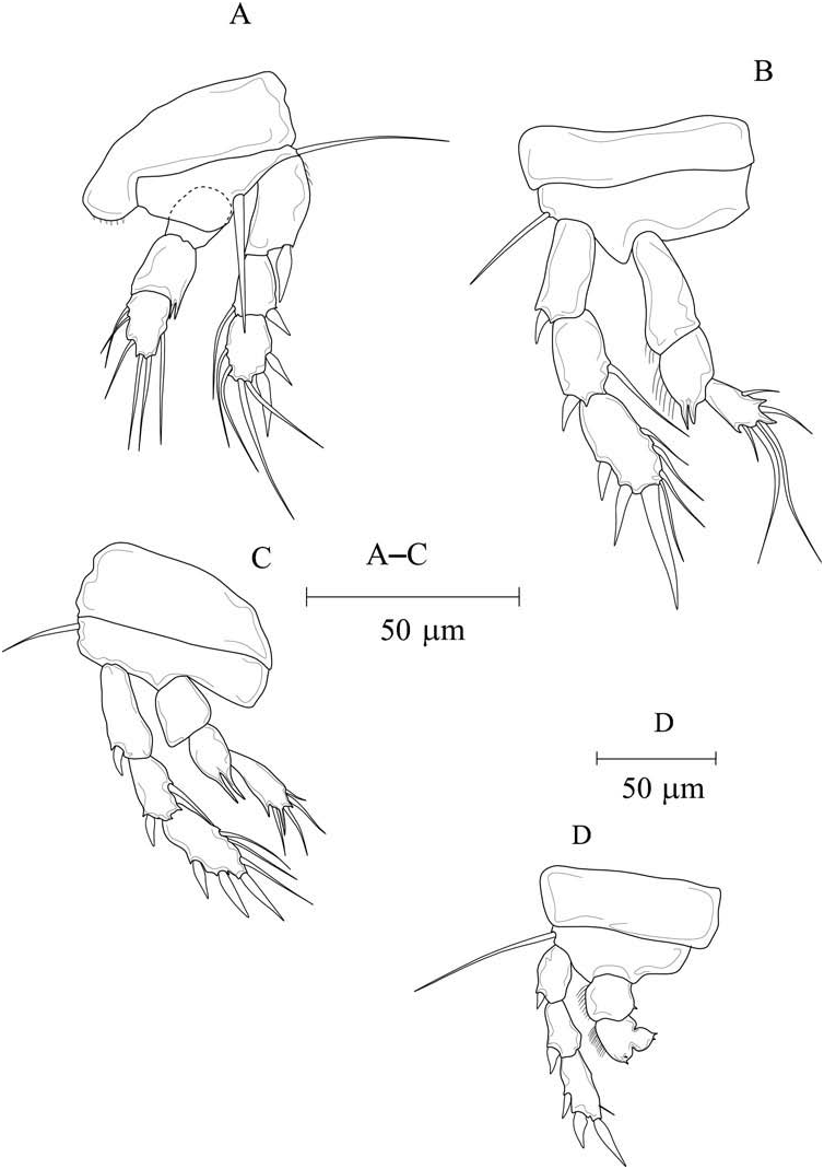
Antennule, antenna, oral cone, mandible, maxillule, maxilla and maxilliped similar to P. laetitiae . Swimming legs 1–4 ( Figure 8A–D View Figure 8 ) biramous, with 3-segmented rami, with exception of endopod of leg 4.
Spine and seta formulae are given in Table 2.
Leg 1 ( Figure 8A View Figure 8 ) with basal medial seta and setiform terminal exopodal element. Leg 2 ( Figure 8B View Figure 8 ) second endopodal segment with 2 acute outer processes. Leg 3 ( Figure 8C View Figure 8 ) second endopod with 2 long setiform processes on outer margin. Leg 4 ( Figure 8D View Figure 8 ) first endopodal segment with 1 inner small spiniform process, second segment with inner projection bearing 2 small spiniform processes, plus 2 spiniform processes on outer margin, lateral margins of endopodal segments each with row of setules. Leg 5 ( Figure 7A View Figure 7 ) with elongate free segment, 3 times longer than wide, bearing 1 sub-distal outer and 2 distal setae. Single outer seta present on surface of fifth pedigerous somite.
Leg 6 ( Figure 7A View Figure 7 ) represented by paired opercular plates closing off gonopores on genital somite, each bearing small seta.
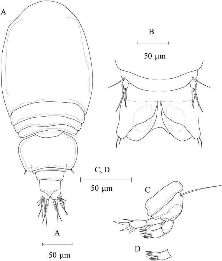
Adult male. Body ( Figure 9A, B View Figure 9 ) 0.44 mm long. Mean body length 0.45 mm, with range of 0.44–0.46 mm (based on three specimens). Body form in general similar to female, except urosome 5-segmented. Prosome elongate, about 1.4 times longer than wide. Genital somite about 1.6 times wider than long. Genital apertures paired, located on ventral surface near posterior margin of somite; paired ovoid spermatophores visible within somite through body wall ( Figure 9B View Figure 9 ). Anal somite 1.8 times wider than long. Caudal rami as for female.
Appendages as for female except antennules, legs 4 and 6.
Antennule 17-segmented, geniculate on both sides, segmental fusion model as follows: 1 (I), 2 (II), 3 (III), 4 (IV), 5 (V), 6 (VI), 7 (VII), 8(VIII), 9 (IX–XII), 10 (XIII), 11 (XIV), 12 (XV–XVI), 13 (XVII), 14 (XVIII), 15 (XIX–XX), 16 (XXI– XXIII), 17 (XXIV–XXVIII). Geniculation located between segments 15 and 16. Segments 1–8 each with 2 setae; segment 9 with 8 setae; segments 10 and 11 each with 2 setae; segment 12 with 4 setae; segment 13–15 each with 2 setae; segment 16 with 2 setae plus one aesthetasc; segment 17 with 10 setae.
Leg 4 endopod ( Figure 9C View Figure 9 ) as for P. laetitiae male, with number of setiform processes variable from 3 to 4, ( Figure 9D View Figure 9 ) on inner projection of second segment; exopod with reduced chaetotaxis, first to third segment with 0, 0, 1 seta, respectively. Leg 6 ( Figure 9A, B View Figure 9 ) represented by paired opercular plates closing off genital apertures, armed with 2 thin setae.
Etymology
The specific name sarcotragusicola is derived from the generic name of the host Sarcotragus spinosulus .
Remarks
Psilomyzon sarcotragusicola sp. nov. differs from P. laetitiae sp. nov. in total body length in both sexes. In the former, female and male mean lengths are 0.65 and 0.45 mm, respectively; whereas in the latter they are 0.70 and 0.64 mm, respectively. The cephalothorax of P. sarcotragusicola sp. nov. is more similar to that of Inermocheres quadratus Boxshall, 1990 ; it is narrow and with acute epimeral margins in dorsal aspect, compared to expanded posteriorly with rounded epimeral margins in P. pauciseta and P. laetitiae sp. nov. The swimming legs of P. sarcotragusicola have a reduced leg 4 endopod such as P. laetitiae , and the same setation to that present in the type species P. pauciseta . In addition there are significant differences in the setal formula between P. sarcotragusicola and P. laetitiae ; the characters of P. laetitiae sp. nov. are in parenthesis. In P. sarcotragusicola sp. nov. leg 2 first exopodal segment is I-0 (I-1); leg 3 first and third exopodal segments are I,0 and II,I,3 (I,1 and II,I,4); third endopodal segment is 1,1,2, (1,2,1); leg 4 exopod from first to third segments are I-0; I-0; II,I,1 (I-1; I-1; II,I,3). In P. sarcotragusicola sp. nov. the reduction of the exopodal setation of the swimming legs has proceeded further than in P. laetitiae sp. nov. in possessing a total of 15 versus 21 setae, respectively.
Ecological traits
Although S. spinosulus is common in the Mediterranean, it is reported here for the first time as a host to asterocherid copepods. The majority of the P. laetitiae and P. sarcotragusicola specimens were found in the washing water of the sponge a considerable time after sampling, when the oxygen concentration in the water would have been low. So, we may infer that the new species inhabits the internal canal system of the host sponge. In P. laetitiae and P. sarcotragusicola , the acute angle in the joining between basis and rami of the swimming legs and the small body size may be considered to be adaptations to life within narrow internal canals of the sponge. P. laetitiae and P. sarcotragusicola copepodites and adults, inhabiting the insides of the aquiferous canals, avoid predation by small fishes living among the sponges, while the diffusion of the species in other sponges is probably entrusted to naupliar forms. Mariani and Uriz (2001) concluded in their study on copepod feeding that the species of the genus Asterocheres spend their entire life cycle at the sponge surface. In this study, on S. spinosulus were found numerous invertebrates belonging to Copepoda Harpacticoida, Siphonostomatoida ( Psilomyzon , Asterocheres ), Cyclopinidae , hyperbenthic Calanoida (Pseudocyclops) , Amphipoda and Decapoda, that probably live at the sponge surface, being found in the washing water immediately after sampling. So, the sponges may represent a hotspot of cryptic biodiversity in respect to the neighbouring environment. Harpacticoid abundance outnumbered all others taxa. The average abundance of the P. laetitae and P. sarcotragusicola specimens, adults and copepodids, per host (about 120 cm 2) was 25 and 10 individuals, respectively. Asterocherid copepods are reported to live in sponges in high abundance. Ivanenko (1997) reported inside a single sponge host, 5 × 8 cm in size, over 3000 copepods. The female to male sex ratio, for both species, was about 4:1. During in situ observations, with repeated samplings in autumn, females of the two new species generally showed 4– 6 eggs at once in the egg sacs.
| ZMC |
Deptment of Biology, Zunyi Medical College |
No known copyright restrictions apply. See Agosti, D., Egloff, W., 2009. Taxonomic information exchange and copyright: the Plazi approach. BMC Research Notes 2009, 2:53 for further explanation.
|
Kingdom |
|
|
Phylum |
|
|
Class |
|
|
Order |
|
|
Family |
|
|
Genus |