
Fulvidius thailandicus (Chérot & Gorczyca) Cherot & Gorczyca, 2016
|
publication ID |
https://doi.org/ 10.11646/zootaxa.4184.2.6 |
|
publication LSID |
lsid:zoobank.org:pub:0B2C4021-BA8E-4FC2-91A4-C85442EF62DF |
|
DOI |
https://doi.org/10.5281/zenodo.6057959 |
|
persistent identifier |
https://treatment.plazi.org/id/5775879A-FFB6-751F-E895-8A313FDEF9F5 |
|
treatment provided by |
Plazi |
|
scientific name |
Fulvidius thailandicus (Chérot & Gorczyca) |
| status |
comb. nov. |
Fulvidius thailandicus (Chérot & Gorczyca) comb. nov.
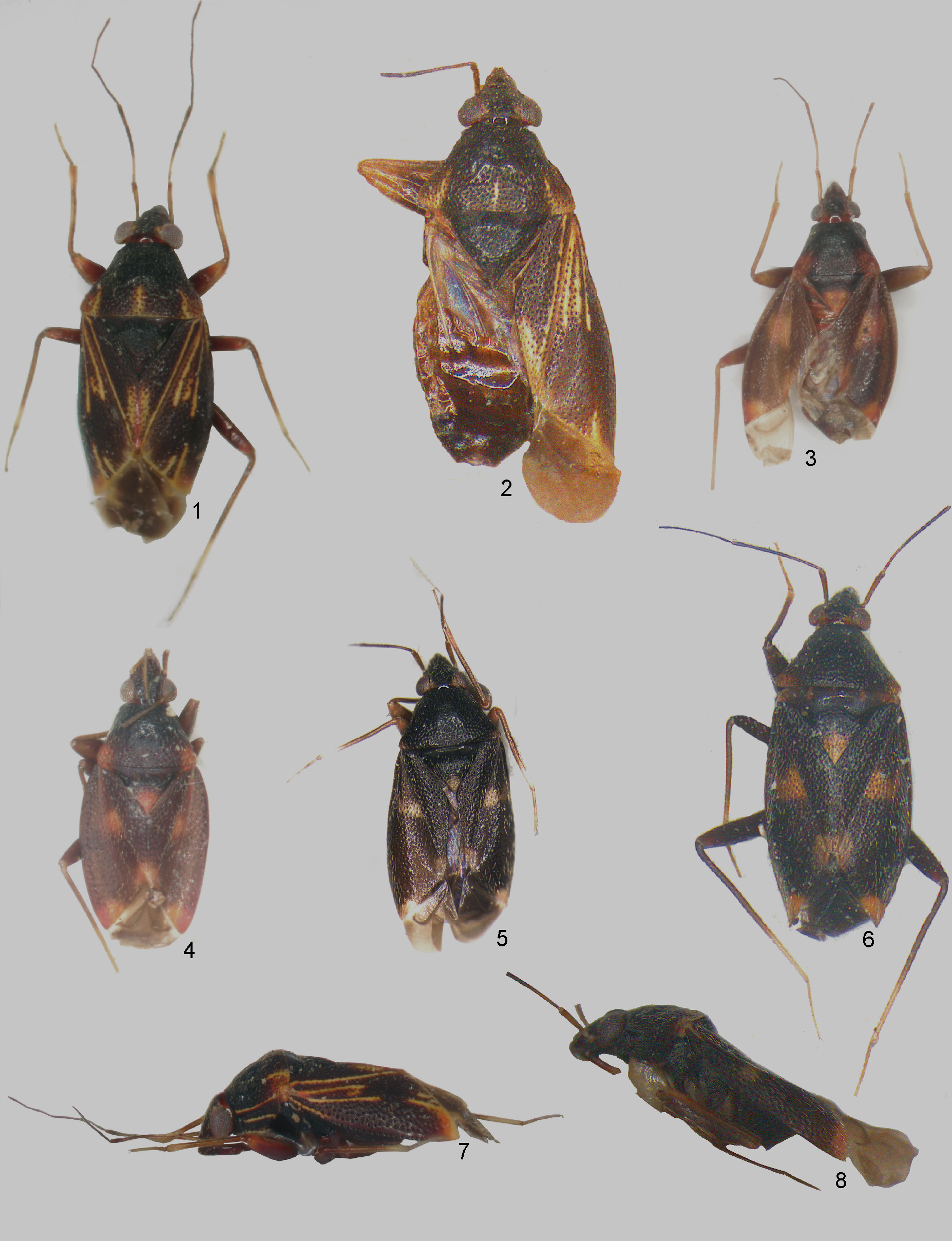
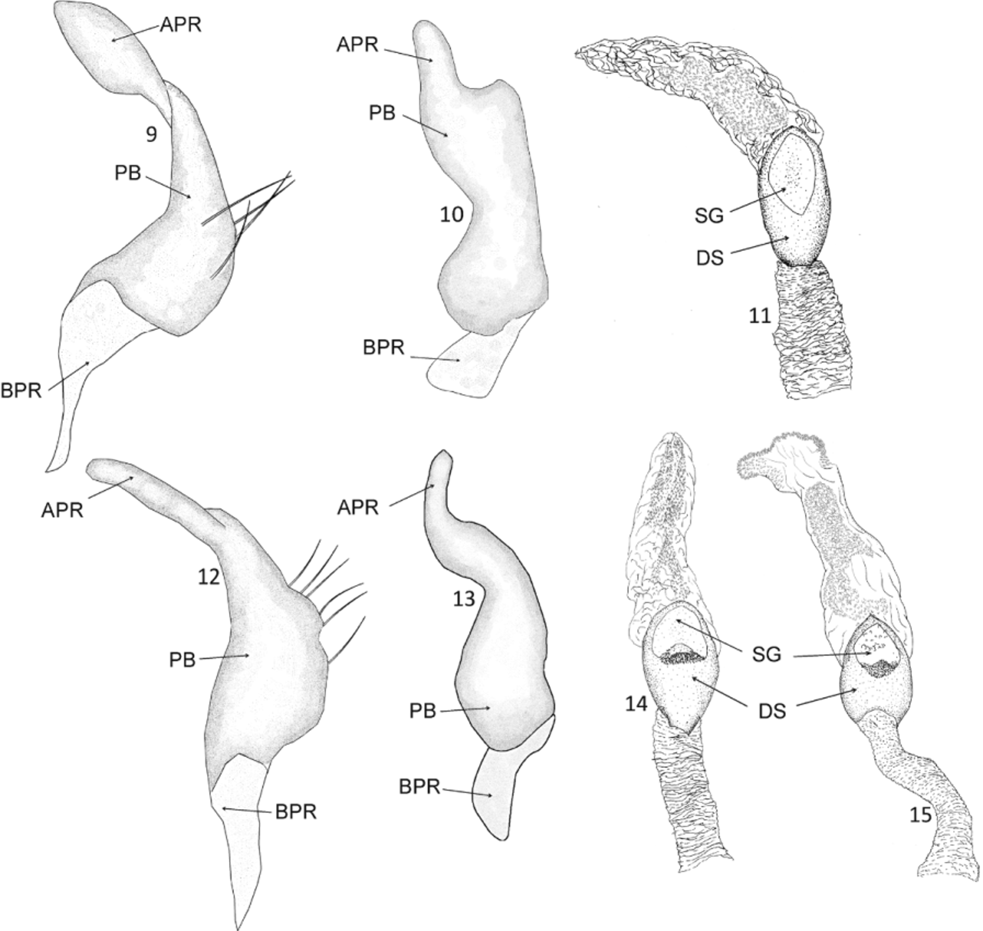
( Figures 3–4 View FIGURES 1 – 8 , 12–14 View FIGURES 9 – 15 )
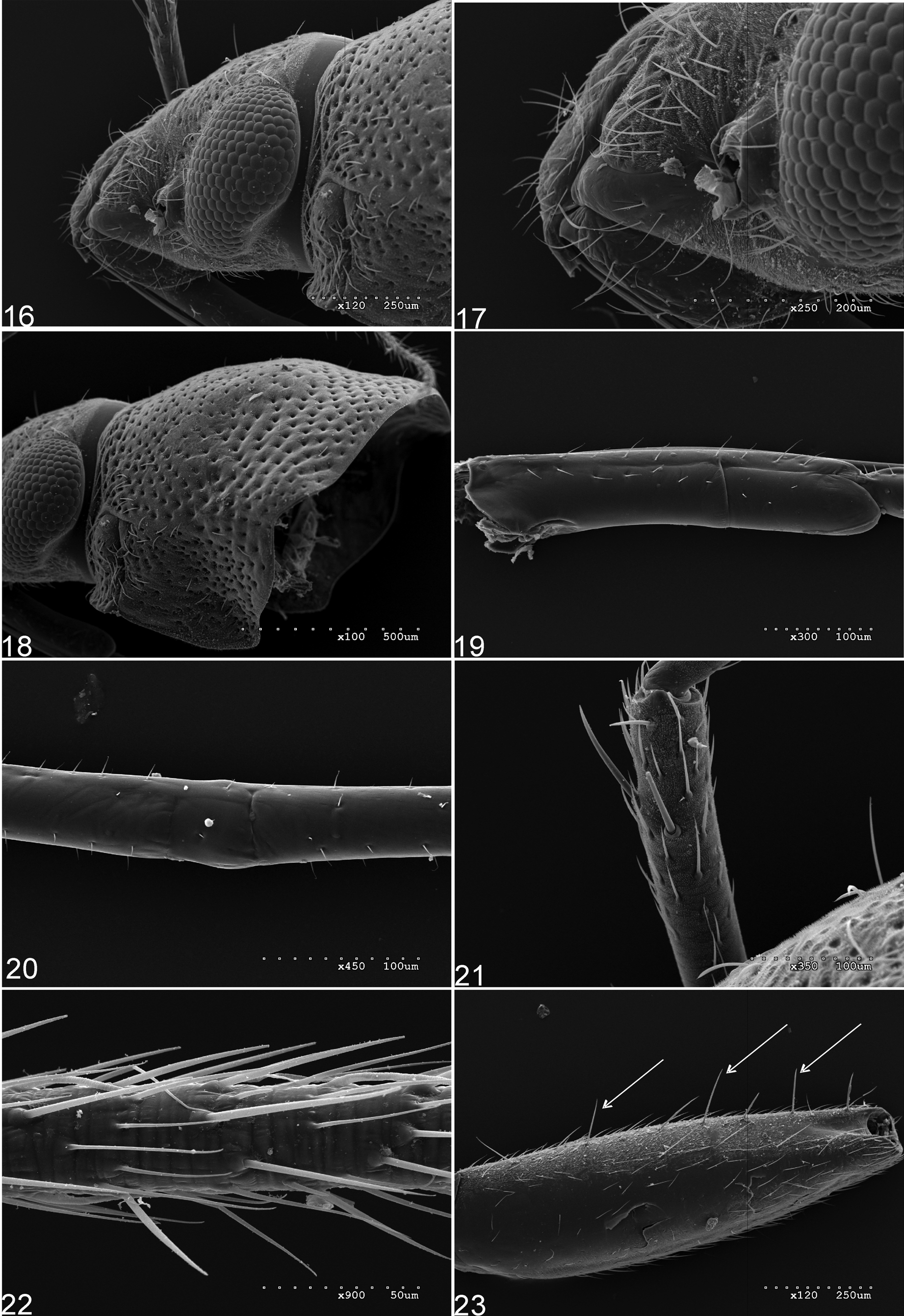
Cylapofulvidius thailandicus Chérot & Gorczyca 2000: 223 View in CoL , 224, 226, Figs. 20 View FIGURES 16 – 23 , 24 View FIGURES 24 – 29 (n. sp.); Gorczyca 2006: 29 (catalog).
Diagnosis. Fulvidius thailandicus is recognized by the following set of features: body relatively small (3.12); proepimeron uniformly dark brown, without any pale stripe (as on Fig. 8 View FIGURES 1 – 8 ); pronotum with humeral angle and apex of scutellum with large, orange patch ( Figs. 3–4 View FIGURES 1 – 8 ); hemelytron with four, relatively large, orange patches ( Figs. 3– 4 View FIGURES 1 – 8 ); male similar to the female, the genital structures as described below ( Figs. 12–14 View FIGURES 9 – 15 ).
Most similar to C. webbi in sharing similar body size and coloration of the dorsum. This species can, however, be distinguished by the yellow patches near antennal insertion and structure of the female (see Chérot & Gorczyca 2000: Figs. 17–20 View FIGURES 16 – 23 , 24 View FIGURES 24 – 29 ) and male ( Figs. 12–14 View FIGURES 9 – 15 ) genitalic structures.
Male genitalia ( Figs. 12–14 View FIGURES 9 – 15 ). Left paramere ( Figs. 12–13 View FIGURES 9 – 15 ). Apical process relatively long and thin, straight in lateral view, sinuate in dorsal view; paramere body with a few long, protruding, thick setae. Endosoma ( Fig. 14 View FIGURES 9 – 15 ). Sclerotized portion of ductus seminis (DS) ovoid; secondary gonopore semicircular.
Biology. Unknown.
Distribution. Thailand (Yala Province, Bang Lang National Park).
Type material. Holotype ♀: Thailand: Yala province, Bang Lang National Park , (6°04' N, 101° 11' E.), on dead tree, 18–20.x.1991, J. Martin leg. ( ZMUC). GoogleMaps
Additional examined material. 3 ♀ ♀ and 2 Ƌ Ƌ: Malaysia: Sarawak, Kapit District, Sebong env., Baleh river , 10–19.iii. 1994, P. B í l ek leg. ( Fig. 3 View FIGURES 1 – 8 ) ( ZJPC) ; 1 ♀ and 1 Ƌ Malaysia: Sarawak, Kapit district, Sebong, Baleh river , 6.–21.iii.1994, S. Bílý leg. ( Fig. 4 View FIGURES 1 – 8 ) ( NMPC) .

No known copyright restrictions apply. See Agosti, D., Egloff, W., 2009. Taxonomic information exchange and copyright: the Plazi approach. BMC Research Notes 2009, 2:53 for further explanation.