
Ocelliemesina sinica Wang, Wang, Cao & Cai
|
publication ID |
https://doi.org/ 10.11646/zootaxa.3936.3.8 |
|
publication LSID |
lsid:zoobank.org:pub:53784CB4-2CFF-4486-AE6F-726BE0FDAB67 |
|
DOI |
https://doi.org/10.5281/zenodo.6100201 |
|
persistent identifier |
https://treatment.plazi.org/id/58174866-BD76-B34D-0F86-FCFC3DB457EF |
|
treatment provided by |
Plazi |
|
scientific name |
Ocelliemesina sinica Wang, Wang, Cao & Cai |
| status |
sp. nov. |
Ocelliemesina sinica Wang, Wang, Cao & Cai View in CoL , sp. nov.
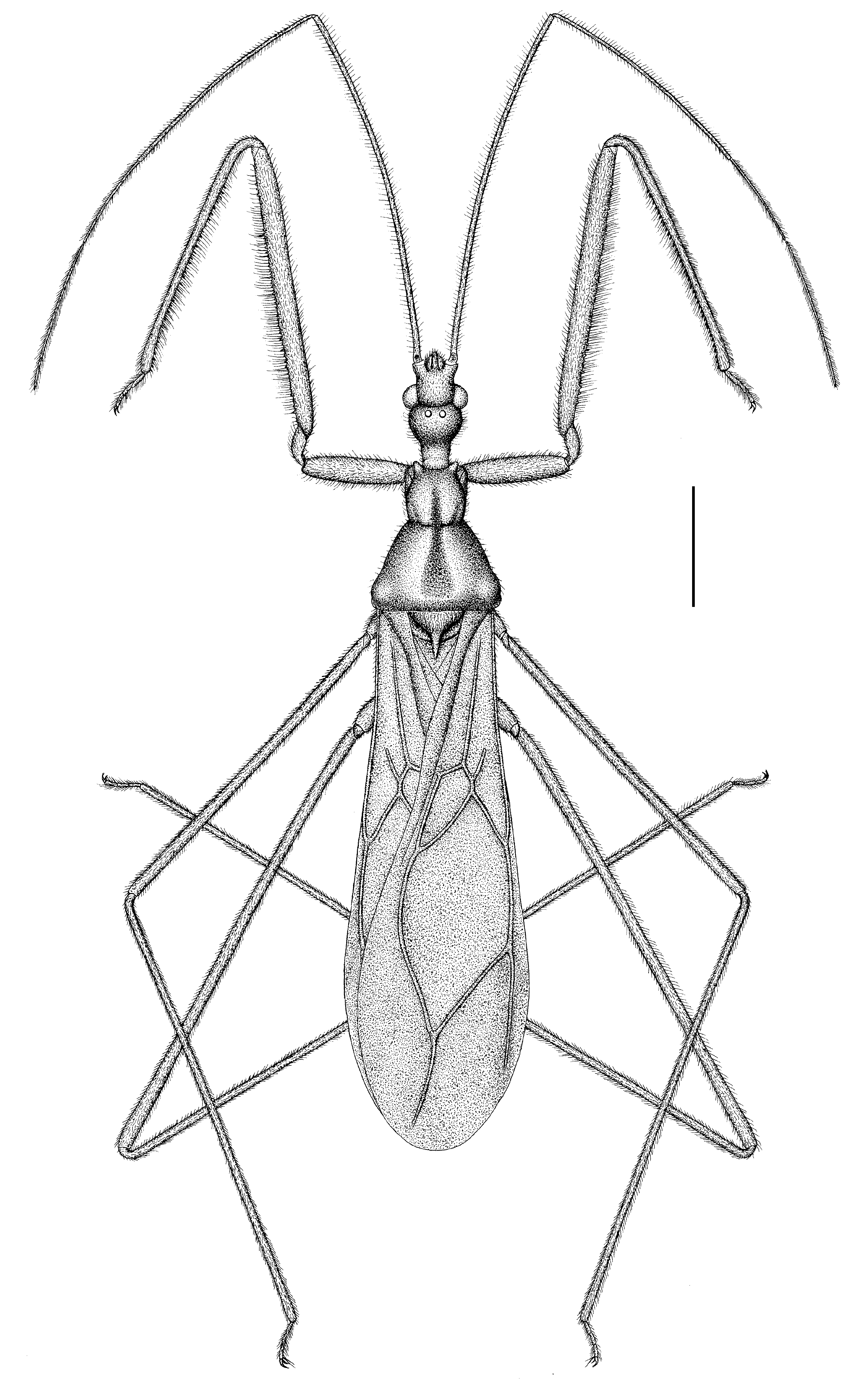
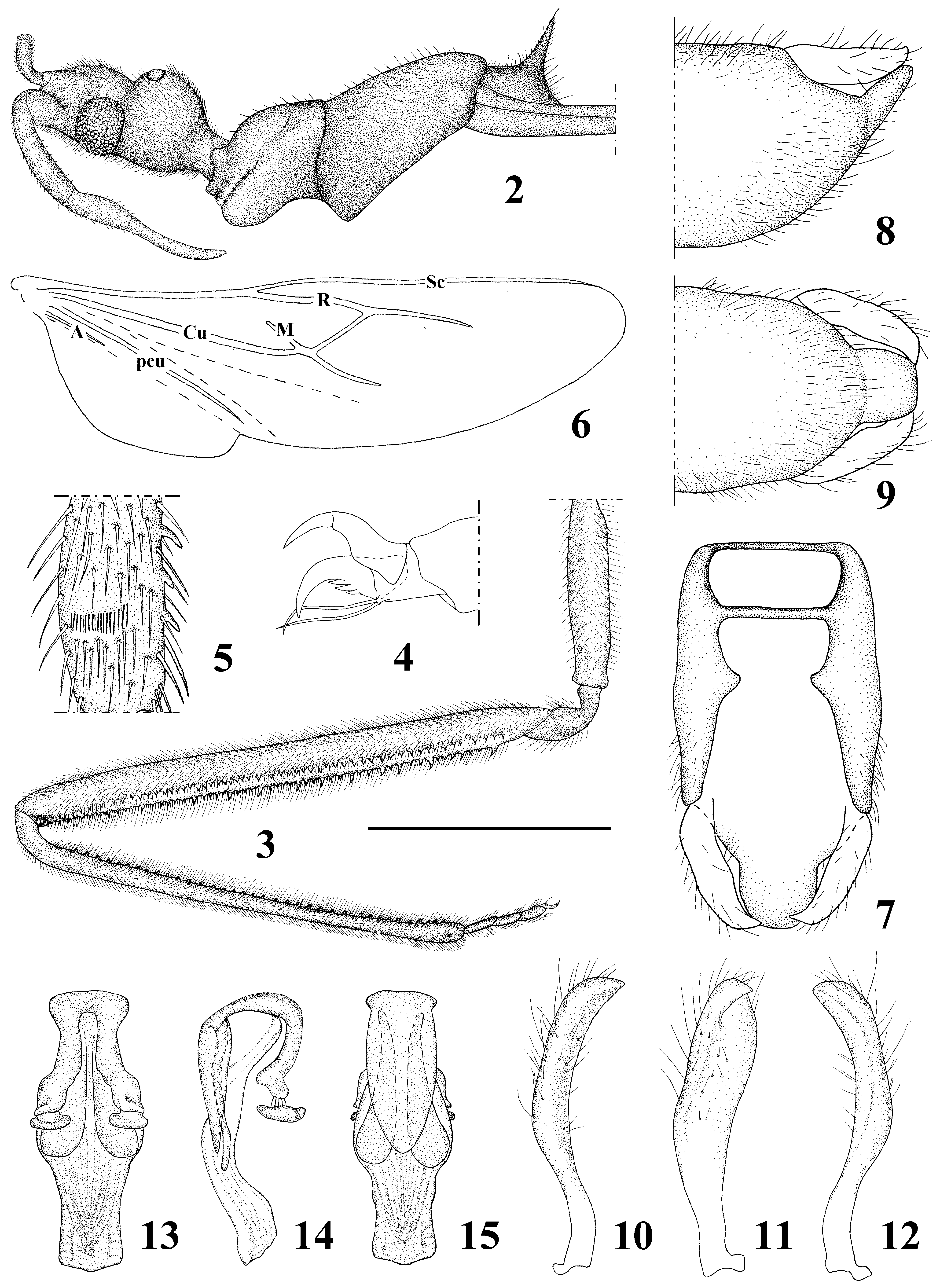
( Figs. 1–15 View FIGURE 1 View FIGURES 2 – 15 )
Description. Colour. Body generally dark brown. Head, labium, legs and abdomen beneath yellowish brown; antennae, thorax fuscous; fore and hind wings brown; legs without annulation.
Structure. Head densely pubescent, thorax obviously polished, shiny, with semi-erect setae. First and second visible labial segments with short semi-erect setae. First antennal segment with erect setae, these setae 1–1.5 times as long as diameter of first segment, other antennal segments densely covered with short, semi-erect setae. Pronotum with sparse erect setae; thoracic pleura, sternites, and abdomen covered with short, procumbent setae. Fore legs with short setae and long bristles ventrally and dorsally, femora covered with several long erect bristles on ventral surface and with semi-erect long setae on dorsal surface; tibiae with strong bristles on ventral surface and with semi-erect setae on dorsal surface; tarsi with semi-erect setae, these setae as long as diameter of corresponding tarsal segment; femora, tibiae and tarsi of mid and hind legs with short, procumbent to semi-erect setae. Abdominal segments with short semi-erect setae.
Head elongated in dorsal view, bilateral margins of anteocular portion subparallel; postocular portion swollen, almost globular in lateral view, not abruptly constricted before neck ( Figs. 1–2 View FIGURE 1 View FIGURES 2 – 15 ). Eyes prominent, ovate. Interocellar space as long as diameter of ocellus; distance between ocellus and ipsilateral eye about as long as distance between ocelli. Antennae gracile; first segment longest and slightly shorter than remaining segments combined. Labium slender, first and second visible segments curved; first visible segment longest, reaching level of middle of eye, slightly longer than third visible segment; median part of second visible segment swollen; third visible segment bent, with apex sharp ( Fig. 2 View FIGURES 2 – 15 ). Anterior pronotal lobe shorter than posterior lobe, nearly foursquare in dorsal view, as wide at maximum width as head, narrower posteriorly, with a median longitudinal furrow extending to posterior lobe, but not reaching anterior nor posterior pronotal margin; posterior lobe slightly winkled; longitudinal furrow with transverse crinkles; humeral angles rounded; posterior margin nearly straight. Scutellar spine large, subequal to second visible labial segment in length. Basal part of fore wing narrow and widest in subapical part, wing apex surpassing tip of abdomen; basal cell pentagonal, area about 1/8 of discal cell; Pcu+1A meeting basal cell near its base ( Fig. 1 View FIGURE 1 ); hind wing extending beyond apex of abdomen; free Sc present; Cu extending distinctly beyond cross vein; anal lobe lacking 2A and less than half long of hind wing ( Fig. 6 View FIGURES 2 – 15 ). Fore legs somewhat robust; coxa cylindrical; femur about three times as long as coxa, ventrally armed with two series of linage spines; posteroventral series starting from base of femur, composed of two types of spiniferous tubercles, both conspicuously shorter than diameter of femur; anteroventral series beginning at base of the segment, composed of spiniferous processes similar to those of posteroventral series; fore tibia shorter than femur, with one series of small, equal-sized spines ( Figs. 3, 5 View FIGURES 2 – 15 ); fore tarsus with two well-developed claws, outer one with three small processes, inner one divided by an incision into a longer basal and a shorter apical portion ( Fig. 4 View FIGURES 2 – 15 ). Mid and hind legs elongate, hind leg longest. Abdomen long-ovate; sixth segment widest. Seventh tergite protruding backwards; 1/3–1/2 of eighth tergite and posterior part of pygophore exposed. Pygophore ovate in dorsal view, almost semicircular in lateral view; median pygophore process broad, flattened, lingulated ( Figs. 7–9 View FIGURES 2 – 15 ). Parameres club-shaped, with apex pointed and bent inwards ( Figs. 10–12 View FIGURES 2 – 15 ). Pedicel shorter than basal plate; basal plate curved; basal plate bridge absent; struts of phallus long, separated each other, apically wide and flat ( Figs. 13–15 View FIGURES 2 – 15 ).
Measurements. [in mm, ♂ (n=1), holotype]. Length of body: to apex of fore wings 7.53; to apex of genital capsule 6.67; length of head 0.98; length of anteocular portion 0.31; length of postocular portion 0.45; width across eyes 0.38; interocellar space 0.05; length of synthlipsis 0.35; lengths of antennal segments I–IV= 3.52, 2.31, 0.84, 1.10; lengths of visible labial segments I–III= 0.51, 0.28, 0.49; length of anterior pronotal lobe 0.55; length of posterior lobe 0.83; width of thorax 1.28; length of scutellum 0.33; length of hemelytron 5.36; greatest hemelytron width 1.76; length of abdomen 3.42, greatest abdomen width 1.35; length of fore femur 2.90, width of fore femur 0.21, length of fore tibia 2.52; length of mid femur 3.73, length of mid tibia 4.52; length of hind femur 4.57, length of hind tibia 6.67.
Type material. Holotype, ♂, China, Yunnan Province, Lvchun County, Huanglian Mountain, 6.V.2011, 1800 m, N22.894270°, E102.296580°, Yang Hailin & Wang Jianyun leg, by light trap. ( CAU).
Etymology. The specific name refers to the locality of the holotype.
Distribution. China (Yunnan).
Remarks. The new species resembles the Australian endemic Armstrongocoris singularis in having ocelli, however it differs from the latter in shorter anteocular portion, flattened postclypeus and only two celled hemelytron as mentioned in the generic diagnosis. In addition, the posterior pronotal lobe of new genus nearly as two times wide as the anterior, while the posterior pronotal lobe is just about 1.5 times as wide as the anterior. The biology of this unique species is unknown except it was collected at light trap.
The ocelli-bearing species in “ocelli-less” subfamilies of Reduviidae have been briefly discussed by Wygodzinsky (1946, 1947, 1949, 1950). Presently only five cases have been reported in the Reduviidae : Lentia corcovadensis Wygodzinsky in the subfamily Chryxinae from Brazil ( Wygodzinsky 1946), Mirambulus morio Breddin (= Megavescia cazieri Wygodzinsky) and M. niger Breddin in the subfamily Vesciinae from South America ( Breddin 1901, 1903; Wygodzinsky 1947), and two cases in the Emesinae . It seems that more ocellibearing emesines can be found in the future as the emesines are widely distributed, greatly diversified, and poorly studied despite the great monograph published by Wygodzinsky (1966). The origin and evolution of the ocellibearing emesine bugs remain to be studied further.
| CAU |
China Agricultural University |
No known copyright restrictions apply. See Agosti, D., Egloff, W., 2009. Taxonomic information exchange and copyright: the Plazi approach. BMC Research Notes 2009, 2:53 for further explanation.