
Indotherium pranhitai Yadagiri, 1984
|
publication ID |
https://doi.org/ 10.5281/zenodo.5375708 |
|
persistent identifier |
https://treatment.plazi.org/id/59258795-FFE2-FFAB-1328-FB6A741BFA82 |
|
treatment provided by |
Marcus |
|
scientific name |
Indotherium pranhitai Yadagiri, 1984 |
| status |
|
Indotherium pranhitai Yadagiri, 1984 ( Figs 6 View FIG ; 7 View FIG )
HOLOTYPE. — GSI type No. SR /PAL/11, left upper molar.
REFERRED MATERIAL. — VPL /JU/ KM /11, right upper molar.
HORIZON AND LOCALITY. — Mudstones associated with the limestone bands of Kota Formation, west of Paikasigudem village, Rebbana Mandalam, Adilabad District, Andhara Pradesh (state), India.
DIAGNOSIS. — Same as for the genus.
DESCRIPTION OF VPL/JU/KM/11
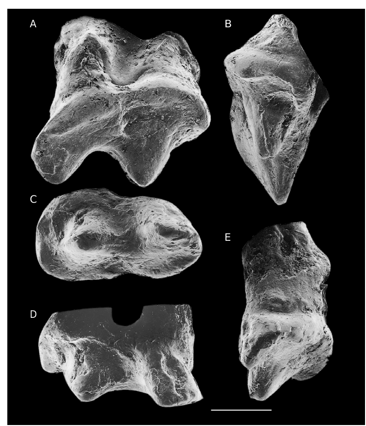
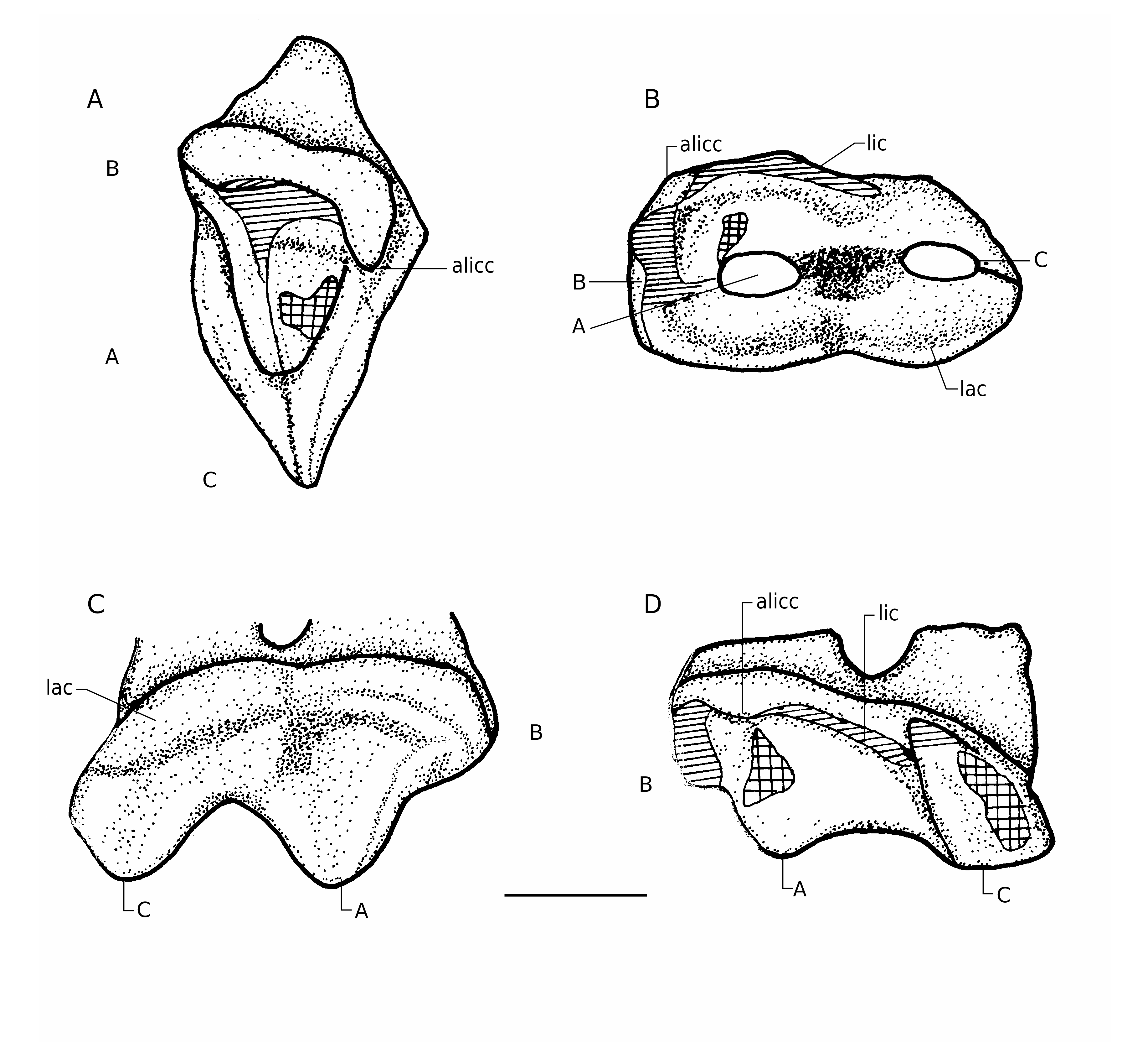
This is an eroded right upper molar (maximum length = 1.437 mm, anterior width = 0.77 mm, posterior width = 0.626 mm). The enamel on the labial surface of the crown is completely spalled off ( Fig. 6A View FIG ). The crests joining A with B and C are completely worn. The cusp formula for this tooth is A> C> B. In occlusal view, the tooth is roughly oval in outline, with slight indentations labially and lingually opposite the posterior limit of A ( Figs 6C View FIG ; 7B View FIG ). The molar has two major cusps and one minor cusp ( Figs 6A View FIG ; 7C, D View FIG ). The principal cusp A is located anterior to the midpoint of the tooth; cusp C, posterior to it, is about the same size. Cusp C is strongly reclined, cusp A is nearly vertical, and the two cusps are separated from each other by a broad, deep groove ( Figs 6A View FIG ; 7C, D View FIG ). A small anterior cusp B, slightly offset from the line of A and C, arises from the anterior cingulum. A broad groove separates this cusp from A. The labial faces of cusps A and C are flat and slope steeply labioventrally. The labial cingulum arises from the mid-height of cusp C ( Figs 6A View FIG ; 7C View FIG ), slopes down anteriorly and joins cusp B at a lower level. This cingulum appears to be narrow opposite cusp A and relatively wide and swollen opposite the groove separating cusp A from C. The highly eroded surface of this cingulum does not provide any evidence for cingular cuspules. Anterior to A, a wide shelf is present both labially and lingually. The labial cingulum is interrupted anteriorly by cusp B. It continues lingually and posteriorly as a narrow worn ridge to the anterior base of C. This cingulum bears a small cingular cuspule at the anterolingual base of A ( Figs 6D View FIG ; 7D View FIG ).
The anterior root is only partly preserved, whereas the posterior root is relatively better preserved. The posterior root is dilated distally in labial view and its transverse width is greater than its length. Wear facets are visible on the lingual face of the crown. The lingual cingulum is quite worn ( Fig. 7B, D View FIG ). The lingual tip of B, anterolingual cingulum between B and anterolingual cingular cuspule, and the valley between A and B exhibit moderate wear ( Figs 6C View FIG ; 7B, D View FIG ). The anterolingual cingular cuspule also exhibits some abrasion. The valley between the posterior base of A and anterior base of C is deeply grooved. The lingual face of A is chipped in its middle part. Therefore, it is difficult to say whether there was any wear facet in this region or not. The enamel on the lingual face of cusp C is chipped particularly near its ventral part, but its dorsal part shows a flat surface pointing to the presence of a possible wear facet here.
COMPARISONS
Yadagiri (1984) described GSI type No. SR/PAL/11 as a lower molar, placing it in the then new genus and species. He made it a type of Indotherium pranhitai , family incertae sedis (order Symmetrodonta ). Prasad & Manhas (1997) described an additional molar (VPL/JU/KM/11) referring to Indotherium pranhitai . They transferred I. pranhitai from the order Symmetrodonta to the order Triconodonta because the principal cusps of the crown are arranged more or less in straight line. But they also mistook the new tooth for a lower molar, as Yadagiri (1984) did for the type. The two molars (GSI type No. SR/PAL/11 and VPL/JU/KM/11), appear to be very similar in their crown morphology. From the photographs and figures provided by Yadagiri (1984: pl. 2a-e, fig. 2a-f), the type specimen appears to be better preserved.
VPL/JU/KM/11 is considered to be an upper molar because of the presence of labial and lingual cingula and because of its similarity to maxillary teeth of morganucodontids. Within all known taxa of Triconodonta as discussed earlier in the section on Dyskritodon , Austrotriconodon and Gobiconodon differ from SR/PAL/11 and VPL/JU/KM/ 11 in the cusp formula and the arrangement of cusps on the crown. Any relationship with the family Triconodontidae can also be ruled out for VPL/JU/KM/11, because in all known taxa of this family, cusps B, A, and C of upper molars are sub-equal in size. The upper molars of Jeholodens are distinguished from VPL/JU/KM/ 11 in having a relatively large cusp A, cusp B excluded from cingulum, and the presence of cusp D.
It is only in Morganucodon Kühne, 1949 (Morganucodontidae) that cusp B is very small and occurs anterior to cusp A or slightly labial to the A -C line, and cusps A and C are equal or nearly equal in size. Some important morphological features common to Morganucodon and VPL/JU/KM/11 are: the presence of an anterior root that is circular or oval in cross section, with an anteroposterior long axis and transversely wide posterior root; balcony-like projection of crown over the posterior root ( Figs 6A, D View FIG ; 7C, D View FIG ); B developed as a small cusp more or less in line with A and C; wide anterior shelf; posteriorly wide labial cingulum, lingual cingulum narrower than labial cingulum; tranversely wide and dilat- ed posterior root. The occlusal pattern in Morganucodon was studied by Crompton & Jenkins (1968) and Mills (1971). According to these works, in the early stage of wear, a concave facet develops in the anterior part of lingual cingulum of Morganucodon upper molars. Subsequently, it extends from the cingulum to the bases of A and B and another facet develops on the lingual cingulum adjacent to cusps A and C because of the occlusion of cusp c of corresponding lower molar between cusps A and C. The wear facets observed on VPL/JU/KM/11 are more or less similar to those observed in Morganucodon in the early stages.
Of the known Morganucodon teeth, the morphology of Indian specimens approaches that of BMNH No. M26014 (Early Jurassic of Wales) the most. This specimen is a right maxillary fragment bearing two teeth possibly the antepenultimate and penultimate molars (because there is an alveolus behind the posterior most preserved tooth). Most of the morphological features observed in VPL/JU/KM/11 are also present in BMNH M26014. Cusp C is reclined in BMNH M26014 as well as in VPL/JU/KM/11 and GSI type No. SR/PAL/11. In the latter specimen, C is slightly smaller than cusp A, as it is in M26014. On the penultimate molar of M26014 and on VPL/JU/KM/11, the posterior crown overhangs the root like a balcony.
However, in Morganucodon , the groove between A and C is not as broad as in VPL/JU/KM/11. This character and the anterior crown morphology might be cited against the inclusion of the Indian specimens in the family Morganucodontidae . But in SR/PAL/11, this groove is nearly at the same level of development as in BMNH M26014 and uncatalogued specimens of Morganucodon from Pant Quarry (Early Jurassic), Glamorganshire, England, housed in University College London. Therefore, the deep groove between A and C might be reflecting the highly eroded nature of VPL/JU/KM/11. On VPL/JU/KM/11, cusp C is stubby in appearance and cusp D is absent. The latter character is not very significant in distinguishing these taxa as cusp D is present on some specimens of M. watsoni ( Crompton, 1974) and absent on others ( Parrington 1971). The labial face of cusps A and C is steeply sloping labioventrally in VPL/JU/KM/11, whereas it is not so in Morganucodon . Another important distinction between the Indian specimens and the molars of Morganucodon lies in the anterior part of the crown. On the upper molars of Morganucodon with significant wear, there is a broad transverse notch separating cusps A and B. Because of this the lingual cingulum slopes dorsally from the anterolingual base of cusp A, whereas in VPL/JU/KM/11, this margin is horizontal and bears a small cingular cuspule at the anterolingual base of cusp A. However, it needs to be emphasised here that VPL/JU/KM/11 appears to be in the early stage of wear. The lingual cingulum of Morganucodon , unlike in VPL/JU/KM/11, bears a variable number of cuspules ( Mills 1971). Moreover, B is not a cingular cusp in Morganucodon (see Mills 1971).
Ventrally sloping steep labial faces of A and C, absence of cuspules on the lingual cingulum except for the anterolingual cuspule, and presence of B as a cingular cusp are the morphological differences sufficient enough to distinguish the Indian specimens at generic level from Morganucodon .
No known copyright restrictions apply. See Agosti, D., Egloff, W., 2009. Taxonomic information exchange and copyright: the Plazi approach. BMC Research Notes 2009, 2:53 for further explanation.
|
Kingdom |
|
|
Phylum |
|
|
Class |
|
|
Order |
|
|
Family |
|
|
Genus |