
Chilicola unicarinata Packer
|
publication ID |
https://doi.org/ 10.5281/zenodo.176627 |
|
DOI |
https://doi.org/10.5281/zenodo.6249369 |
|
persistent identifier |
https://treatment.plazi.org/id/59368781-A455-FFF5-FF7D-FD06E6F1FD55 |
|
treatment provided by |
Plazi |
|
scientific name |
Chilicola unicarinata Packer |
| status |
sp. nov. |
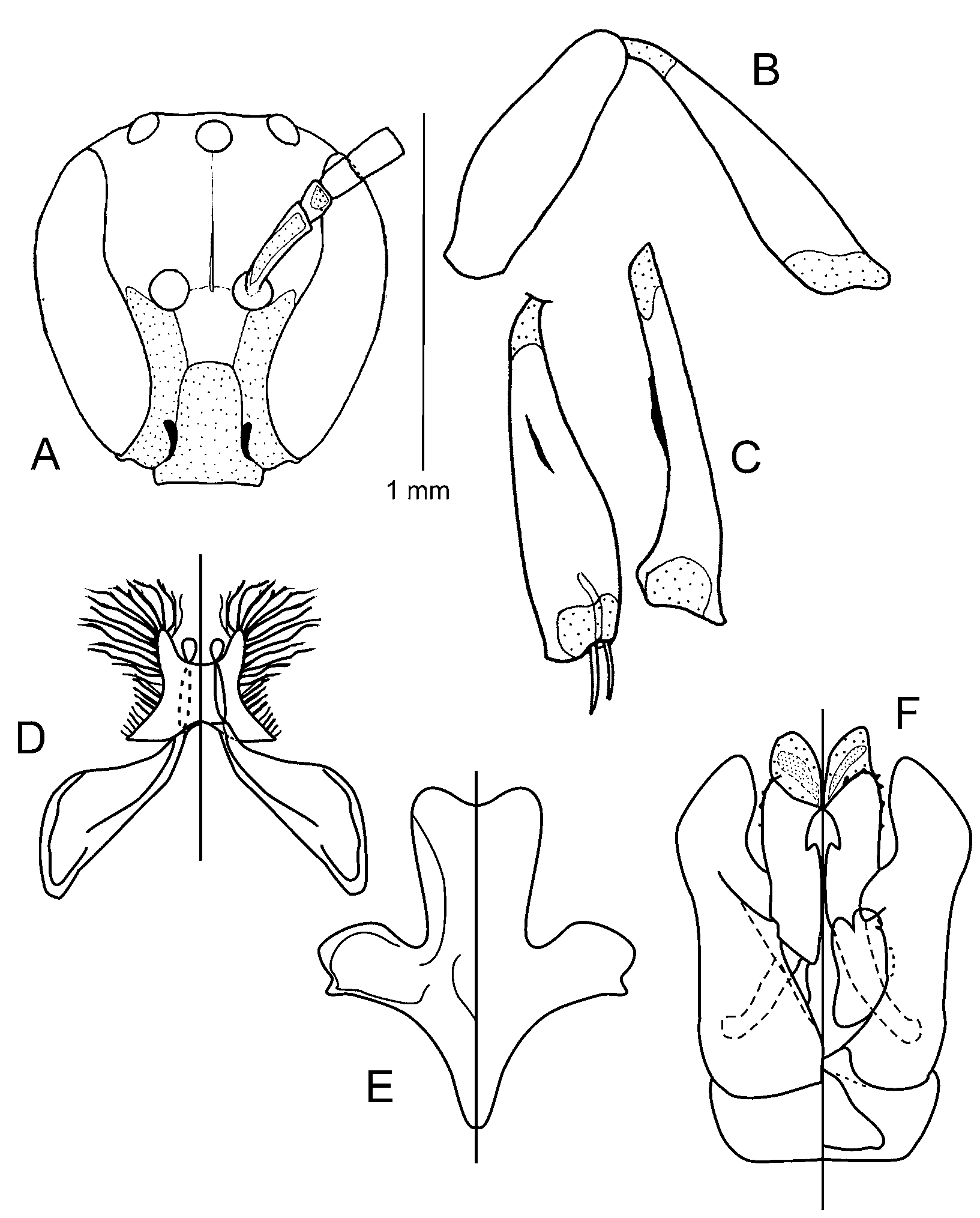
Chilicola unicarinata Packer View in CoL , n. sp.
( Figs. 5A–O View FIGURES 5 A – O )
Diagnosis: As with the previous species pair, phylogenetic analyses (Packer, in press) suggest that a new subgenus is required for this and the following species. This group has the following combination of characteristics that is unique among the Xeromelissinae. In the males, the form of the hind tibia, which is very narrow in dorsal view, somewhat expanded laterally towards the apex and with a single oblique carina on the inner surface ( Fig. 5F View FIGURES 5 A – O ); the numerous long hairs on the ventral lobes of S7 ( Fig. 5G View FIGURES 5 A – O ), the deep cleft in the penis valves from which the large membranous lobes arise and the crenulate lateral margins of the penis valves ( Fig. 5I View FIGURES 5 A – O ). The short distance between lateral ocellus and compound eye, less than the diameter of lateral ocellus ( Figs. 5A and B View FIGURES 5 A – O ), is found uncommonly elsewhere in the subfamily; the lack of long hairs on the lateral surface and dorsolateral area of the propodeum is also unusual. For females, the restriction of branches on the scopal hairs of S2 to the apex of the hairs is rarely found elsewhere among Chilicola species and with the orange markings on the lower paraocular area form a unique combination.
Males of C. unicarinata can be differentiated from those of C. chubutense Packer , n. sp., described below, by the more extensive pale colouration on the legs, the carina on the inner surface of the hind tibia approximately centrally positioned along the length of the tibia ( Fig. 5F View FIGURES 5 A – O ) as opposed to being more basal in C. chubutense and the presence of only one membranous lobe to the penis valve ( Fig. 5I View FIGURES 5 A – O ) whereas the latter species has two ( Fig. 6F View FIGURES 6 A – F ). In addition to the next described species, there are several additional species from northern Argentina and Bolivia that are considered to belong to a new subgenus of Chilicola . Except for colouration characteristics of the females and minor details of the male genitalia, these bees appear difficult to distinguish from C. unicarinata and additional study is required. The entirely orange legs of the females of C. unicarinata are diagnostic, other species have at least some dark patterning on several leg segments.
Description. Male: Body length 4.4mm, wing length 3.3mm, head width 1.0mm.
Colouration: Blackish brown, with following parts yellow: mandible (except apex red), clypeus, paraocular area to antennal socket ( Fig. 5A View FIGURES 5 A – O ), anterior surface of scape and pedicel, broad apical ring on forefemur, foretibia except for brown spot on posterior surface, apical ring on midfemur, basal and apical rings on mid tibia, narrow apical ring on hind femur, narrow basal and apical rings and complete ventral longitudinal line and incomplete dorsal longitudinal line on hind tibia, all tarsi except hind basitarsus (whitish) and pretarsi. Following parts orange: Anterior surface of flagellum, all trochanters and all pretarsi. Posterior surface of antenna red-brown. T1, T2 and T6 brown, T3 dark brown, T4 and 5 blackish. Tegula translucent colourless. Wing veins pale testaceous. Apical impressed areas of all terga translucent straw.
Surface Sculpture: Labrum deeply and moderately densely punctate (i~d) on shiny background. Clypeus, supraclypeal area and lower paraocular area with irregular, very weak punctures (i=1–3d) and entirely dull due to microsculpture; supraclypeal area somewhat shinier. Frons densely, finely and shallowly punctate (i<d); interspaces moderately shiny. Vertex obscurely punctate. Hypostomal and genal areas shiny, punctures largely effaced. Pronotal collar, mesoscutum and scutellum moderately shiny with fine, shallow punctures (i=1–2d). Metanotum shiny with sparser punctures except denser on anterior and posterior margins. Mesopleuron sparsely punctate (i=2–5d), microsculpture stronger, surface moderately dull. Dorsal surface of propodeum weakly and irregularly reticulostriate, reticulations almost reaching apex; lateral surface very weakly rugulose; dorsolateral area narrowly wrinkled, wrinkles subvertically oriented. Metasomal terga with few very shallow and obscure punctures, surface shiny, microsculpture weak; apical impressed areas with very weak microsculpture.
Pubescence: Whitish, generally short, sparse and not especially plumose, hairs on lower surface of thorax longer (1MOD). Genal beard present, longer basally (2MOD) than apically (0.5MOD). Metasomal terga with few short hairs, lacking apicolateral hair patches. Metasomal sterna with patches of erect hairs laterally on S2–S5, those on S2 sparse and short (at most 0.5MOD in length), those on S3–S5 short anteriorly, increasing in length posteriorly, longest hairs (1MOD) on posterior of S4.
Structure: Head: Longer than broad (62:57) ( Fig. 5A View FIGURES 5 A – O ). Labrum 2X as broad as median length; apical margin slightly convex. Mandible less than 2X as long as basal depth (19:11). Clypeus slightly longer than broad (21:20) ( Fig. 5A View FIGURES 5 A – O ), lower approximately one quarter extending beyond lower ocular tangent, weakly protuberant in profile, with weak broad median longitudinal depression; lateral margin of clypeus almost straight dorsal to anterior tentorial pit, abruptly bent laterad beneath pit ( Fig. 5B View FIGURES 5 A – O ); anterior tentorial pit situated in elongate depression slightly laterad of epistomal suture separated from it by less than pit diameter. Subantennal sutures strongly convergent below ( Fig. 5A View FIGURES 5 A – O ); supraclypeal area sharply defined above, length to apical breadth 13:7 ( Fig. 5A View FIGURES 5 A – O ). Frons without swellings or depression to house scape. Frontal line weak, but entire. Facial fovea absent. Eyes emarginate, strongly converging below ( Fig. 5A View FIGURES 5 A – O ) (UOD:LOD 37:20). Lateral ocelli much closer to compound eye than each other (OOC:IOC 13:35) and OOC less than LOL, 9:13. Vertex slightly longer behind lateral ocellus than LOL (9:8). Head not strongly developed above summit of eye, upper ocular tangent crosses lower tangent of median ocellus. Occipital margin sharp but not carinate. Scape 3.5X as long as apical breadth, shorter than pedicel + F1 + F2 combined; F1 shorter than F2, 12:15; all flagellomeres cylindrical, longer than broad, (for F8, 20:18), F10 and F11 subequal in length; flagellomeres lacking modifications in structure or setation; flagellum slightly broader towards apex. Malar space linear such that presence or absence of malar suture indetectable. Gena slightly greater than 0.5X width of eye in lateral view (12:22) ( Fig. 5B View FIGURES 5 A – O ).
Mesosoma: Elongate , length to greatest depth: 106:55. Pronotal collar short ( Fig. 5D View FIGURES 5 A – O ), medial length 0.75 LOL, anterolateral margins rounded. Episternal groove distinct and complete; scrobal groove weakly developed posterior to scrobe, absent anteriorly. Propodeum slightly longer than scutellum (ratio of scutellum:metanotum:dorsal surface of propodeum 17:9:18) slightly shorter than its posterior depth (20); propodeal sulcus shallow but distinct, weakly pitted. Hind trochanter slightly swollen apically, lacking spines, teeth or angulations ( Fig. 5E View FIGURES 5 A – O ). Hind femur weakly expanded with ventral surface slightly sinuate, length to greatest depth 70:24 ( Fig. 5E View FIGURES 5 A – O ). Hind tibia laterally compressed, but with apical one sixth abruptly, but not very strongly expanded, length to apical depth 85:21, inner surface with strong oblique internal carina for middle 1/5th of tibia, distance from base of tibia to base of carina equal to distance from apex of carina to apex of tibia ( Figs. 5E and F View FIGURES 5 A – O ); apex of tibia attaining base of trochanter when folded; hind tibial spurs unmodified. Hind basitarsus 6X longer than greatest depth, parallel sided. Hind tarsal claws bifid. Basal vein evenly curved; distal stigmal perpendicular traversing second submarginal cell near middle; stigma shorter than length of marginal cell on wing margin (28:34); stigmal margin in marginal cell angularly convex; first recurrent vein interstitial with first submarginal crossvein on vein Rs+M.
Metasoma: Subpetiolate, T1 longer than apical breadth (40:34). Apical impressed areas of terga ~1/4 as long as tergum. S1–S6 unmodified; gradulus of S2 with long posteriorly directed lateral portion, S3 with very small lateral gradular mark, S4–S6 lacking gradular marks.
Terminalia : S7 with one pair of well developed lateral lobes; dorsal lobe, narrowly rectangular but expanded laterad anteriorly; bearing very long, thick, apically branched hairs on posterior and lateral margins; remnants of second (ventral) lobe restricted to short longitudinal lamella ( Fig. 5G View FIGURES 5 A – O ). S8 with apical process large, truncate and broadest apically ( Fig. 5H View FIGURES 5 A – O ). Apicoventral process of gonobase narrow, apex comparatively deeply emarginate. Volsella U-shaped; outer margin deeply concave before apex. Gonoforceps with strongly reflexed angulate mesal margin, ventromedial lobe long and rounded, oriented anteromesad; gonostylus not clearly demarcated from gonocoxite. Penis valve extremely broad towards apex, bearing one long, broadlybased membranous lobe on outer surface, merging imperceptibly with rest of penis valve, arising within deep and broad groove and curved laterad ( Figs. 5I and J View FIGURES 5 A – O ).
Female: Body length 4.6mm, forewing length 3.1mm, head width 1.0mm.
Colouration: Black, with following parts orange: labrum, apical half of mandible, antenna (except for brownish markings on scape, pedicel and F1), all trochanters, femora, tibiae and tarsi (femora and tibia sometimes suffused with reddish-brown). Base of mandible yellowish. Following parts orange-brown: posterior surface of F8–F11, part of posterior surface of scape, most of clypeus and lower paraocular area below anterior tentorial pit.
Surface Sculpture: As in male but with punctures generally somewhat finer and microsculpture more distinct. Clypeus dull to apex. Dorsal surface of propodeum with irregular reticulations not attaining posterior margin.
Pubescence: As in male but hairs on ventral surface of thorax shorter and finer. Scopal hairs of hind tibia not plumose. Metasomal terga lacking apical marginal hair patches. Scopa of S2 forming corbicula of long (up to 3MOD) but sparse, hairs either lacking branches or with few short branches apically on anterior surface of rhachis.
Structure: Maxillary palpus 0.5X as long as prementum (13:26). Prementum 5X as long as greatest breadth; fovea covering most of ventral surface, margins carinate. Lacinia triangular, 3X as long as greatest breadth. Lorum membranous except for two small longitudinal apical straps. Rest of body as in male except for usual sexual differences and as follows: Labrum with apical margin somewhat more angularly produced medially, very slightly concave either side of middle. Head longer than wide (75:64) ( Fig. 5C View FIGURES 5 A – O ). Clypeus with length and breadth subequal, extending below lower ocular tangent for less than one third of its length ( Fig. 5C View FIGURES 5 A – O ); UOD:LOD 40:27. Lateral ocelli much closer to compound eye than each other ( Fig. 5C View FIGURES 5 A – O ), OOC:IOC 6:18 and closer to eye than in male; ratio of OOC to LOL 12:14. Occipital margin rounded. Ratio of length of gena to eye 16:22. Propodeal sulcus extremely weak. Apical lunule of S5 almost an equilateral triangle.
Sting apparatus: As in Figs. 5K–O View FIGURES 5 A – O . Hemitergite 7 with lateral portion of marginal ridge thickened from apodemal region to lateral process; lateral process short and broad; lateral lamella rounded anteriorly narrowing posteriorly; medial portion of marginal ridge concave; spiracle close to posterior margin of lamina spiracularis, equidistant from lateral and medial portions of marginal ridge; posterior margin of lamina spiracularis concave medially ( Fig. 5K View FIGURES 5 A – O ). Hemitergite 8 with anterior ridge very slightly concave continuing as medial ridge to junction with plate, plate parallel-sided ( Fig. 5L View FIGURES 5 A – O ). First valvifer with dorsal process much shorter than ventral one, ventral margin concave. Second valvifer with apodemal ridge sinuate, apical process moderately developed, its lower margin strongly convex such that apical cleft is narrower apically than at mid-length; area of plate basal to gonostylus membranous; rostral process almost parallel-sided; pars articularis weakly expanded to acute apex; incisura postarticularis broadest at base abruptly narrowing to basal third and then approximately parallel-sided to apex ( Fig. 5M View FIGURES 5 A – O ); gonostylus narrow in lateral view but strongly converging to apex from broad base in ventral view ( Fig. 5N View FIGURES 5 A – O ). Sting shaft with ventral surface almost straight, processus muscularis and processus medianus poorly developed (Fig. M). Furcula with ventral arms comparatively straight and parallel-sided, unevenly curved in lateral view (Fig. O).
Material studied. Holotype male, allotype female, three male and three female paratypes: ARGEN- TINA, Catamarca; Andalgala, 1510km marker on Andalgala-Belen Highway, 18.x.1973; J.L. Neff, ex Prosopis chilensis (Fabaceae) . Additional paratypes as follows: same locality as holotype but 15.x.1973 ex P. nigra , one male; remaining paratypes all from ARGENTINA: Catamarca: Andalgala, 25.x.1972, G.E. Bohart, ex Prosopis alba , two males; Punta Balasto, 30km from Santa Maria, 24.xi.1966, Willink coll. two males; Colpes, upper bajada, 19.x.1973, J.L. Neff, ex P. n i g r a, one female. All specimens in CTMI except for two male and two female paratypes in PYU, the two males from Punta Ballasto, which are at IML and one male and one female paratype at MACN.
Etymology. The specific epithet refers to the single oblique carina on the hind tibia of the male.
Comments. Some females of this species possess labels stating Pseudiscelis sufferugineus or Pseudiscelis sufferuginia, and one male bears the label Oediscelis xanthopoda. These are Moure manuscript names. This species is not a member of either of the subgenera suggested by Moure’s attributions (Packer, unpublished data).

No known copyright restrictions apply. See Agosti, D., Egloff, W., 2009. Taxonomic information exchange and copyright: the Plazi approach. BMC Research Notes 2009, 2:53 for further explanation.
|
Kingdom |
|
|
Phylum |
|
|
Class |
|
|
Order |
|
|
Family |
|
|
Genus |