
Chilicola obesifrons Packer
|
publication ID |
https://doi.org/ 10.5281/zenodo.176627 |
|
DOI |
https://doi.org/10.5281/zenodo.6249365 |
|
persistent identifier |
https://treatment.plazi.org/id/59368781-A45A-FFF3-FF7D-F911E7DAFE17 |
|
treatment provided by |
Plazi |
|
scientific name |
Chilicola obesifrons Packer |
| status |
sp. nov. |
Chilicola obesifrons Packer View in CoL , n. sp.
( Figs. 2, 3A–M View FIGURES 3 A – M )
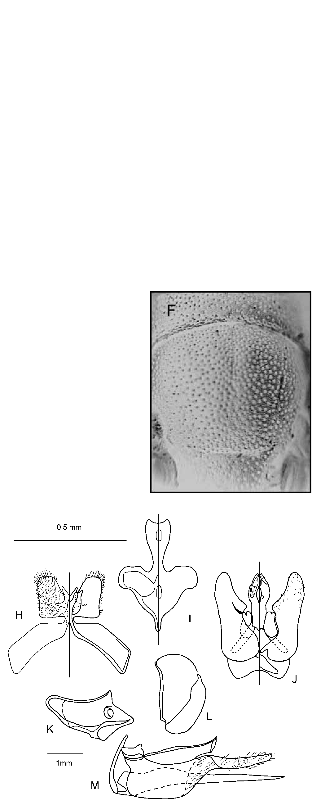
Diagnosis. Chilicola obesifrons and its close relatives (all of which are undescribed, but include this and the following species) are among the smallest bees in the genus. As a group, they can be differentiated from any of the named subgenera of Chilicola on the basis of the shape of the pronotum, the collar of which is moderately long, not strongly convergent anteriorly but angulate at the anterolateral corners ( Fig. 3E View FIGURES 3 A – M ). A new subgenus is being described for this group of species (Packer, in press). Other unusual characteristics are the lack of an emargination on the inner margin of the eye ( Figs. 2, 3A and C View FIGURES 3 A – M ) (shared only with some species of the subgenus (Prosopoides) ) and the comparatively coarse punctation of the thoracic dorsum ( Fig. 3F View FIGURES 3 A – M ). The frons is at least slightly expanded around the median ocellus and just mesad of the upper portion of the compound eye ( Figs. 2, 3A and C View FIGURES 3 A – M ) in all of the species in the group with the exception of the next species to be described ( C. catamarcense Packer , n. sp.). These expanded areas are comparatively impunctate but bear strongly imbricate microsculpture appearing almost granular, some species of the subgenus Prosopoides also share this condition. The membranous lobes of S7 are unique ( Fig. 3H View FIGURES 3 A – M ) in form, colouration and setation among related species, including the subgenus Prosopoides. These other taxa have much more robust lobes that usually bear long and/or thick, capitate setae.
Chilicola obesifrons can be differentiated from other members of its group by the following characters: the head of both sexes in profile has the median ocellus entirely hidden by frontal swellings and the vertex is flat around the lateral ocellus ( Figs. 3B and D View FIGURES 3 A – M ). This feature combined with the anterolateral corners of pronotum approximately right-angled serve to identify the male ( Fig. 3E View FIGURES 3 A – M ). For the female, the comparatively small mesoscutal punctures, with space for approximately 12 between the median and parapsidal lines, separate this species from others with a swollen frons and flat vertex. Other species with the median ocellus hidden in profile either have males with the head angularly produced in front of the lateral ocellus in profile (an undescribed species known only from the male from Neuquen, Argentina) or anterolateral corners of pronotum acutely angled (a Bolivian species) and females with larger mesoscutal punctures such that at most 9 would fit in the space between admedian and parapsidal lines (both an additional northern Argentinian species and the Bolivian one).
Description. Male: Body length 3.2mm, forewing length 2.0mm, head width 0.8mm.
Colouration: Black, with following parts yellow: labrum, mandible (except apex red-brown), most of clypeus, spot on lower paraocular area, anterior mark on scape. Pedicel and flagellum orange. Legs with yellow-orange as follows: anterior surface of foretibia, outer surface of forebasitarsus, apical rings on all femora; basal and apical rings on all tibiae. Tarsi pale brown. Tegula and apical impressed areas of metasomal terga pale amber. Wing veins orange-brown.
Surface Sculpture: Microsculpture strongly imbricate almost throughout. Labrum deeply, coarsely and densely punctate (i<d) on shining background. Clypeus dull with sparse, irregular and obscure punctures, i=1– 4d. Supraclypeal and lower paraocular areas somewhat shiny with punctures more distinct and somewhat more dense, i=1–3d. Frons with larger, denser punctures, i~d, with impunctate swellings around median ocellus and laterad of lateral ocellus. Vertex with transverse wrinkles among punctures. Hypostomal area irregularly punctate, i=1–4d. Pronotum, mesoscutum and scutellum ( Fig. 3F View FIGURES 3 A – M ) dull with dense punctures that are large for size of insect, i<d; sparser on scutellum, i~d; metanotum duller than mesoscutum, punctures i<d. Mesopleuron somewhat shiny, irregularly punctate, i<1–3d. Dorsal surface of propodeum with disk slightly depressed, weakly rugosoreticulate on either side of median carina, dorsolateral area roughened. Metasomal terga with weak microsculpture, sparse shallow obscure punctures, anterior portions of terga not differently sculptured from disks, apical impressed areas impunctate with very weak microsculpture.
Pubescence: White, short and sparse, not especially plumose; lacking long erect hairs on hypostomal area. Without apicolateral hair patches on metasomal terga. No areas of specialized pubescence on metasomal sterna or legs.
Structure: Head: Longer than broad, length to width 62:53 ( Fig. 3A View FIGURES 3 A – M ). Labrum 2X as broad as long, apex almost straight. Mandible short and broad, length:basal depth ~2:1. Clypeus with length and breadth subequal, lower one third extending beyond lower ocular tangent, lacking median longitudinal groove; epistomal suture expanded below anterior tentorial pit almost to laterally reflexed portion of suture, pit not separated from suture ( Fig. 3A View FIGURES 3 A – M ). Subantennal suture curved inward from near origin on antennal socket, otherwise straight; supraclypeal area ( Fig. 3A View FIGURES 3 A – M ) long and narrow, length to breadth <2:1, weakly produced and poorly demarcated from frons above. Frons greatly swollen around median ocellus and mesad of upper inner margin of eye such that median ocellus not visible in profile; swellings causing head to be flat dorsally in profile ( Fig. 3B View FIGURES 3 A – M ). Frontal line distinct to median ocellus. Facial fovea broadly oval, shiny and shallow ( Fig. 2). Inner margin of compound eye not emarginate ( Fig. 3A View FIGURES 3 A – M ) making UOD difficult to assess, but eyes strongly convergent below ( Fig. 3A View FIGURES 3 A – M ); OOC subequal to IOC. Lateral ocellus separated from compound eye by more than 2X its diameter. Vertex slightly longer than lateral ocellus, abruptly rounded onto occipital region. Upper ocular tangent passing well below lower margin of median ocellus by more than MOD. Scape 3X longer than greatest breadth, much longer than pedicel ( Fig. 3A View FIGURES 3 A – M ) and F1–F3 combined; F1 broader than long, F2 4X broader than long ( Fig. 2); middle flagellomeres with length and breadth subequal; F11 slightly longer than F10; flagellum gradually increasing in breadth from F1 to F11; flagellomeres lacking unusual patterns of setation or structural modifications. Genal area approximately one third as long as eye ( Fig. 3B View FIGURES 3 A – M ). Malar space linear such that presence or absence of malar suture indetectable.
Mesosoma: Elongate , length more than 2X its greatest depth (17:7). Pronotal collar long, slightly more than ½ as long as scape and approximately 1.5 LOL, laterally weakly concave, anterolateral corners forming right angle ( Fig. 3E View FIGURES 3 A – M ). Episternal groove complete, sharply curved anteriorly below. Scrobal groove weakly defined posterior to scrobe. Propodeum elongate, dorsal surface as long as posterior depth and subequal to length of scutellum (scutellum:metanotum:propodeum 24:15:25); propodeal sulcus marked by shallow pits becoming more distinct posteriorly. Hind leg not strongly modified; trochanter lacking modifications; femur 3X as long as greatest depth, convex ventrally; tibia gradually expanding from base to apex, somewhat more strongly so in basal half, length 3.5X greatest depth, lacking angles, carinae or ridges ( Fig. 3G View FIGURES 3 A – M ); hind tibial spurs long and not strongly curved or sclerotised; hind basitarsus 6X longer than greatest depth, parallel sided; hind tarsal claws bifid. Basal vein evenly curved; distal stigmal perpendicular crossing near apex of second submarginal cell, stigma shorter than length of marginal cell on wing margin, stigmal margin in marginal cell convex; first recurrent vein and first submarginal crossvein approximately interstitial on Rs+M.
Metasoma: Length and apical width of T1 subequal. T2 and T3 with weak basal depressions; apical impressed area approximately 0.33X length of tergum. Metasomal sterna unmodified except S1 slightly swollen at apex, gradulus of S2 with long posteriorly directed lateral portion, gradulus missing on S3–S6.
Terminalia : S7 with one pair of lateral lobes, ventral lobe broad, membranous, dusky pigmented except for basal and apical extremities; dorsal lobe reduced to narrow lamella with acute dorsolaterally oriented angulation ( Fig. 3H View FIGURES 3 A – M ). S8 with apical process elongate; widest at apex; emarginate apically ( Fig. 3I View FIGURES 3 A – M ). Ventroapical process of gonobase broad with comparatively long lateral projections. Volsella kidney-shaped; gonostylus not clearly demarcated from rest of gonoforceps. Gonoforceps with medioventral lobe approximately right-angled. Penis valve with pair of subapical membranous lobes, both oriented dorsally ( Fig. 3J View FIGURES 3 A – M ).
Female: Body length 3.3mm, wing length 2.0mm, head width 0.8mm.
Colouration: As in male except as follows: Labrum, mandible and anterior surface of flagellum orange. Clypeus and lower paraocular area lacking pale markings. Legs with marking darker orange; rings on femora and tibiae narrower. Metasomal sterna dark brown. Wing veins pale amber.
Surface Sculpture: As for male except as follows: Somewhat less dull throughout due to slightly weaker microsculpture. Spacing of punctures on supraclypeal area less regular, i=1–5d. Scutellum with space for approximately 12 punctures between admedian and parapsidal line. Metanotum no duller than scutellum. Mesopleural punctures finer. Lateral sulcus of propodeum very weak.
Pubescence: As in male except as follows: Comparatively sparse scopa on hind leg, with hairs on femur and tibia <3MOD. Metasomal scopal hairs with short branches on anterior side of rhachis, well developed corbicula on S2, <5MOD; scopal hairs on S3 <4MOD; apical row of hairs on S4 <3MOD;
Structure: Maxillary palpus unmodified, somewhat less than 0.5X as long as prementum. Prementum 4X longer than greatest width; premental fovea large, carinate laterally. Lacinia an elongate triangle, more than 3X as long as greatest breadth. Lorum weakly sclerotised except towards apex, 0.33X as long as cardo. Rest of body as in male except for usual secondary sexual characteristics and as follows: Facial fovea larger ( Fig. 3C View FIGURES 3 A – M ). Frons less strongly swollen around median ocellus and dorsally along inner margin of eye, median ocellus not visible in profile, area around it somewhat flattened (as in Figs. 3C and D View FIGURES 3 A – M ), region between medial and lateral swellings concave. Supraclypeal area shorter than in male ( Fig. 3C View FIGURES 3 A – M ). Gena more than 0.5X as long as width of compound eye ( Fig. 3D View FIGURES 3 A – M ). Apical lunule of S5 forming approximately equilateral triangle.
Sting apparatus: Hemitergite 7 ( Fig. 3K View FIGURES 3 A – M ) with lateral portion of marginal ridge thick almost to apex with obtuse angle at base of lateral process; lateral lamella approximately triangular, medial portion of marginal ridge concave; spiracle closer to lateral portion of marginal ridge than to apex of lamina spiracularis, set in shallow depression; apodeme poorly developed; posterior margin of lamina spiracularis obtusely excised. Hemitergite 8 ( Fig. 3H View FIGURES 3 A – M ) with anterior ridge strongly developed to apex, straight except slightly produced anteriorly at apex; margin of plate and junction of apodeme and plate both slightly sinuate. First valvifer comparatively long and parallel-sided with short dorsal and ventral processes. Second valvifer with apodemal ridge slightly convex, apical process weakly developed, pars articularis narrowly rounded, incisura postarticularis moderately broad, portion of plate basal to gonostylus membranous, gonostylus somewhat parallel-sided. Sting shaft with basal bulb 2/3 as long as stylet, ventral surface slightly concave; processus muscularis and processus medianus not strongly developed ( Fig. 3M View FIGURES 3 A – M ). Furcula with ventral arms narrow apically considerably broadened near basal one third, in lateral view strongly reminiscent of a cheese knife with ventral margin of dorsal arm strongly convex.
Material studied. Holotype male and allotype female, ARGENTINA, Catamarca, 17km N. of Andalgala, 14–15.ii.2003, L. Packer, pan traps; all paratypes are also from Catamarca province and are as follows: 25km N. of Andalgala, 14.ii.03 L. Packer, six females (one in glycerin); 20km N. of Andalgala, 27 o29’477”S, 0 66 o 23’006”W, 1736m, 14.ii.03, L. Packer, three females; Los Nacimientos de Abajo, 16–31.i.1969 Willink, Torán & Stange, malaise trap, [Entomofauna Subandina], one female; 6km N. of Belén, 1240m, 16–31.i.1969 Willink, Torán & Stange, malaise trap, [Entomofauna Subandina], one female; Punta Balasto, i.1997, Arriagada, one female; San Fernando, 7.iii.1990, Rozen and Roig, on Sclerophyllax gilliesii (Sclerophyllaceae) , six females. The holotype and allotype will be housed at MACN pending completion of revisionary studies of the group; the Willink, Torán, Stange females are at IML, Arriagada’s and Rozen and Roig’s specimens are at the AMNH, the remaining paratype females are at PYU.
Etymology. The specific epithet refers to the large swellings around the median ocellus and just mesad of the upper portion of the eye.
Comments. The type series collected by the senior author was found on the roadside between Andalgala and Capillitas in an area that was substantially moister than were the surrounding areas. The two specimens collected by Duckworth have the propodeal sulcus more strongly developed than in the others and approach several undescribed species in the group in this regard.
Two females from Sumalao, Salta Province, Argentina (AMNH), collected by Fritz in January 1996 may be attributable to this species.
No known copyright restrictions apply. See Agosti, D., Egloff, W., 2009. Taxonomic information exchange and copyright: the Plazi approach. BMC Research Notes 2009, 2:53 for further explanation.