
Shaanxispira linxiaensis, Shi & He & Chen, 2014
|
publication ID |
https://doi.org/ 10.11646/zootaxa.3794.4.1 |
|
publication LSID |
lsid:zoobank.org:pub:844C9EF5-3A66-4F3D-B4FC-DD8081F21FEA |
|
DOI |
https://doi.org/10.5281/zenodo.5082771 |
|
persistent identifier |
https://treatment.plazi.org/id/593887E9-424F-FC0F-FF64-E15E9EF0FB77 |
|
treatment provided by |
Felipe |
|
scientific name |
Shaanxispira linxiaensis |
| status |
sp. nov. |
Shaanxispira linxiaensis nov. sp.
( Figs. 3–4 View FIGURE 3 View FIGURE 4 ; Tables 1–3 View TABLE 1 View TABLE 2 View TABLE 3 )
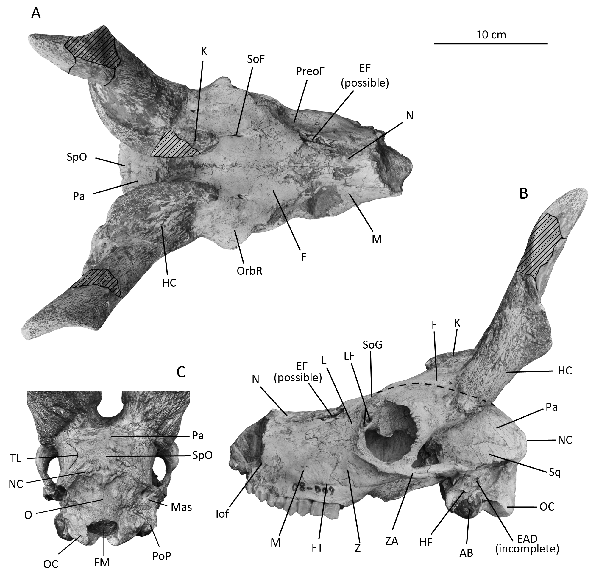
Holotype. HMV 1922 ( Figs. 3–4 View FIGURE 3 View FIGURE 4 ). An almost complete and undeformed skull.
Etymology. The species name comes from the locality of the holotype, Linxia Basin.
Geological distribution. Lower part of the upper Miocene, Liushu Formation, Linxia Basin, Gansu Province, China. The age of the associated fauna is equivalent to European MN9.
Diagnosis. Horn-core with only one keel. Antero-medial keel large and wing-shaped, extending anteriorly intensively at the base and protruding above the frontal. Cross section of the horn base sickle-shaped. Horn-core inserted uprightly and caudally. Parietal slightly inclined in side view. Braincase short and broad. Paroccipital process strong. Depression medial to paroccipital process shallow, but without accessory articular surface. Supraorbital foramen small, facing mostly forwards, with distinct supraorbital groove extending to lachrymal. Preorbital fossa wide and shallow. Infra-orbital foramen above P3. Nasals domed. Muzzle low and narrow. Teeth mesodont, without strong ribs or styles. Premolar row much shortened. Basal pillars absent or weak.
Description. The skull, HMV 1922, is nearly complete, with a well-preserved right horn-core. The tips of the horn-cores are broken, but skillfully restored with plaster. The tip of muzzle is missing. The right maxilla and left squamosal are slightly damaged. The tooth row is well preserved apart from the left P2 that is missing. The skull belongs to an adult specimen.
The muzzle is relatively low, with the nasal aperture being about as broad as high. The nasal is narrow and domed, and slightly widens caudally. The caudal end of the nasal is pointed, extending behind the level of the anterior rim of the orbit ( Fig. 3A, N View FIGURE 3 ). The naso-incisival notch is anterior to the level of P3. The infra-orbital foramen opens near the tooth row, at the level of P3 ( Fig. 3B View FIGURE 3 , Iof). The facial tuberosity is caudally positioned, close to the maxillo-jugal suture ( Fig. 3B View FIGURE 3 , FT). There is no facial crest. The facial part of the jugal is large and bilobed ( Fig. 3B, Z View FIGURE 3 ). The lachrymal is small, quadrangular, long and low ( Fig. 3B, L View FIGURE 3 ). The pre-orbital fossa is wide and shallow, without clear margins ( Fig. 3A View FIGURE 3 , PreoF). The maxillae close to the ethmoidal fissure are broken on both sides. However, the ethmoidal fissure is probably present because the nasal and lachrymal bones remain wide apart, and their margins are smooth, but size and shape of the ethmoidal fissure are uncertain ( Fig. 3B View FIGURE 3 , EF).
The frontal is wide and flat in front of the horn-core, rising slightly above the orbit, and declining at the horncore. The bending of the frontal is weak ( Fig. 3B View FIGURE 3 , dashed lines). The mid-frontal suture is somewhat complex and elevated as a sagittal ridge before the horn bases, whereas between the horn-cores, the suture is flat, but more complex ( Fig. 3A, F View FIGURE 3 ). The fronto-parietal suture points forwards in the centre, with the midpoint positioned between the horn-cores. The supra-orbital foramen is small, deeply sunken into the frontal, and facing mostly forwards, making the opening nearly round in anterior view, but tear-shaped in dorsal view ( Fig. 3A View FIGURE 3 , SoF). The internal opening of the supra-orbital foramen faces mostly downwards, and is more caudal than its external opening. The supra-orbital groove is long and distinct ( Fig. 3B View FIGURE 3 , SoG). There is no post-cornual fossa. The orbit is large with wide orbital rims; its dorsal border is lower than the frontal surface; its anterior border is posterior to the back of M3 ( Fig. 3B View FIGURE 3 ). The lachrymal foramen is large, and is partitioned into two foramina by a thin bone layer ( Fig. 3B View FIGURE 3 , LF). The zygomatic arch is weak.
The horn-core is straight, homonymously twisted, making a little more than one whorl, and diverging at an angle of about 35˚ ( Fig. 3C View FIGURE 3 ). In lateral view, the horn-core is inserted far behind the orbit and is almost perpendicular to the dorsal border of the cranium, and only slightly inclined backwards ( Fig. 3B View FIGURE 3 , HC). The anteromedial keel is large and wide, protruding like a wing ( Fig. 3A, K View FIGURE 3 ). The keel twists homonymously, following the twist of the horn-core. At its base, the keel occupies an antero-medial position on the horn-core, extending anteriorly to the level of the posterior rim of the orbit, and being strongly prominent above the frontal ( Fig. 3B, K View FIGURE 3 ). As the keel ascends from the base to the tip, the size decreases gradually. In the basal part, the keel has a thickened margin (approximately 2 cm wide at the base, and becomes thinner upwards) with porous surface, whereas the surface of the horn-core is covered by many small longitudinal grooves. The cross section of the horn base is sickle-shaped because of the prominent keel. There is no posterior keel.
The braincase is short and wide, with parallel lateral sides ( Fig. 3C View FIGURE 3 ). The lateral part of the parietal is long, separating the frontal from the squamosal. The temporal lines are wide apart ( Fig. 3C View FIGURE 3 , TL). The occipital is low and broad. The angle between the parietal and occipital planes is about 120˚ ( Fig. 3B View FIGURE 3 ). The nuchal crest is thick, with a coarse and large external occipital protuberance below ( Fig. 3C View FIGURE 3 , NC). The mastoid exposure is long and broad; its dorsal part faces caudally and the ventral part faces laterally. The mastoid exposure is separated from the parietal by the squamosal and the occipital.
a Measurements taken from Zhang (2003); b Estimated measurement
a Measurements taken from Zhang (2003)
b The longest distance from antero-medial point to posterior-lateral point
c The diameter perpendicular to the antero-posterior diameter
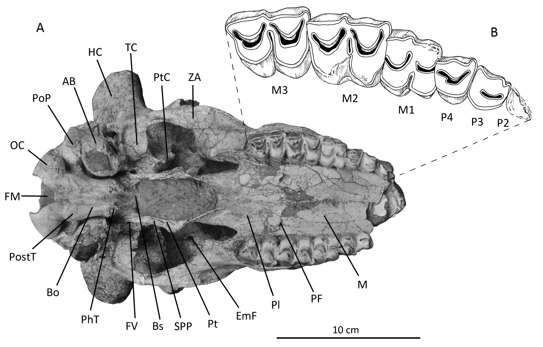
The foramen magnum is large ( Fig. 3C View FIGURE 3 , FM). The occipital condyle is not as strong as in other “ovibovines” like Hezhengia or Plesiaddax ; it is divided into dorsal and ventral articular surfaces by a clear ridge, and the dorsal articular surface forms a notable angle with the occipital surface above ( Fig. 3B View FIGURE 3 , OC). The depression between the condyle and paroccipital process is shallow and small, but no accessory articular surface is visible ( Fig. 4A View FIGURE 4 ). The paroccipital processes are broken on both sides, with their bases considerably robust and converging anteriorly ( Fig. 4A View FIGURE 4 , PoP).
The basioccipital is rectangular and thick. A deep and broad longitudinal groove runs along its midline ( Fig. 4A View FIGURE 4 , Bo). Both the posterior tuberosity and the pharyngeal tuberosity are well developed and of similar size ( Fig. 4A View FIGURE 4 , PhT, PostT). The basioccipital makes an angle with the palate of about 20˚. The auditory bulla is large and round. The posterior tip of the auditory bulla is laminar, positioned laterally to the paroccipital process. A triangular muscular process is visible anterior to the auditory bulla ( Fig. 4A View FIGURE 4 , AB). The hyoid fossa is deep and small, and hardly observable in ventral view ( Fig. 3B View FIGURE 3 , HF). The external auditory duct is small and short, pointing postero-laterally ( Fig. 3B View FIGURE 3 , EAD). The oval foramen is moderate in size, facing mainly laterally ( Fig. 4A View FIGURE 4 , FV). The pterygoid crest is robust ( Fig. 4A View FIGURE 4 , PtC). The ventral border of the pterygoid process of the sphenoid is thick. A small groove is observed along the spheno-pterygoid suture in ventral view ( Fig. 4A View FIGURE 4 , SPP, Pt). The palatine is long, and the anterior edge of the choanae is much more caudal than the tooth row ( Fig 4A View FIGURE 4 , Pl).
The cheek teeth are mesodont. The premolar row is short, and the length ratio of premolar to molar row is 54% ( Fig. 4B View FIGURE 4 , Table 3 View TABLE 3 ). P2 is broken, but was certainly small. Its lingual border is straight. P3 is larger than P2, with a round lingual border. The width of P3 is similar to its length. The protocone is more distal than the paracone. The fossette of P3 is simple but deep. P4 is similar to P 3 in morphology but is a little larger. The protocone is more mesial, making P4 almost symmetrical. The fossette is long and narrow, with its anterior and posterior ends close to the border of the tooth. A spur is observed in the posterior part of the fossette, pointing forwards.
M1 is almost square, being only slightly wider than long. The first lobe is shorter than the second lobe. The fossettes are U-shaped, and lack spurs. The lingual valley is narrow but deep, reaching almost 1/3 of the width of M1. The enamel island is thin and long. There is a tiny basal pillar on the posterior lobe of M1. M2 is much larger and longer than M1. The parastyle and mesostyle are moderately strong in M2, whereas the metastyle is not developed, and the buccal ribs are weak. The fossettes are simple, with the anterior and posterior ends close to the buccal wall. There are small folds along the border of the fossettes, but no distinct spur. The enamel island is also thin and long in M2. M3 is similar to M 2 in size and morphology, but is a little longer and narrower. The ribs and styles are even weaker in M3 than in M2, with only a moderately developed parastyle. There is no enamel island in M3, and the lingual valley penetrates deeply towards the buccal wall.
No known copyright restrictions apply. See Agosti, D., Egloff, W., 2009. Taxonomic information exchange and copyright: the Plazi approach. BMC Research Notes 2009, 2:53 for further explanation.