
Pungentus intertextus ( Thorne & Swanger, 1936 ) Thorne 1939
|
publication ID |
https://doi.org/ 10.5281/zenodo.203129 |
|
DOI |
https://doi.org/10.5281/zenodo.5621377 |
|
persistent identifier |
https://treatment.plazi.org/id/593A87D3-FF9C-C63F-FF2F-FBAEFF6D6D9F |
|
treatment provided by |
Plazi |
|
scientific name |
Pungentus intertextus ( Thorne & Swanger, 1936 ) Thorne 1939 |
| status |
|
Pungentus intertextus ( Thorne & Swanger, 1936) Thorne 1939
( Figs. 1 View FIGURE 1 & 2 View FIGURE 2 )
Material examined. Two females, two males and 13 juveniles; in variable states of preservation since most specimens have become flattened and lack cuticle.
Measurements. See Tables 2 View TABLE 2 & 4.
Description. Adult: Very slender nematodes of large size, 3.99–4.92 mm long. Body cylindrical, distinctly tapering towards the anterior end, less so towards the posterior one because the caudal region is rounded. Habitus curved ventrad after fixation, especially in posterior body region. Cuticle 2 μm thick in anterior region, 4 μm (n=1) at mid-body and 8.0, 11.5 μm (n=2) on tail; with fine but perceptible transverse striation throughout the entire body. Two ventral and three dorsal pores are present usually at level of odontostyle, one ventral and one dorsal pore at level of odontophore base, other body pores inconspicuous. Lateral chord 19 μm (n=1) wide at mid-body, occupying about one-fourth (24%) of mid-body diameter. Lip region rounded, offset by constriction, 2.2–2.4 times as wide as high and about one-fifth (19, 20%; n=2) of body diameter at neck base; lips amalgamated; labial papillae hardly protruding. Amphid fovea funnel-shaped, its aperture 5.5 μm (n=1) or about two-fifths (41%) of lip region diameter. Cheilostom nearly cylindrical, with a thin perioral, weakly sclerotized ring perceptible in several specimens. Odontostyle comparatively long and slender, 10.2–11.6 times as long as wide, 1.9 times as long as lip region diameter, and 0.51–0.64% of body length; aperture 3.0–4.5 μm long or occupying one-eighth to one-sixth (12–17%) its length. Guiding ring difficult to observe, but apparently double. Odontophore linear, rod-like, 0.9–1.0 times as long as odontostyle. Anterior region of pharynx enlarging very gradually; basal expansion 7.1, 7.4 (n=2) times as long as wide, 4.3, 4.5 (n=2) times as long as body diameter, and occupying 51–55% of total neck length. Pharyngeal gland nuclei located as follows: DN = 49, 52 (n=2); S1N1 = 69 (n=1); S1N2 = 75 (n=1); S2N = 86, 88 (n=2). Nerve ring located at 177, 180 μm (n=2) from anterior end or 29, 31% (n=2) of total neck length. Cardia rounded, almost as long as wide, 18.0–19.5 x 19.5–22.0 μm, and surrounded by intestinal tissue. Prerectum 0.9–2.7, rectum 0.8–1.4 anal body widths long.
Female: Genital system didelphic-amphidelphic, with both branches equally and well developed, the anterior 417, 447 μm long or 9, 11% of body length, and the posterior 415, 446 μm long or 9, 11% of body length. Ovaries moderately developed, not surpassing the sphincter level in the two specimens examined; the anterior 103, 115 μm, the posterior 92, 114 μm long; oocytes arranged first in two or more rows, then in a single row. Oviduct consisting of slender part with prismatic cells and well developed pars dilatata, the anterior 134, 153 μm long or 1.8 (n=1) times the corresponding body diameter, and the posterior 137, 144 μm long or 1.7 (n=1) times the corresponding body diameter. Oviduct-uterus junction marked by a sphincter. Uterus a long tube-like structure, apparently (not very well preserved) with two sections of nearly the same length, the proximal one of which is wider and with more perceptible lumen; the anterior 270, 278 μm long or 3.2 (n=1) times the corresponding body diameter, and the posterior 247, 250 μm long or 3.0 (n=1) times the corresponding body diameter. Vagina not observed in lateral view, but pars refringens is certainly present as seen in frontal view. Vulva an equatorial, short, ovoid, transverse slit. Tail short and rounded. Caudal pores two pairs at the middle of tail, one dorsal, another lateral.
Male: Genital system diorchic, with opposite testes. In addition to the adcloacal pair, situated at 9.5 μm from cloacal aperture, there is a series of 19 ventromedian supplements slightly irregularly spaced (10.5–15.5 μm apart), the two most posterior of which are situated within the range of spicules and the posteriormost one at 25 μm from adcloacal pair. Spicules curved ventrad and relatively slender, about 6.2 times as long as wide and 1.7 times as long as anal body diameter. Lateral guiding pieces 17.5 μm long and 7.0 times as long as wide. Tail rounded conoid, ventrally almost straight, dorsally convex. Caudal pores three pairs at the middle of tail, one dorsal, two lateral.
Juveniles: Only two juvenile stages (J2 and J3) are present in the material herein studied. Their general morphology is similar to that of adults.
Diagnosis (based on specimens examined). This species is characterized by its body 3.99–4.92 mm long, lip region offset by constriction and 14 μm broad, odontostyle 25.5–26.5 μm long and with aperture occupying 12– 17% of its length, neck 568–610 μm long, pharyngeal expansion 297–327 μm long or 51–55% of total neck length, uterus apparently bipartite and 247–278 μm long or 3.0–3.2 times the corresponding body diameter, pars refringens vaginae present, V = 49–51, tail rounded (32–40 μm, c = 100–129, c’ = 0.6–0.8), spicules 82–86 μm long, and 19 shortly spaced ventromedian supplements.
Remarks. Slide “83. Dorylaimus intertextus ” contains part of type material originally studied by Thorne and Swanger (1936) since the information provided in its corresponding label perfectly agrees with the type locality of the species as mentioned by the American authors: “Foothill soil, Riverside, California. 1923”. Slides “83a. Dorylaimus intertextus ” and “83b. Dorylaimus intertextus ” certainly contain the material from Riverside, California, later examined by Thorne (1939). There is no doubt then about the original identity of this material. The above description fits well with the original one, although many new morphological details and morphometrics are now given for the first time. The few differences observed, such as the shorter odontostyle (25.5–26.0 vs 28–29 μm) and longer female tail (39 vs 28 μm), might be due to intraspecific variation and/or original errors in taking measurements.
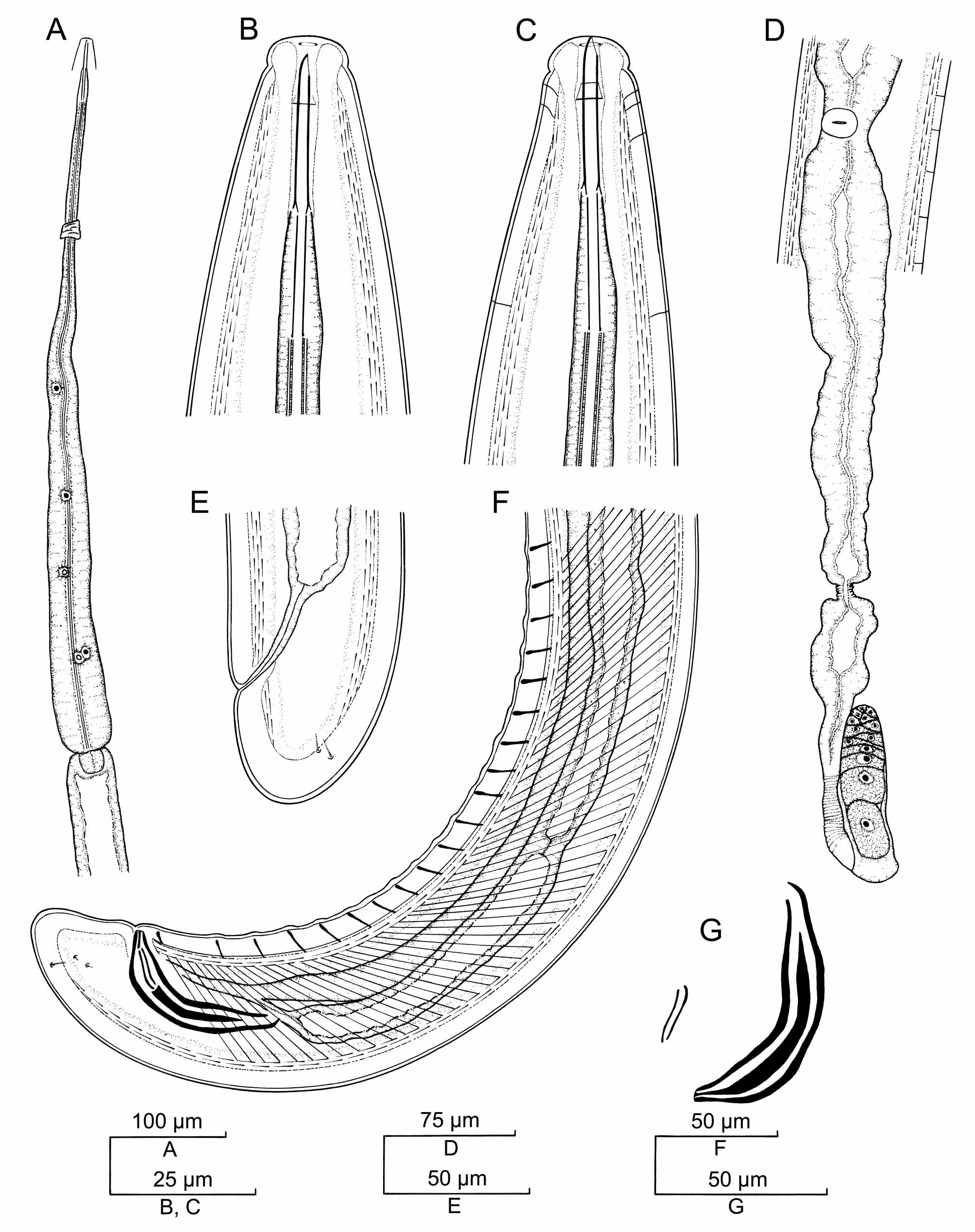

TABLE 2. Morphometric data of Pungentus intertextus (Thorne & Swanger, 1936) Thorne, 1939 (adult specimens). Measurements in μm (except L, in mm).
| Population | Riverside, California. Foot-hill soil | Riverside, California. |
|---|---|---|
| Character L a b | n 2ƤƤ 3 4.02, 4.51* 3.99?, 53.3 51.5 7.1, 7.4 6.8 | 3 4.92?? |
| c c' | 128.6, 114.1 99.7 0.6, 0.7 0.8 | ?? |
| V Lip region diameter Odontostyle length Odontophore length | 51.3, 49.1 - 14, 14 14 25.5, 26.5 26 22.5, 25.0 25 | -? 25.5 25 |
| Guiding ring from anterior end Neck length | 11.5, 12.0 11.5 568, 610 583 | ?? |
| Pharyngeal expansion length Diameter at neck base at mid-body at anus/cloaca Prerectum length Rectum length | 310, 327 297?, 73 69?, 84 77 50, 53 49 46, 56 133 45.5, 42.5 66 | ?????? |
| Tail length Spicule length Ventromedian supplements | 31.5, 39.0 40 - 82 - 19 | ? 86? |
| *Specimen broken in two pieces. |
No known copyright restrictions apply. See Agosti, D., Egloff, W., 2009. Taxonomic information exchange and copyright: the Plazi approach. BMC Research Notes 2009, 2:53 for further explanation.