
Asterolepis alticristata, Downs & Daeschler & Lo & Carey & Shubin, 2019
|
publication ID |
https://doi.org/ 10.5252/geodiversitas2019v41a19 |
|
publication LSID |
urn:lsid:zoobank.org:pub:C34F475B-6315-400C-ACE3-B67B1667C033 |
|
DOI |
https://doi.org/10.5281/zenodo.3703489 |
|
persistent identifier |
https://treatment.plazi.org/id/D6EAF712-9D00-4A37-9612-93CF17333564 |
|
taxon LSID |
lsid:zoobank.org:act:D6EAF712-9D00-4A37-9612-93CF17333564 |
|
treatment provided by |
Valdenar |
|
scientific name |
Asterolepis alticristata |
| status |
sp. nov. |
Asterolepis alticristata n. sp.
Asterolepis View in CoL sp. – Daeschler et al. 2006: 759.
“Large asterolepidid” – Downs et al. 2011a: 994.
“Asterolepid placoderm” – Downs et al. 2013: 48.
“Asterolepid antiarch” – Downs et al. 2018: 1.
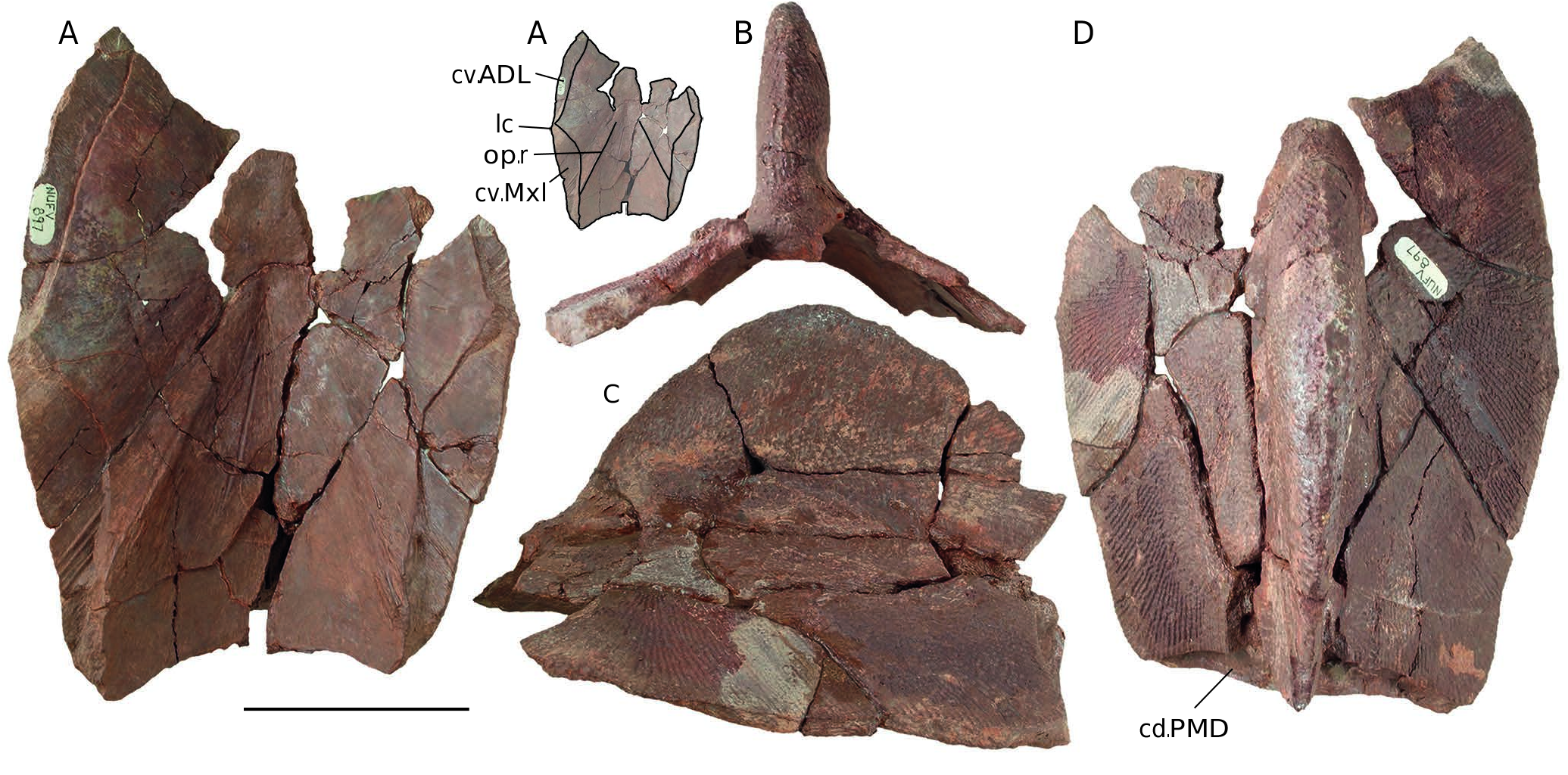
HOLOTYPE. — NUFV 897 , anterior median dorsal plate of thoracic skeleton ( Fig. 2 View FIG ).
ETYMOLOGY. — From the Latin ‘altus,’ high, and ‘crista,’ crest, in reference to the tall midline crest of the anterior median dorsal plate that helps to diagnose the species.
TYPE LOCALITY AND HORIZON. — NV 2K17 locality (77˚09’59.1”N, 86˚16’9.42”W), Fram Formation near the eastern arm of Bird Fiord on southern Ellesmere Island, Nunavut Territory, Canada. Palynological data indicate an early Frasnian age (medius and maclarenii zones of Chi & Hills 1976).
REFERRED MATERIAL. — NUFV 1265, nuchal plate; NUFV 812, 813, premedian plates; NUFV 822, partial anterior median dorsal plate; NUFV 814, 815, 823, 828, 834, 835, 842, 845, 849-851, 854, 856, 860-861, 864, 867, 870-873, 876-878, 884-886, 889, 890, 896, 1270-1277, crests of anterior median dorsal plates; NUFV 796, 806, 874, 879, 898, posterior median dorsal plates; NUFV 802.1, 802.2, 821, 848, posterior median dorsal fragments; NUFV 892, articulated partial thoracic and pectoral skeleton including median ventral, right and left anterior dorsolateral plates, anterior ventrolateral plates, dorsal central 1 and ventral central 1 plates; NUFV 895, right mixilateral plate .
DIAGNOSIS. — Antiarch referred to Asterolepis by the presence of an anterior median dorsal plate that broadly overlaps the mixilateral plates ( Moloshnikov 2008). Distinguished from previously described species by the following combination of anatomical features: 1) a tall, rounded midline crest that rises from a position near the tergal angle of the anterior median dorsal plate and loses most of its height before reaching the posterior median dorsal plate and (2) a midline crest of the posterior median dorsal plate that decreases in height cranial to caudal.
REMARKS
Because the new species is diagnosed by a crest of the anterior and posterior median dorsal plates, we use spatial association with diagnostic plates or plate fragments to refer additional material to the species (including isolated premedian, mixilateral and nuchal plates and an articulated partial skeleton [NUFV 892] that lacks the median dorsal plates). All of these referred materials also share a size category and dermal ornament with the Asterolepis alticristata n. sp. type specimen.
DESCRIPTION OF CEPHALIC SKELETON
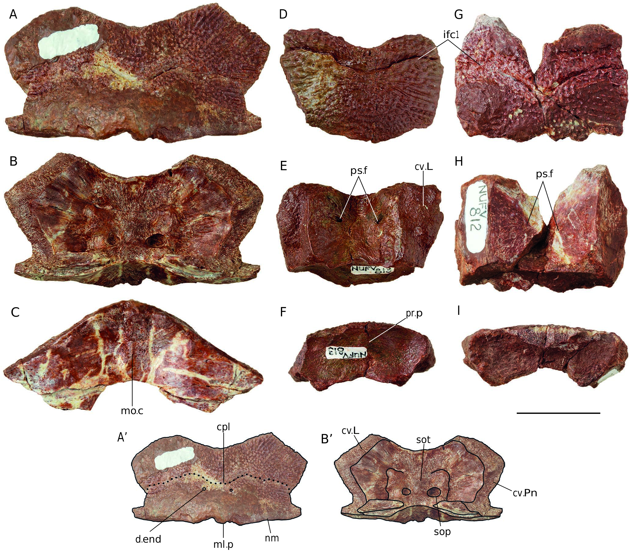
Nuchal plate ( Fig. 3 View FIG A-C)
The nuchal plate (NUFV 1265: Fig. 3 View FIG A-C) is short and wide; the width/length index (maximum transverse width × 100 divided by maximum sagittal length) is 195. The maximum reported index for Asterolepis is 210 in Asterolepis scabra ( Young 1987). Several smaller, unassignable nuchals from NV2K17 (NUFV 1247, 1248, 1252, 1254, 1263) have an index that is comparable with, though none higher than, this reported maximum (See Description of Other Asterolepidoid Specimens from NV2K17). As in Asterolepis ornata, the width across the lateral corners is slightly greater than the width across the caudolateral corners; this is despite the presence of pronounced caudolateral processes. As is true of all known species of Asterolepis , the notch for the postpineal plate is shallow, reaching no further caudal than the lateral corners of the nuchal plate. The central pit-line groove ( Fig. 3 View FIG A’[cp1]) extends across the width of the nuchal; it exhibits a pronounced median dip and meets the lateral margins of the plate well caudal to the lateral corners. Tiny, parasagittal foramina just caudal to the pit-line are weakly visible among the tubercles of the ornament. Stensiö (1931) presumed these to be the external openings of the endolympatic ducts ( Fig. 3 View FIG A’[d.end]).
The nuchal is a thick plate with broad and deep zones of overlap onto lateral ( Fig. 3 View FIG A’[cv.L]) and paranuchal ( Fig.3 View FIG A’[cv. Pn]) plates. There is a very short obtected area ( Fig. 3 View FIG A’[nm]) along the nuchal’s caudal margin that is unornamented though not depressed. The caudal margin of the plate is notched in parasagittal positions close to the plate’s midline. These notches border a short caudal midline process ( Fig. 3 View FIG A’[ml.p]) of the plate similar to the one described in A. ornata ( Upeniece 2011).
The visceral surface of the nuchal exhibits a supraotic thickening ( Fig. 3 View FIG B’[sot]) that surrounds the large supraoccipital pits (containing the internal openings of the endolymphatic ducts; Stensiö 1931; Fig. 3 View FIG B’[sop]). The caudal surface of the nuchal plate exhibits a median occipital crista ( Fig. 3C View FIG [mo.c]) and, on either side of it, parasagittal fossae that were interpreted by Stensiö (1931) to be attachment areas for cranial levator muscles. In caudal view, the visceral surface of the plate has a tight, nearly semicircular arch to it. The areas lateral to the visceral arch are depressed relative to the area external to it such that a pronounced horizontal ledge runs across the plate at the most external reach of the visceral arch. The caudal surface of the nuchal is the thickest part of the plate and there is a dramatic decrease in nuchal thickness rostrally from the depressed visceral extensions of the plate’s caudal surface.
The ornament of the nuchal plate is coarsely tuberculate. Tubercles are fused into ridges close to the rostral and lateral margins of the plate; these ridges radiate from the center.
Premedian plate ( Fig. 3 View FIG D-I)
The two premedian plate specimens referred to Asterolepis alticristata n. sp. (NUFV 812 [maximum sagittal length of 29.85 mm] and 813 [same length = 22.02 mm]: Fig. 3 View FIG D-I) share a size category and an ornament type but differ from one another in some specific details. The premedian plate has been recognized as a source of intraspecific variation throughout Antiarchi ( Young 1984; Downs et al. 2011b). Both specimens exhibit the generalized premedian plate shape that has been well described in Asterolepis ornata ( Lukševičs 2001). Both are thick plates, especially at the caudal end, and are arched across the midline. Both have unornamented zones in lateral positions along the depressed rostral margin of the plate; the rostral margin exhibits a midline notch. In both, the tall caudal surface is depressed rostrally (analogue of the bothriolepidid preorbital recess; Lukševičs 2001) and this surface has a prerostral process ( Fig. 3F View FIG [pr.p]) across the midline with a small depression on the midline (the nasal notch of Lukševičs 2001). The prerostral process separates the orbital cavities. Both premedian specimens are widest at the lateral corners and these corners are closer to the rostral margin than they are to the caudal one.
The two premedian specimens referred to A. alticristata n. sp. have a width/length index (as calculated for the nuchal above) of 134 (NUFV 812) and 157 (NUFV 813). These values are within the range of variation that Upeniece (2011) reported for adult Asterolepis ornata (120-233). This broad range of values for adult A. ornata again highlights the variability of this plate within a single antiarch species.
The principal section of the infraorbital pit-line groove ( Fig. 3D View FIG [ifc1], G[ifc1]) extends across the width of the premedian but follows a different path on each of the two specimens. In NUFV 812 ( Fig. 3G View FIG ), the groove forms a caudally directed point at the midline and strongly arches rostrally on either side of the midline. It is positioned at about mid-length along the plate, such that the midline dip of the groove reaches into the caudal half of the plate. In contrast, the groove on NUFV 813 ( Fig. 3D View FIG ) has a shallow, rounded dip at the midline and is much straighter across its entire width. The groove is situated close to the rostral margin of the premedian such that no part of it reaches into the plate’s caudal half.
Tiny foramina ( Fig. 3E View FIG [ps.f], H[ps.f]) penetrate the visceral surface of the premedian; the two most prominent are parasagittally located in the rostral half of the plate. The external ornament of the premedian is similar to that of adult Asterolepis ornata in that rows of tubercles radiate out from the center of ossification. Close to the lateral margins of the plate, the tubercles within a row fuse together to form ridges .
DESCRIPTION OF THORACIC AND PECTORAL SKELETONS Anterior median dorsal plate ( Fig. 2 View FIG )
The anterior median dorsal plate of Asterolepis alticristata n. sp. features a tall rounded crest on the midline. The remarkable height (c. 60 mm in the largest specimen, NUFV 897, maximum anterior median dorsal plate width = 112.55 mm) and
thickness (c. 18 mm in NUFV 897) of the crest is unlike any previously described for Asterolepis ( Fig. 2 View FIG ). The crest rises sharply from a position just cranial to the tergal angle and reaches its maximum height just caudal to the center of the anterior median dorsal plate, nearly in line with the plate’s greatest width. The long dorsal margin of the crest caudal to the maximum height is slightly convex to deeply concave. The closest point of comparison among antiarchs, for a crest in this position and of this height, is in Bothriolepis cristata Traquair, 1895 ( Scotland). A tall dorsal crest also appears in Bothriolepis gippslandiensis Hills, 1929 (Victoria, Australia); Bothriolepis cullodenensis Long, 1983 (Victoria Australia); and Bothriolepis zadonica Moloshnikov, 2004 (Central European Russia). In each of these three examples, however, the crest is formed nearly equally by anterior median dorsal and posterior median dorsal plates; the crest rises from near the anterior median dorsal plate’s tergal angle and reaches its maximum height only at the contact between anterior median dorsal and posterior median dorsal plates ( Moloshnikov 2004). In addition to the three partial anterior median dorsal plates with crests (NUFV 822, 896, 897 [holotype]), the A. alticristata n. sp. referred material includes thirty-seven isolated crests from anterior median dorsal plates. No other part of the A. alticristata n. sp. anatomy is better represented in the sample. The size and shape variation among these crests does not fit a single growth trend (See Discussion: Variation in anterior median dorsal plate crests of Asterolepis alticristata n. sp.).
At the caudal end of the anterior median dorsal plate, the midline crest continues onto the posterior median dorsal plate. In NUFV 897 (maximum crest height c. 60 mm), the height of the anterior median dorsal plate crest at its caudal terminus is c. 14 mm. The total length of the anterior median dorsal plate crest on NUFV 897, from where it first rises to its contact with the crest of the posterior median dorsal plate is 108.10 mm. The anterior median dorsal plate of A. alticristata n. sp. is steeply vaulted in the transverse plane such that the dorsal surfaces lateral to the crest face distinctly craniolaterally. The posterior median dorsal plate overlaps an unornamented, depressed zone along the caudal end of the anterior median dorsal plate’s dorsal surface ( Fig. 4D View FIG [cd.PMD]). None of the available material allows description of the anterior median dorsal plate cranial to the crest. In NUFV 897, the tergal angle is cranial to the lateral corners ( Fig. 2 View FIG A’[lc]) of the anterior median dorsal plate and caudal to the most cranial position of the midline crest.
The visceral surface of the anterior median dorsal plate shows the depressed surfaces where the plate overlaps the anterior dorsolateral ( Fig. 2 View FIG A’[cv.ADL]) and mixilateral ( Fig. 2 View FIG A’[cv.Mxl]) plates cranial and caudal to the anterior median dorsal lateral corners. On each side, these overlap surfaces are continuous with one another across the lateral corner. Obliquely oriented, low, wide ridges (oblique posterolateral ridge of Karatajūte-Talimaa 1963; Fig. 4 View FIG A’[op.r]) extend and widen from the center of the anterior median dorsal plate’s visceral surface to the mixilateral underlap close to the caudolateral corners of the plate.
The ornament of the dorsolateral faces of the anterior median dorsal plate is densely tuberculate centrally and ridged closer to the lateral and caudal margins of the plate. The ridges retain the appearance of fused tubercles and radiate from the center. The ornament of the crest is densely tuberculate especially at the base. The tubercles are disorganized at the base, continuing the ornament type of the plate’s dorsolateral surfaces, and are organized into rows more distally. In one specimen (NUFV 872), these organized tubercles are fused into high ridges. The rows of tubercles or ridges radiate from a cranioproximal position on the crest.
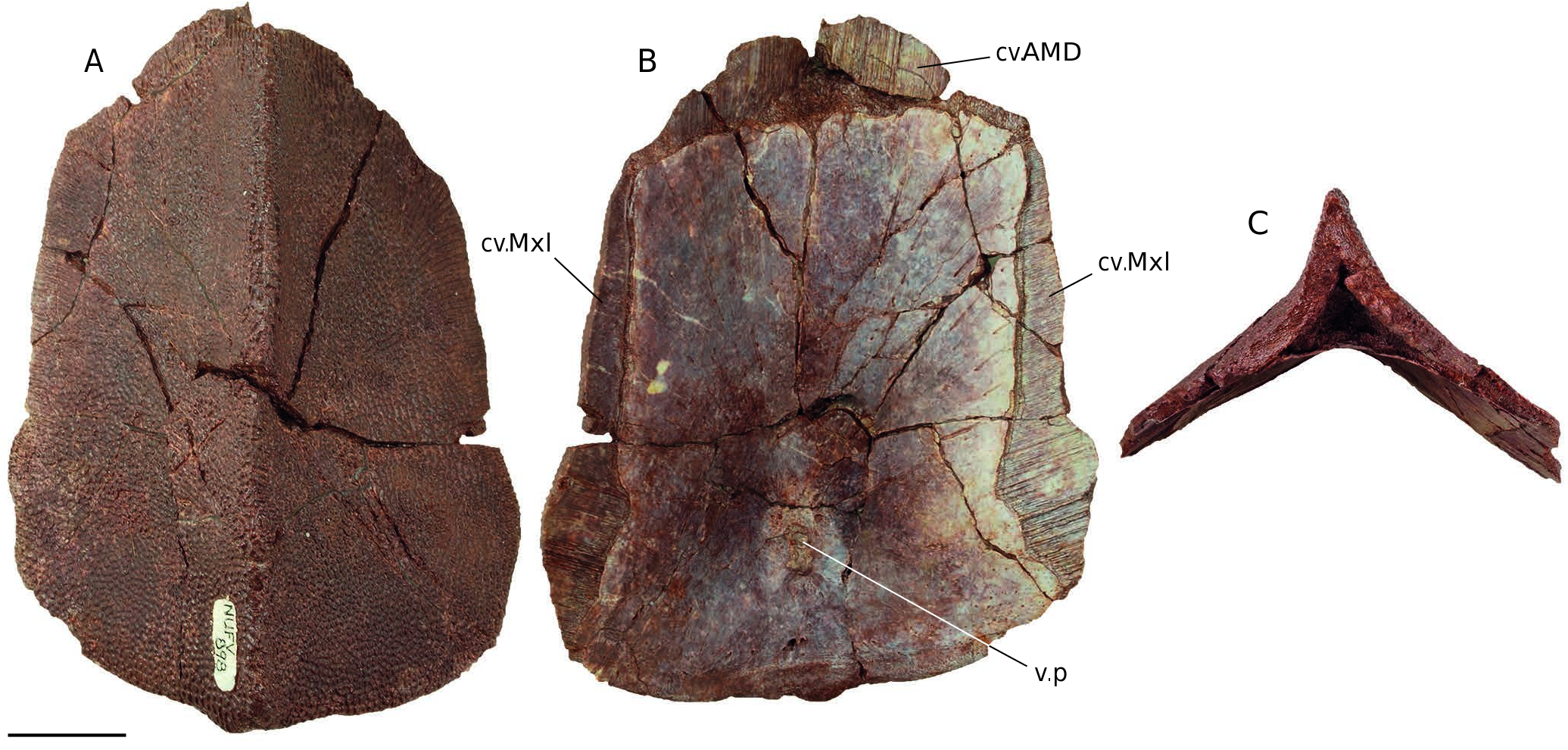
Posterior median dorsal plate ( Fig. 4 View FIG )
The posterior median dorsal plate of Asterolepis alticristata n. sp. is elongate and narrow. The greatest width of the plate is across the lateral corners; these corners are located close to the caudal margin of the plate. There is little increase in width from craniolateral to caudolateral corners and much of that increase occurs abruptly just cranial to the caudolateral corner. With nearly parallel lateral margins and craniolateral margins that angle toward the midline, the posterior median dorsal plate of A. alticristata n. sp. is more obelisk-shaped in dorsal view than the bulging diamond-shaped posterior median dorsal plate of Asterolepis ornata ( Lyarskaja 1981: pl. 30, figs 1, 3). As is typical for Asterolepis , the posterior median dorsal plate broadly overlaps both anterior median dorsal ( Fig. 4B View FIG [cv.AMD]) and mixilateral ( Fig. 4B View FIG [cv.Mxl]) plates with the deepest zones of overlap at the rostral midline (onto the posterior median dorsal plate) and in the positions of the caudolateral corners (onto the mixilateral plates).
A low midline crest extends along the entire length of the plate that shallows from its cranial to its caudal end. There is a sharp division between the narrow peak of the posterior median dorsal plate’s external tissue layers and the more shallow vaulting of the plate’s visceral surface. Between the two there is a mass of bone tissue that fills the vaulted core of the peak ( Fig. 4C View FIG ). The division between external and internal tissue layers results in a dramatic thickening of the plate along the midline, from the peak of the midline crest to the smooth visceral surface. The non-overlapping portion of the visceral surface ( Fig. 4B View FIG ) is generally rectangular but for a broadening at its caudal end. On the visceral midline, near the caudal end, there is a small circular pit ( Fig. 4B View FIG [v.p]) that is surrounded by parasagittal depressions cranial to it and a single midline depression caudal to it.
The ornament is densely tuberculate. The tubercles are tightly-spaced and arranged into rows in locations close to the plate’s margin; these rows are parallel to the plate’s lateral edges in a small specimen (NUFV 796) and perpendicular to the cranial and lateral margins in large specimens (NUFV 874, 898; Fig. 4 View FIG ). In positions along the lateral margin of the largest known posterior median dorsal plate (NUFV 898; Fig. 4 View FIG ), the tubercles are fused together to form ridges .
Anterior dorsolateral plate ( Fig. 5 View FIG )
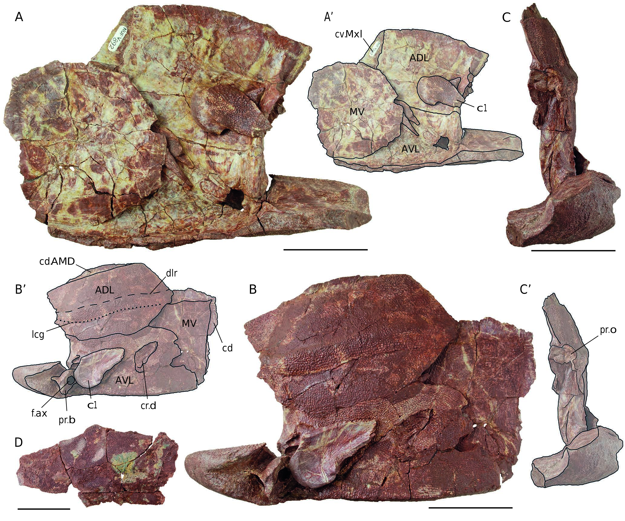
The anterior dorsolateral plate of Asterolepis alticristata n. sp. expectedly overlaps the mixilateral plate ( Fig. 5 View FIG A’[cv. Mxl]) and is overlapped by the anterior median dorsal plate ( Fig. 5 View FIG B’[cd.AMD]). The dorsolateral ridge ( Fig. 5 View FIG B’[dlr]) separates the dorsal lamina (maximum transverse width of 47.60 mm in NUFV 892 [left side]) from the lateral lamina (maximum transverse height of 38.69 mm in NUFV 892 [left side]). The angle between dorsal and lateral laminae is impossible to measure in NUFV 892 due to preservational flattening of the specimen that has deformed those laminae into nearly the same plane. The thickest part of the plate is in the position of the processus obstans ( Fig. 5 View FIG C’[pr.o]) which consists of a short, medial, cranially-pointing process and a cranially-facing facet that likely accommodated the postmarginal plate of the cephalic skeleton. The ornament is densely tuberculate with those tubercles close to the dorsolateral ridge, on both dorsal and lateral laminae, appearing in rows parallel to that linear feature. Along the ventral margin of the lateral lamina, tubercles are conjoined into ridges oriented perpendicular to the contact between anterior median dorsal and anterior ventrolateral plates. The main lateral line groove ( Fig. 5 View FIG C’[lcg]) is on the lateral lamina of the anterior dorsolateral plate and follows a path that is roughly parallel with the dorsolateral ridge ( Fig. 5 View FIG C’[dlr]).
Anterior ventrolateral plate ( Fig. 5 View FIG )
The anterior ventrolateral plate forms a broad thick platform for the submarginal plates of the Asterolepis cephalic skeleton. Along with the anterior dorsolateral plate, the cranial end of the anterior ventrolateral plate’s lateral lamina forms a wide, crested backwall for the cephalic skeleton. One specimen (NUFV 892; Fig. 5 View FIG ) preserves the lateral lamina, pectoral articulation, and only a short section of the ventral lamina of both anterior ventrolateral plates of Asterolepis alticristata n. sp. As in adult Asterolepis ornata, the lateral lamina of the anterior ventrolateral plate is tall across its entire length and reaches its maximum height close to the caudal end. The anterior ventrolateral plate is remarkably thick at the pectoral articulation and, at 38.87 mm in NUFV 892, presents the position of greatest skeletal thickness known for the species. The processus brachialis ( Fig. 5 View FIG B’[pr.b]) resides in a fossa that is deeply excavated from the ornamented surface. This fossa narrows to a point cranially. The processus brachialis is penetrated by an oval-shaped (long axis dorsal-ventral) foramen axillaris ( Fig. 5 View FIG B’[f.ax]). The most cranial margin of the plate’s ventral platform exhibits a tall concave, cranial-facing facet that likely accommodated the semilunar plate. As in A. ornata, this facet terminates laterally in a cranial-pointing process (the antero-mesial process of Upeniece 2011); further lateral is the most cranial point of the plate (the anterior lateral corner of Upeniece 2011).
The ornament of the anterior ventrolateral plate’s lateral lamina is densely tuberculate; the tubercles are arranged in rows that radiate from a position just caudal to the pectoral articulation. The preserved portion of the ventral lamina, just ventral to the pectoral articulation, is unornamented as is a depressed zone just dorsal to the fossa that surrounds the processus brachialis.
Note. On the lateral lamina of the left anterior ventrolateral plate of NUFV 892, there is a crescent-shaped depression ( Fig. 5 View FIG B’[cr.d]) that interrupts the surrounding ornament pattern. The ornament within the depression consists of larger, isolated and fused tubercles that are more widely spaced and chaotically-organized than those of the surrounding lateral lamina. This depression may represent bone repair of puncturetype damage or an instance of skeletal pathology.
Median ventral plate ( Fig. 5A, B View FIG )
The one median ventral plate (NUFV 892; Fig. 5A, B View FIG ) is best preserved in visceral view. The outline of the median ventral plate is generally circular although it appears to pinch to form blunt points in at least three corners. What is exposed of the external surface ( Fig. 5B View FIG ) shows a densely tuberculate ornament with evidence for rows of fused tubercles in some areas. The median ventral plate is overlapped by anterior ventral lateral and posterior ventral lateral plates. Two such overlap surfaces are continuous with one another and visible on the dermal surface of the median ventral plate of NUFV 892. It is impossible to determine which plates overlap these visible surfaces because the plate is out of articulation in NUFV 892.
Mixilateral plate ( Fig. 5D View FIG )
Only one specimen (NUFV 895; Fig. 5D View FIG ) is complete enough to both recognize it as a mixilateral plate and assign it to Asterolepis alticristata n. sp. NUFV 895 is incomplete and lacks most of the zones of overlap that accommodate the anterior median dorsal and posterior median dorsal plates. But for a short segment, the specimen also lacks the plate’s lateral lamina. The ornament is densely tuberculate with the tubercles arranged in rows close to cranial and caudomedial edges. These rows of tubercles meet each of these two ornamented margins at steeply oblique angles. On the visceral surface, oblique ridges extend caudolaterally from the caudal end of the mixilateral’s contact with the anterior median dorsal plate. These continue the path of the ridges on the visceral surface of the anterior median dorsal plate that are described above.
Posterior ventrolateral plate
Several fragments (NUFV 802.1, 802.2, 821, 848) are identifiable as the caudoventral corner of the posterior ventrolateral plates and are here attributed to Asterolepis alticristata n. sp. They are not complete enough to support an anatomical description but for the ornament pattern of rows of isolated or conjoined tubercles that is consistent with much of the rest of the known skeleton.
Pectoral appendage ( Fig. 5A, B View FIG )
The identifiable plates of the Asterolepis alticristata n. sp. pectoral appendage include dorsal and ventral central 1 plates ( Fig. 5A View FIG [c1], B[c1]) and a lateral marginal 2 plate, but the material also includes additional unidentified central and marginal plates. The dorsal and ventral central 1 and lateral marginal 2 plates appear as they do in the type species. On the central plates, the ornament is tuberculate with larger, more widely spaced tubercles at the proximal end. On the marginal plate, the ornament is densely tuberculate with no obvious pattern to their arrangement.
No known copyright restrictions apply. See Agosti, D., Egloff, W., 2009. Taxonomic information exchange and copyright: the Plazi approach. BMC Research Notes 2009, 2:53 for further explanation.
|
Kingdom |
|
|
Phylum |
|
|
Class |
|
|
Order |
|
|
Family |
|
|
Genus |
Asterolepis alticristata
| Downs, Jason P., Daeschler, Edward B., Lo, Nathanael, Carey, Emily N. & Shubin, Neil H. 2019 |
Asterolepis
| DAESCHLER E. B. & SHUBIN N. H. & JENKINS JR F. A. 2006: 759 |