
Kentisuchus astrei
|
publication ID |
https://doi.org/ 10.1111/zoj.12357 |
|
publication LSID |
lsid:zoobank.org:pub:724315E1-6D20-450A-AD40-76E4C5F8D767 |
|
DOI |
https://doi.org/10.5281/zenodo.5670337 |
|
persistent identifier |
https://treatment.plazi.org/id/59667E71-FFC0-7C7C-4AD5-D83B9518D52A |
|
treatment provided by |
Plazi |
|
scientific name |
Kentisuchus astrei |
| status |
sp. nov. |
KENTISUCHUS ASTREI SP. NOV.
Etymology
Specifically named after Gaston Astre (1896–1975), a French geologist and palaeontologist who first described the holotype of the species.
Holotype
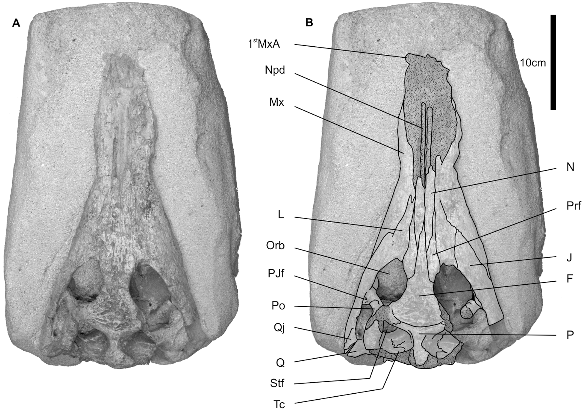
MHNT.PAL.2010.0.49, skull lacking premaxillae, and exposed in dorsal view ( Figs 3 View Figure 3 , 4 View Figure 4 ).
Nomenclatural act
This published work and the nomenclatural acts it contains have been registered in ZooBank. The Life Sciences Identifier (LSID) for this publication is: [urn:lsid:zoobank.org:pub:724315E1-6D20-450A-AD40- 76E4C5F8D767].
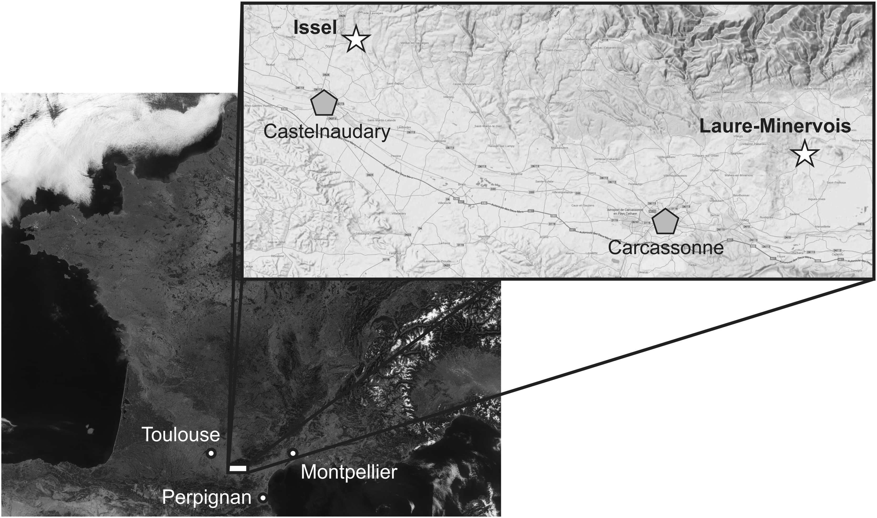
Type locality
Issel, Aude, France ( Fig. 1 View Figure 1 ).
Age View in CoL and stratigraphic horizon
Rough sandstone, ‘ grès d’Issel ’ formation (Issel sandstone formation), latest Lutetian (MP13–14).
Diagnosis
Differs from the only known species from the same genus, K. spenceri , in having a more robust snout; the constriction of the snout at the level of the seventh– eight teeth is more significant, being only 80% of the largest maxillary width; its snout does not bear an anteroposterior shallow fossa at the level of the lacrimomaxillary suture.
Description
State of preservation
The holotype consists of a damaged partial skull ( Figs 3 View Figure 3 A, 4A), with its ventral surface embedded in the matrix. The premaxillae are not preserved, and the dorsal surface of the anterior portion of the snout is eroded. The posterior area of the skull is strongly damaged, the posterior margin of the supratemporal fenestrae being strongly abraded, and the lateral side of the right fenestra is not preserved.
General shape
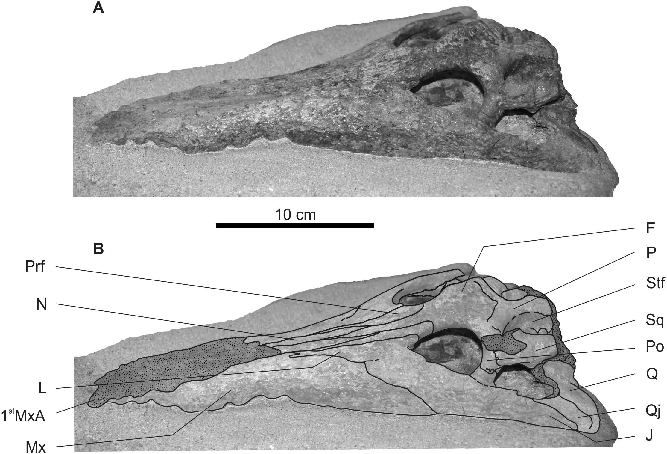
The skull is elongated in shape, with a relatively welldeveloped snout. The snout proportion is estimated to have probably been around 70% of the skull length (from the tip of the snout to the posteromedial margin of the parietal; Fig. 3 View Figure 3 ). The snout seems to be nearly rounded in cross section, slightly wider than high. Its lateral margin has a sinusoidal lateral margin in dorsal view, and a concave dorsal margin in lateral view ( Fig. 4 View Figure 4 ).
The alveoli are not visible, but the lateral waves of the maxilla allow us to estimate their number and variation in size. The fifth and sixth alveoli seem to be large and not separated by a notch for the lower tooth, whereas notches are present from the first to the fifth and from the sixth to the ninth alveoli. The seventh and eighth teeth seem to be more widely separated than the other teeth on both sides ( Fig. 4 View Figure 4 ).
Cranial openings
The external nare is not preserved, as premaxillae are missing, but as the dorsal surface of the snout is strongly eroded, the cast of the nasal duct is exposed ( Fig. 3 View Figure 3 ).
The orbits are bordered medially by the frontal and prefrontal, anteriorly by the lacrimal, ventrally by the jugal, and posteriorly by the postorbital ( Figs 3 View Figure 3 , 4 View Figure 4 ). They are separated by a narrow and concave interorbital space, formed largely by the frontal, and by the prefrontal anteromedially.
The supratemporal fenestra is damaged, but seems to have been slightly wider than long, and subcircular. The fenestra is bordered by the postorbital anterolaterally, and by the parietal anteriorly, medially, and posteromedially. The squamosal is poorly preserved and its participation to the lateral and posterior margin of the fenestra cannot be evaluated ( Figs 3 View Figure 3 , 4 View Figure 4 ).
The temporal canal, within the supratemporal fenestra, opens in a large fenestra, lateromedially oval in shape ( Fig. 3 View Figure 3 ). As the posterior margin of the supratemporal fenestra is missing, the dorsal margin of the temporal canal is not preserved. The posterior wall of the supratemporal fenestra does not preserve the sutures. So, the participation and proportion of the bones in the temporal canal margin cannot be evaluated.
The left infratemporal fenestra is partially preserved ( Fig. 4 View Figure 4 ). It is short and smaller than the orbit. It is anteriorly limited by the postorbital bar, which is formed by the postorbital and the jugal. The jugal forms most of the ventral margin of the bar. The dorsal margin of the fenestra is strongly damaged, therefore the participation of the quadrate and quadratojugal cannot be evaluated. The quadratojugal seems to contribute at least slightly to its ventral margin ( Fig. 4 View Figure 4 ).
Maxilla
The maxilla is large, moderately sculptured, with deep pits and anteroposterior furrows ( Figs 3 View Figure 3 , 4 View Figure 4 ). Its lateral margin is sinusoidal in dorsal view, with a lateral concavity at the level of the eighth tooth. The ventral side of the maxilla is not available, so the alveoli cannot be seen. The shape and size of the alveoli can be evaluated in lateral view according to the waves of the lateral margin of the maxilla ( Fig. 4 View Figure 4 ). Each maxilla may bear 14–16 teeth anterior to the orbits, and as the ventral surface of the maxillae is embedded in the matrix, the exact number of teeth posterior to the anterior margin of the orbit cannot be evaluated. The sizes of the teeth vary along the row, with the fifth and sixth teeth appearing to be the largest.
Nasal
The nasals are formed by paired and unfused elements. The anterior portions are strongly worn ( Figs 3 View Figure 3 , 4 View Figure 4 ). Posteriorly, it ends anterior to the level of the orbits. It is ornamented with anteroposteriorly-directed ridges and furrows.
Prefrontal
The prefrontal is narrow, elongate in shape, and forms less than half of the anterior medial margin of the orbit ( Figs 3 View Figure 3 , 4 View Figure 4 ). It is heavily ornamented with deep pits. Anteriorly, its suture with the lacrimal is anteroposteriorly directed, parallel with the medial plane. It is anteriorly longer than the frontal.
Even if they are damaged, the dorsal half of the prefrontal pillar seems to have been anteroposteriorly expanded.
Lacrimal
The lacrimal is long, relatively narrow, and its contact with the nasal is short ( Figs 3 View Figure 3 , 4 View Figure 4 ). It forms the anterior margin of the orbit. It is strongly ornamented, with deep pits and furrows. There is a short posterior maxillary process within the lacrimal at its anterior edge, just lateral to the maxilla–nasal–lacrimal contact. This process is clear only on the left lacrimal.
Frontal
The frontal forms the posteromedial margin of the orbit ( Figs 3 View Figure 3 , 4 View Figure 4 ). It is thin and long anteriorly, extends far anterior to the jugal, but more posterior than the lacrimal and prefrontal. It penetrates deeply between the nasals. The frontal widens progressively posteriorly from its anteriormost tip to its suture with the postorbital. It is slightly concave and ornamented with deep pits. The single frontal (frontals are fused) and its strong ornamentation suggest that this specimen was mature.
The frontoparietal suture lies entirely on the skull roof, anterior to the supratemporal fenestrae, with a posteriorly convex shape in dorsal view ( Fig. 3 View Figure 3 ).
Postorbital
The postorbitals are very damaged, and their shape and contacts with other bones are largely unavailable. The postorbital forms the posterior margin of the orbit, the anterolateral margin of the supratemporal fenestra, and participates in the dorsal half of the postorbital bar ( Figs 3 View Figure 3 , 4 View Figure 4 ).
The postorbital bar is robust and slightly oval in cross section ( Fig. 4 View Figure 4 ). Its dorsal portion is damaged. Laterally, at the level of the suture with the jugal, it bears a weak rugosity. Only the posterior portion of the postorbital–jugal suture is visible. In the lateral surface of the postorbital bar, it is parallel with the dorsal margin of the jugal, and runs ventrally in the posterior margin of the bar.
Jugal
The jugal broadly extends anteriorly, but remains posterior to the anterior extent of the frontal and prefrontal ( Figs 3 View Figure 3 , 4 View Figure 4 ). Similar to the lacrimal and the prefrontal, it is moderately ornamented with ridges and furrows anteriorly, and more heavily sculptured with deep pits posteriorly. The posterior process of the jugal ends well anterior to the jaw joint, at the level as the posterior margin of the parietal, with the quadratojugal being largely visible in lateral view ( Figs 3 View Figure 3 , 4 View Figure 4 ).
The jugal forms more than half of the lateral height of the postorbital bar. It forms an elevation lateral to the orbit separated from the postobital bar by a shallow gutter. The posterior jugal foramen is very small, posteroventral to the postorbital bar ( Fig. 3 View Figure 3 ).
Quadratojugal
Only the posteroventral portion of the left quadratojugal is preserved ( Figs 3 View Figure 3 , 4 View Figure 4 ). It seems to form the posteroventral margin of the infratemporal fenestra. As a result of its poor preservation, the presence of a quadratojugal spine is unknown.
Parietal
The parietal forms the narrow, and strongly ornamented with deep pits, interfenestral bar ( Fig. 3 View Figure 3 ). Its contact with the postorbital is partially on the skull table, and is continuous with the frontal–postorbital suture. The posterior margin of the parietal is slightly convex posteriorly at the level of the supraoccipital. Its sutures with other bones within the supratemporal fenestra cannot be evaluated.
Squamosal
Part of the left squamosal is preserved. Anteroventrally, it passes beneath the postorbital to form the posterodorsal part of the postorbital bar laterally ( Fig. 4 View Figure 4 ). Its posterior and dorsal margins are too damaged to provide relevant information.
Quadrate
Part of the left quadrate is present but poorly preserved ( Figs 3 View Figure 3 , 4 View Figure 4 ). Overall, it follows the basic eusuchian morphology, but details of the morphology cannot be trustfully interpreted.
Supraoccipital
The supraoccipital is visible in occipital view, but its sutures with the exoccipital and parietal are not visible. Its dorsal portion is damaged, such that its participation in the skull table cannot be evaluated. It seems to bear a small dorsoventral crest.
Exoccipital
The exoccipitals are large and form most of the occipital surface, but their lateral margins are lacking. They surround the foramen magnum that seems to be slightly narrower than the occipital condyle.
Basioccipital
The basioccipital forms the relatively modest occipital condyle.
Laterosphenoid
The capitate process of the laterosphenoid is orient- ed anteroposterolaterally. Its posteroventral portion is too damaged to assess the relathionships with the parietal, basisphenoid, and prootic.
Basisphenoid
The basisphenoid rostrum is poorly preserved. It is lateromedially flattened, and its anterior margin is rounded.
Ectopterygoid
The dorsal portion of the left ectopterygoid is preserved. It seems to participate in the ventromedial portion of the postorbital bar, but the suture with the postorbital is not visible.
COMPARISON AND TAXONOMIC
CONSIDERATIONS
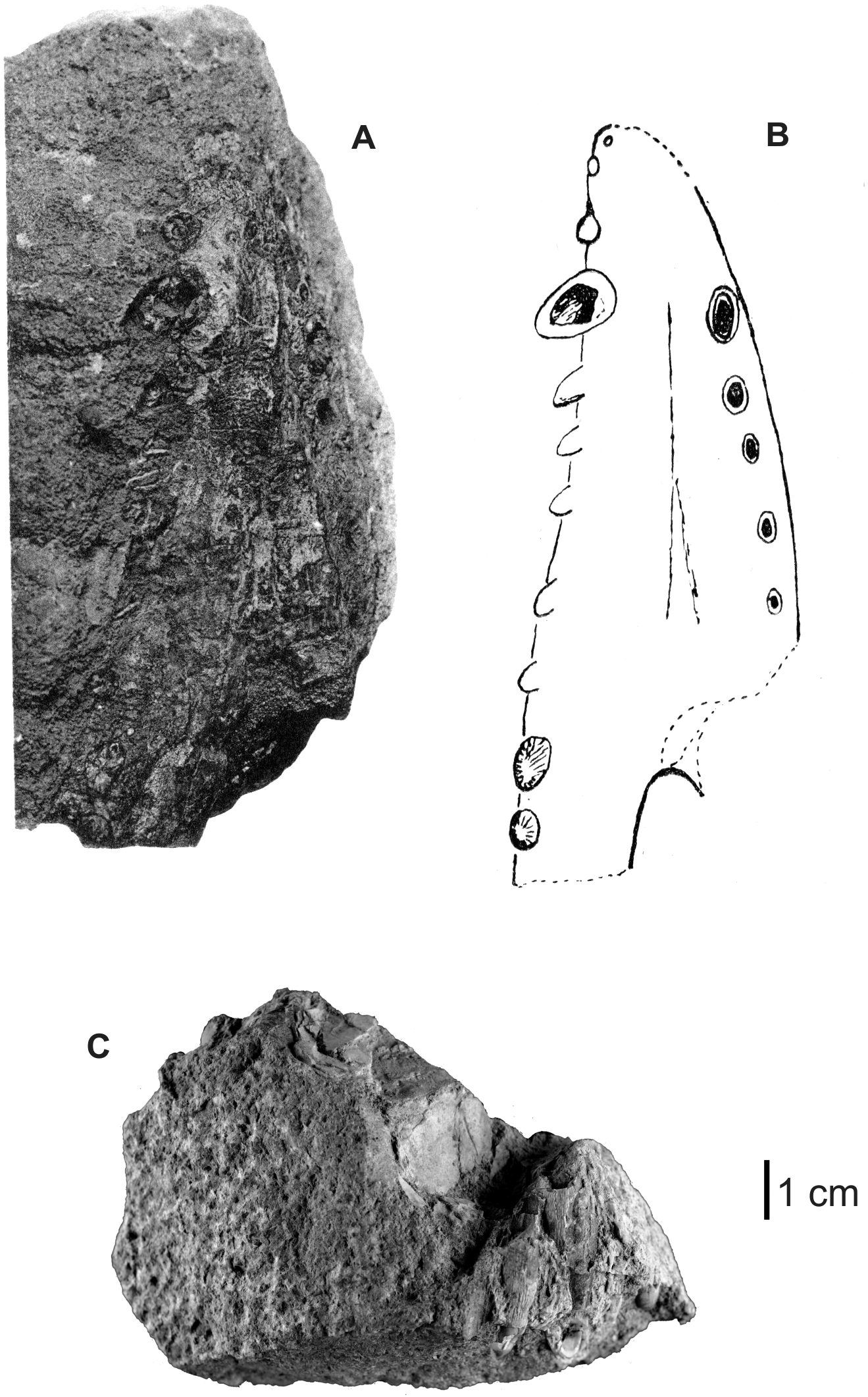
The holotype, noted as preserved in the collection of the ‘Société d’Etudes Scientifiques de l’Aude’ ( Fig. 2 View Figure 2 A, B), is now lost, but the figure and description allow us to exclude that it could be a planocraniid (Brochu, 2013). The length of the symphysis, reaching the level of the tenth tooth posteriorly, and the splenial portion of the symphysis extending forwards to the seventh dentary alveolus (Astre, 1931) suggest a longirostrine crocodyliform, whereas the variation in teeth size suggests a tomistomine. In the most primitive tomistomine Maroccosuchus zennaroi Jonet & Wouters, 1977, the symphysis reaches the level of the ninth tooth and the splenial reaches the level of the seventh tooth. The symphysis in ‘ Atacisaurus glareae ’ is nearly as long as in Dollosuchoides densmorei Brochu, 2007 (reaches the level of the ninth or tenth tooth), in which the splenial stops at the level of the eighth tooth. In a specimen from the Ypresian of La Borie (Southern France), and referred to K. spenceri (Laurent et al., 2010) , the symphysis and the splenial lengths are the same as in ‘ A. glareae ’, reaching, respectively, the tenth and the seventh teeth (S. Jouve, pers. observ.). Unfortunately, the length of the symphysis and participation of the splenial can vary for the same species in crocodyliforms, particularly in longirostrine forms, with a variation of up to three teeth (Vignaud, 1995; Jouve, 2004, 2007). So these characters cannot be considered as diagnostic, and no other characters are diagnosable in the mandibular fragment.
Ortega et al. (1996) suggested that the skull MHNT.PAL.2010.0.49 could be designated as the species lectotype, if a preparation reveals new information to compare it with the diagnosis of the holotype. Unfortunately, even if a new preparation has been conduct- ed, no character allows us to relate the mandible from Laure-Minervois to the skull MHNT.PAL.2010.0.49 from Issel. As a consequence, ‘ Atacisaurus glareae ’ cannot be considered as a valid species, and is a nomen dubium.
Nevertheless, if the skull cannot be confidently attributed to ‘ A. glareae ’, its close examination and comparison allow us to review its taxonomic affinities ( Figs 3 View Figure 3 , 4 View Figure 4 ). The shape of the snout strongly suggests that this skull belongs to a longirostrine taxa, whereas the maxilla sending a posterior process in the lacrimal clearly confirms the hypothesis that this skull belongs to a tomistomine. Compared with other primitive tomistomines, MHNT.PAL.2010.0.49 differs from M. zennaroi, inter alia, in having a more slender snout and narrower lacrimal. Its lacrimal is three times wider than long (four in M. zennaroi), and slightly wider than the prefrontal at the level of the anterior margin of the orbits (more than twice the width in M. zennaroi). The snout is more robust in the Issel skull, and the constriction at the level of the seventh–eighth maxillary teeth is stronger than in Dollosuchoides densmorei Brochu, 2007 , and all more derived tomistomines. The frontal does not reach the anterior level of the prefrontal, whereas it exceeds it strongly in Megadontosuchus arduini (de Zigno, 1880).
MHNT.PAL.2010.0.49 has a long prefrontal and a short lacrimonasal contact, whereas the lacrimonasal contact is much longer in all other tomistomines, except in K. spenceri . It also bears several common characters with this species. The maxillary process in the lacrimal is located at the anteriormost extent of the lacrimal, and the prefrontal is nearly as long as the lacrimal, two diagnostic characters of the until now monospecific genus Kentisuchus (Brochu, 2007) . Unfortunately, the other K. spenceri character, the M-shape of the skull table in occipital view, cannot be observed in MHNT.PAL.2010.0.49 because of the poor preservation of the occipital portion, and, in particular, the poor preservation of the squamosals. But as in K. spenceri , the frontal exceeds the anterior level of the jugal, whereas in other tomistomines it is shorter, or reaches the same level as the frontal.
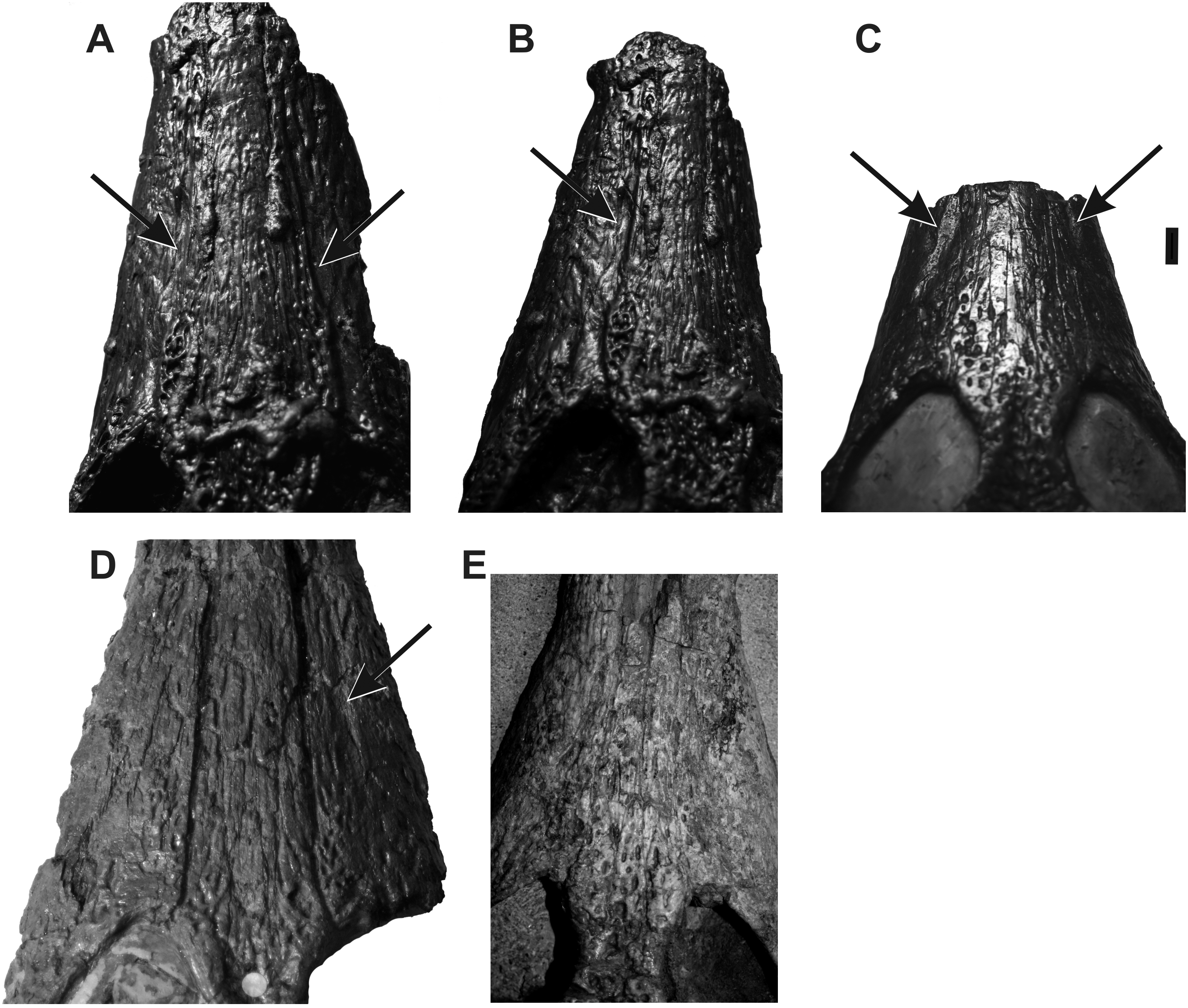
So, MHNT.PAL.2010.0.49 can be considered as a Kentisuchus . With the low number of anatomical characters available on the specimen, the comparison with K. spenceri is limited. The Issel specimen is nearly the same size as the specimen NHM 38974, with a skull length of 33 cm from the first maxillary tooth to the posterior margin of the parietal, 6% larger than the London Clay specimen (31 cm), but the snout at the level of the fifth–sixth teeth is 30% wider in MHNT.PAL.2010.0.49. The lateral margin of the maxilla is more sinusoidal in the Issel specimen, such as the width of the snout at the level of its posterior constriction (seventh–eighth teeth) being 90–93% of the snout width at the level of the fifth–sixth teeth, whereas it is less than 80% in MHNT.PAL.2010.0.49 ( Table 1 View Table 1 ). The relative similarity between the two values observed in the two specimens of K. spenceri , strongly different in size, NHM R1753 being 45% larger than NHM 38974, suggests that this character can be used with confidence. A lateral anteroposterior shallow fossa is also present in K. spenceri , lateroventral to the lacrimomaxillary contact, clear in NHM 19633 and NHM 37717, only discernable on the right side in NHM 38974, and not at all discernable in NHM R1753 because of weathering ( Fig. 5 View Figure 5 ). This shallow fossa is absent in MHNT.PAL.2010.0.49 ( Fig. 5 View Figure 5 E). As the ornamentation is present in this area, the absence of the shallow fossae cannot be related to weathering. So, the Issel specimen differs from K. spenceri by a more sinusoidal lateral margin of the snout and by the absence of a lateral fossa along its lacrimomaxillary suture. It is thus referred to a new species, Kentisuchus astrei sp. nov.
No known copyright restrictions apply. See Agosti, D., Egloff, W., 2009. Taxonomic information exchange and copyright: the Plazi approach. BMC Research Notes 2009, 2:53 for further explanation.