
Phyllodesmium tuberculatum, Moore, Elizabeth & Gosliner, Terrence, 2009
|
publication ID |
https://doi.org/ 10.5281/zenodo.189627 |
|
DOI |
https://doi.org/10.5281/zenodo.6216514 |
|
persistent identifier |
https://treatment.plazi.org/id/5C0D674A-6357-A814-FF77-F3E2366DFE77 |
|
treatment provided by |
Plazi |
|
scientific name |
Phyllodesmium tuberculatum |
| status |
sp. nov. |
Phyllodesmium tuberculatum n. sp.
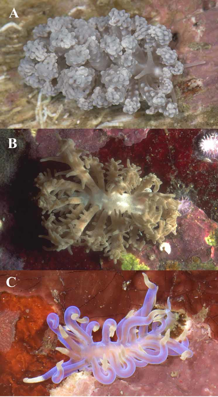
( Figures 1 View FIGURE 1 A, 2A–C, 3A, 4, 5A,B, 6A)
Synonymy: Phyllodesmium sp. 2 Gosliner Behrens & Valdés, 2008: 385.
Material examined: Holotype: CASIZ 177520, not dissected, molecular sample taken from foot, 0–17m depth, Twin Rocks, Mabini, Batangas Province, Luzon Island, Philippines, 21 March 2008, T.M. Gosliner et al. Paratype: CASIZ 106547, dissected, 0–16m depth, Twin Rocks, Mabini, Batangas Province, Luzon Island, Philippines, 19 April 1996, T.M. Gosliner. Paratype: CASIZ 177663, dissected, buccal mass not found, reproductive system poorly preserved, molecular sample taken from foot, 0–16m depth, Sepok, Batangas Province, Luzon Island, Philippines, 19 April 2008, T.M. Gosliner.
Geographic range. This species is known only from southern Luzon Island, Philippines (this study). Etymology. The species is named after the rounded tubercles present on the cerata of these animals.
Natural history. This species is found on coral and rubble near its prey species, a soft coral in the genus Anthelia .
Description. Color and external morphology. Living animals ( Fig. 1 View FIGURE 1 A) are elongate, with cerata obscuring the majority of the dorsum and mantle. The preserved specimens examined were 14mm (CASIZ 177520) and 13mm (CASIZ 106547) in length. Specimen CASIZ 177663 was poorly preserved and not measured. The anterior portion of the foot margin is broad with moderately tentacular foot corners while the posterior end tapers to an elongate point. The body of the living animal, including rhinophores, oral tentacles, cerata and foot are predominantly a bluish gray color. Large, thick, and somewhat dorso-ventrally flattened cerata cover the dorsum with smaller cerata at the lateral margins of the mantle. Large cerata are covered nonuniformly by round, nodulose tubercles that are also bluish gray in color. The tubercles tend to aggregate on, but are not exclusive to, the narrow margins and ends of the cerata, which terminate bluntly with a hardly visible tip. When the animal is preserved, the distal-most point on each ceras is marked by a small but distinctive cnidosac that lacks nematocysts ( Fig. 2 View FIGURE 2. A C). The smallest cerata, near the posterior end of the body and along the mantle margin, have no tubercles and appear cylindrical and smooth with a slightly pointed tip. The digestive gland visibly branches throughout the cerata, with visible secondary, and possibly tertiary, branches that extend to the surface of each ceras ( Fig. 2A View FIGURE 2. A ). Zooxanthellae are present in great numbers in the digestive tissue of the largest cerata (near the dorsum) ( Fig. 2 View FIGURE 2. A B), but not in the small, laterally-located cerata. Zooxanthellae also appear to be located outside the digestive tissue near the surface of each ceras. The ceratal arrangement consists of arches and rows, with arches consisting of two rows of cerata forming in the anterior ceratal groups and rows in the most posterior groups ( Fig. 3 View FIGURE 3 A). The precardiac cerata are grouped into one arch on each side of the body containing approximately 15 cerata. On the arms of the precardiac arches there are double rows of cerata, but on the dorsal curve of each arch there is a single row. The genital aperture is located between the arms of the precardiac arch on the right side of the animal, and there are separate openings for the male and female genital systems. The renal opening is situated slightly toward the posterior between the precardiac arch and the first postcardiac arch on the right side. The postcardiac cerata are grouped on both sides into arches containing approximately 13 cerata for the first two arches. These first two postcardiac arches also contain double rows on the ventral arms, but a single row of cerata on the dorsal curve of each arch. The anal papilla is located within the first postcardiac arch on the right side. The third postcardiac arch contains approximately 8 cerata and appears to be a single-rowed arch. The last three ceratal groups are curved rows containing 4, 3, and 3 cerata respectively. The rhinophores are smooth, with a somewhat wrinkled appearance and are conical in shape with blunt tips. The oral tentacles are also smooth and taper from the anterior edge of the head to bluntly pointed apices. They are approximately the same length as the rhinophores.
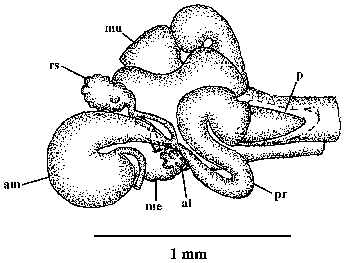
Reproductive System ( Fig. 4 View FIGURE 4 ). Large quantities of ovoid gonad fill the posterior portion of the specimen. The female gland mass is large and consists predominantly of the mucous gland with smaller albumen and membrane glands. The ampulla is large and curved, and it branches to the oviduct and the prostatic portion of the vas deferens. The oviduct connects to a nodulose receptaculum seminis. A second duct extends from near the base of the receptaculum seminis and joins the female glands near the albumen gland. The second branch of the ampulla connects to the vas deferens. The proximal portion is prostatic with the prostate being convoluted and prominent. The prostate increases in size until joining a large, conical-shaped, unarmed penial bulb.
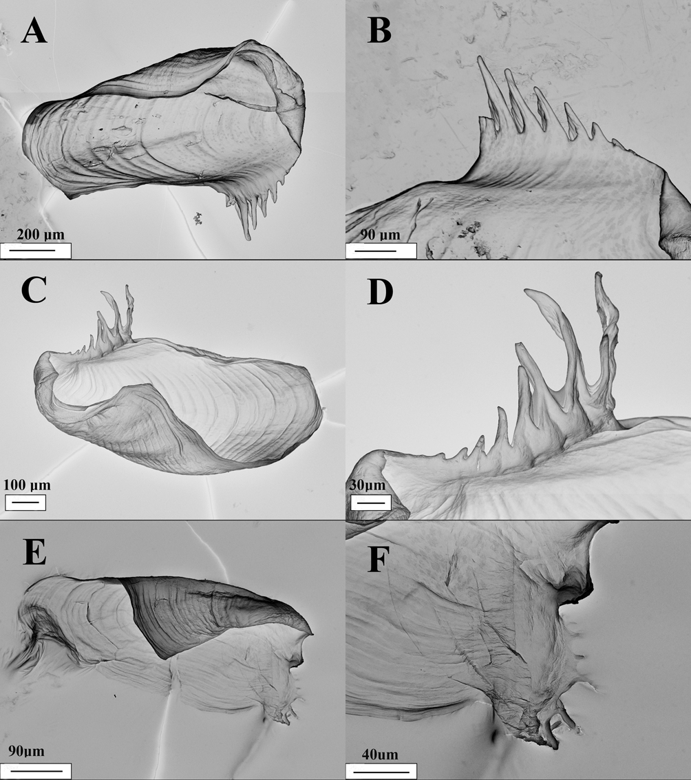
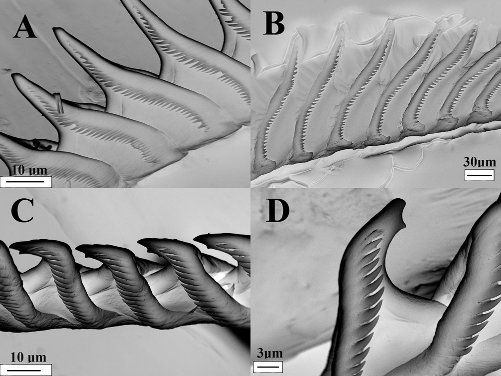
Buccal Armature. The jaws are thin and coriaceous, slightly elongating posteriorly. There are six to eight slender denticles situated along the masticatory border of each jaw. Each denticle has a ribbed projection extending laterally, making them appear wing-like ( Fig. 5A View FIGURE 5. A. and B , B). The radula has a formula of 25 x 0.1.0. The teeth are triangular in shape leading to a long and pointed primary cusp ( Fig. 6A View FIGURE 6. A ). The rib on the ventral side of each tooth extends from the posterior of the tooth to the middle of the cusp. Denticulation extends along the margin from the base of the tooth nearly to the apex. The number of denticles varies between 28–30 per radular tooth. The denticles are slightly elongated with well-separated points that reach slightly under the edge of the tooth. Denticles at the base of the tooth are generally more defined, where denticles near the apex of the cusp become less distinct.
Remarks. Of the described species of Phyllodesmium there are five species, P. crypticum Rudman, 1981 , P. hyalinum , P. lizardensis , P. lembehensis , and P. koehleri with obvious nodules on the external portions of the cerata. Phyllodesmium tuberculatum can generally be distinguished from all of these species by the notably spherical and highly raised nodules that appear upon all parts of the largest cerata. In comparison, P. koehleri has spine-shaped tubercles, while P. crypticum and P. hyalinum have low, often barely raised, conical tubercles. Phyllodesmium lembehensis has tubercles that congregate predominantly on the margins of the flattened cerata, appearing almost like rounded serrations. Lastly, P. lizardensis can be distinguished from P. tuberculatum by the non-uniform shape of blotchy ceratal tubercles, which tend to form only on the distal third of each ceras. Of these five species, P. crypticum and P. hyalinum are perhaps most likely to be mistaken for P. tuberculatum . In addition to ceratal tubercles, P. crypticum is described as a golden brown color rather than blue-gray. Rudman (1981) described P. crypticum as a large animal, up to 60mm long, which is notably different from the smaller size of P. tuberculatum . In addition, the jaws of P. tuberculatum have 6–8 slender, evenly graded denticles. Rudman (1981) did not enumerate the denticles of P. crypticum , but according to the drawings, and to SEM images of a specimen from the CAS collection, they are coarse and irregular, with only 4 prominent denticles. The radular formulae are also noticeably different. Phyllodesmium tuberculatum has 25 teeth on the radula while P. crypticum has 36, and P. tuberculatum has 38–40 denticles per cusp, whereas P. crypticum has up to 30 ( Rudman 1981). Phyllodesmium crypticum has a smooth receptaculum seminis and convoluted ampulla compared to the nodulose receptaculum seminis and curved ampulla in P. tuberculatum . Phyllodesmium tuberculatum differs noticeably from P. hyalinum in color and in the position of the anus. Phyllodesmium tuberculatum is darker in color than P. hyalinum , and has a more ventrally located anus compared to the unique, dorsally located anus in P. hyalinum . The buccal armature of these species also differ, as P. tuberculatum has denticulated cusps with 28–30 denticles per tooth, while P. hyalinum can have up to 56 denticles per tooth.


No known copyright restrictions apply. See Agosti, D., Egloff, W., 2009. Taxonomic information exchange and copyright: the Plazi approach. BMC Research Notes 2009, 2:53 for further explanation.