
Phyllodesmium karenae, Moore, Elizabeth & Gosliner, Terrence, 2009
|
publication ID |
https://doi.org/ 10.5281/zenodo.189627 |
|
DOI |
https://doi.org/10.5281/zenodo.6216518 |
|
persistent identifier |
https://treatment.plazi.org/id/5C0D674A-635F-A81F-FF77-F3F33411FA01 |
|
treatment provided by |
Plazi |
|
scientific name |
Phyllodesmium karenae |
| status |
sp. nov. |
Phyllodesmium karenae n. sp.
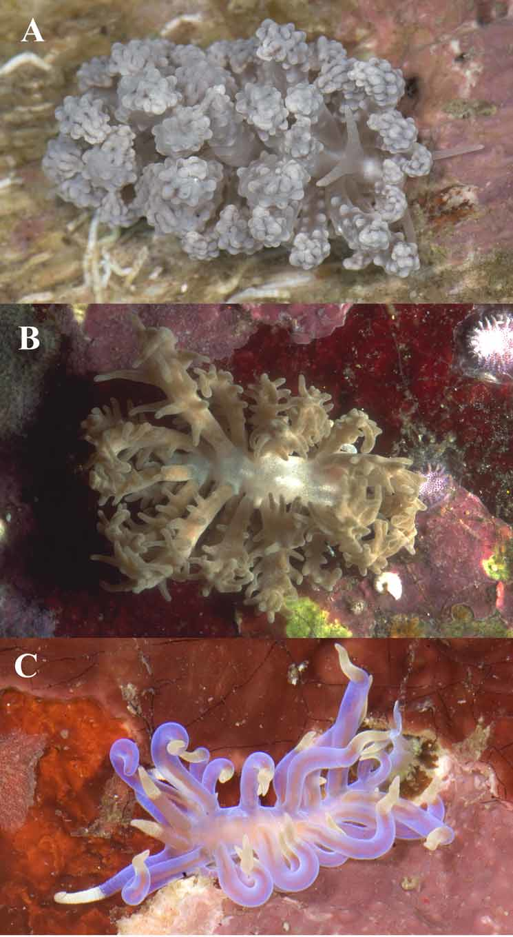
( Figures 1 View FIGURE 1 C, 2F, 3C, 5E, F, 6C,D, 8)
Synonymy. Phyllodesmium sp. 7 Gosliner, Behrens & Valdés, 2008: 390.
Material examined. Holotype: CASIZ 177313, dissected, molecular sample taken from foot, 0–17m depth, Matotonggil Rock, Mabini Batangas Province, Luzon Island, Philippines, 17 March 2008, T.M. Gosliner et al.
Geographic range. This species is known only from southern Luzon Island, Philippines (this study) and Indonesia ( Gosliner et al. 2008).
Etymology. This species is named after the senior author’s mother for her years of endless support and enthusiasm regarding the study of marine organisms.
Natural history. Found among sand and rocks. Prey species is unknown.
Description. Color and external morphology. Living animals ( Fig. 1 View FIGURE 1 C) are elongate, with edges of the foot extending laterally just beyond the mantle. The preserved specimen is 8mm in length. The anterior portion of the foot margin is broad with moderately tentacular foot corners while the posterior end is tapered to a point. The body of the living animal, including oral tentacles and foot, is predominantly transparent with a blue hue. There are no opaque markings on the dorsum and the viscera and gonad are readily visible through the mantle tissue. There are no zooxanthellae present in the epidermal tissues. The cerata are elongate, smooth, and cylindrical, with larger cerata near the medial region of the dorsum. In living animals, the apex of each ceras is curled and prehensile in appearance. The digestive gland is straight and undivided ( Fig. 2 View FIGURE 2. A F) within the cerata and is a uniform brownish-purple color. The tip of each ceras has a yellow-cream colored epithelium, which obscures the apex of the digestive duct. Otherwise the cerata are primarily transparent, with slight blue hue, and lack a distinct cnidosac and lack nematocysts. The ceratal arrangement ( Fig. 3 View FIGURE 3 C) consists of arches and rows, with an arch forming only in the precardiac ceratal group. The precardiac cerata form a single arch on each side of the body containing 7 cerata. The genital aperture is located between the arms, and ventral to the precardiac arch on the right side of the animal. The renal opening is situated in the interhepatic space, slightly toward the posterior between the precardiac arch and the first postcardiac row on the right side. The postcardiac cerata are grouped on both sides into rows containing 3 cerata in the first two rows, and 3–4 cerata in the third row. The anal papilla is located just posterior to the first postcardiac row on the right side. The fourth and fifth postcardiac groups also appear as rows and contain 2 and 1 ceras respectively. The rhinophores are smooth, and elongate in shape and are roughly two thirds as long as the oral tentacles. The rhinophores are primarily a brownish-purple color, leading to a yellow-cream, pointed tip. The oral tentacles are smooth, and taper from the anterior edge of the head to pointed apices. They are transparent or slightly bluish basally, leading to yellow-cream tips.
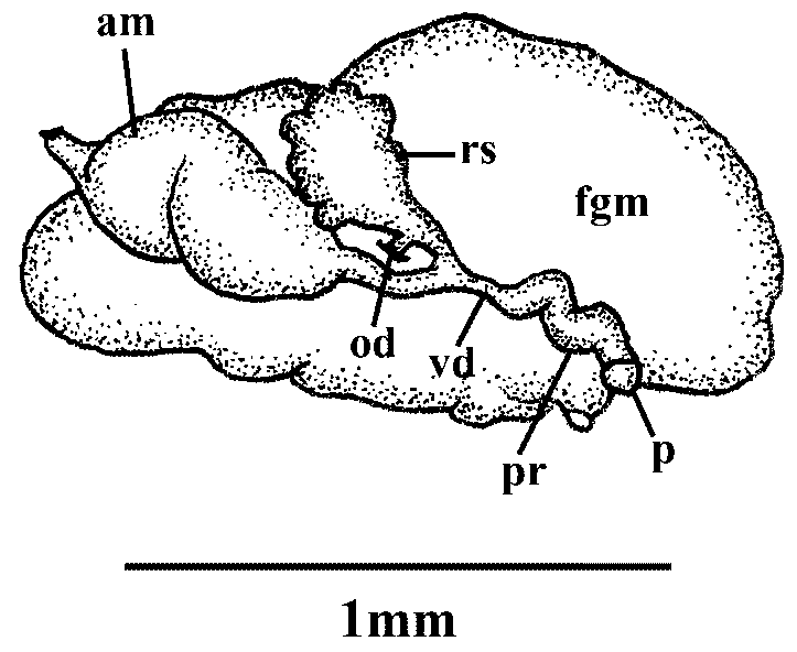
Reproductive System ( Figure 8 View FIGURE 8 ). Large quantities of gonad fill the posterior portion of the specimen. The female gland mass is large and consists predominantly of the mucous gland with smaller albumen and membrane glands. The ampulla is large and bulbous, and it branches to the oviduct and the prostatic portion of the vas deferens. The oviduct connects to a small, nodulose receptaculum seminis. A second duct extends from near the base of the receptaculum seminis and joins the female glands near the albumen gland. The second branch of the ampulla connects to the vas deferens. The proximal portion is prostatic with the prostate being convoluted and small. The convoluted prostate joins a small, conical shaped, and unarmed penial bulb.
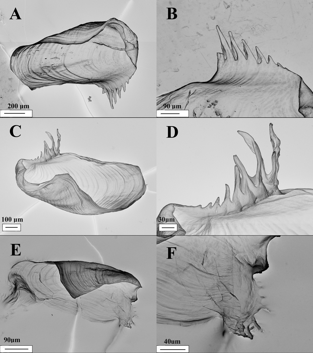
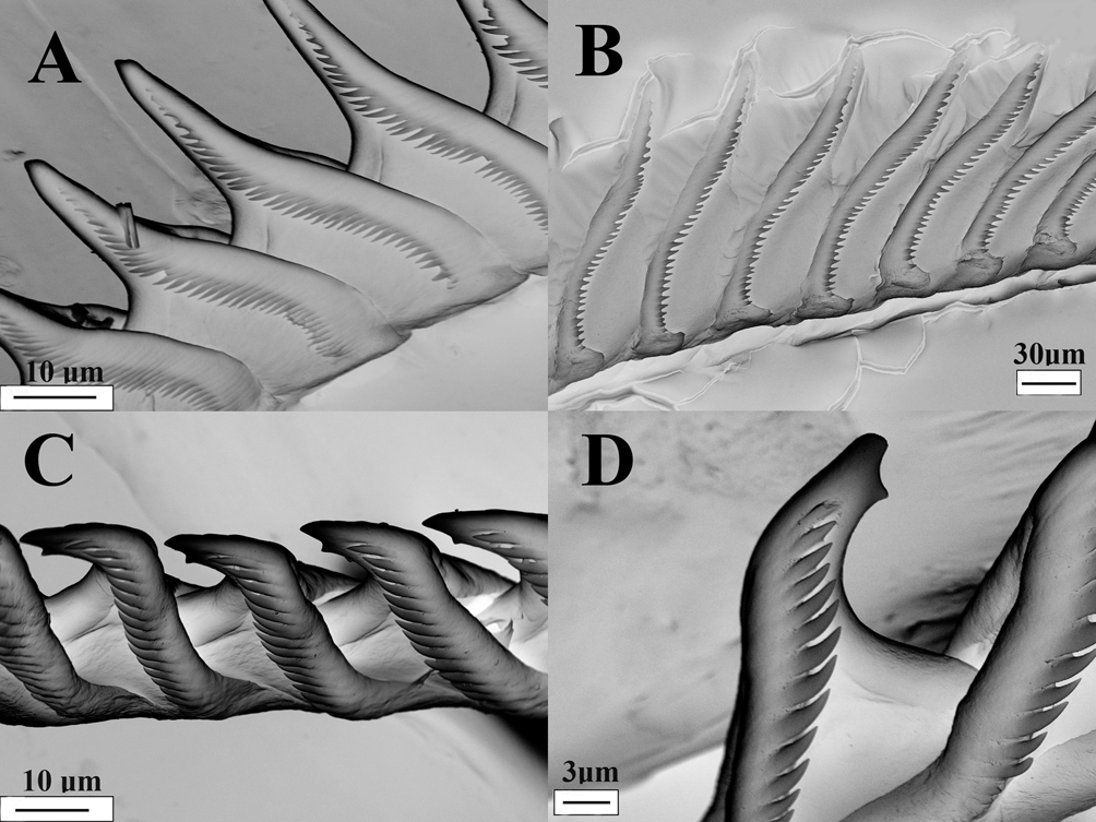
Buccal Armature. The jaws are thin and coriaceous, slightly elongating posteriorly. There are at least seven pointed, and uniformly sized small denticles situated along the masticatory border of each jaw ( Fig. 5 View FIGURE 5. A. and B E, F). The radula has a formula of 21 x 0.1.0. The teeth ( Fig. 6 View FIGURE 6. A C, D) are triangular in shape leading to a long and pointed primary cusp. Near the apex of each cusp, on the ventral side, is a triangle-shaped denticle, which gives the cusp a nearly bifid appearance. The rib on the ventral side of each tooth extends from the posterior of the tooth to the middle of the cusp. Denticulation extends along the margin from the base of the tooth nearly to the apex. The number of denticles varies between 14–16 per radular tooth. The denticles are slightly elongated with well-separated points that reach slightly under the edge of the tooth. Denticles are defined and wellseparated for the entire length of the tooth.
Remarks. Of the previously described species of Phyllodesmium , there are only four, P. horridum ( Macnae, 1954) , P. opalescens Rudman, 1991 , P. sp. 2 Moore & Gosliner, in press and P. sp. 1 Moore & Gosliner, in press, that have undivided digestive tissue within the cerata. Because P. karenae has relatively straight digestive tissue within the cerata, and is predominantly transparent, it is superficially most similar to P. opalescens . However, P. karenae has no opaque white markings on the dorsum, distinguishing it from P. opalescens as well as the other mentioned species. In addition, the radular cusps have a unique denticle located on the ventral part of each pointed tip, which has not been seen in any other species of Phyllodesmium . It is possible that this is variation, or an individual aberration seen within one of the other species, however in all documented accounts of dental structures seen in species of Phyllodesmium , this has never been noted. The small prostate and penis, along with the bulbous ampulla, further distinguish it from the other externally similar species, which have comparatively large prostate glands and penial papillae (Moore & Gosliner in press). The fact that the female glands are large and fully developed indicates the specimen is reproductively mature, although it is possible that the specimen was not fully grown. Comparison with additional material when it becomes available would be highly desirable.


No known copyright restrictions apply. See Agosti, D., Egloff, W., 2009. Taxonomic information exchange and copyright: the Plazi approach. BMC Research Notes 2009, 2:53 for further explanation.
|
Kingdom |
|
|
Phylum |
|
|
Class |
|
|
Order |
|
|
Family |
|
|
Genus |