
Suphis cimicoides Aubé, 1837
|
publication ID |
https://doi.org/10.11646/zootaxa.4619.1.5 |
|
publication LSID |
lsid:zoobank.org:pub:A36CA107-82C5-440C-8ADF-77243E3D38E5 |
|
DOI |
https://doi.org/10.5281/zenodo.4323720 |
|
persistent identifier |
https://treatment.plazi.org/id/5D2187B7-FFA6-FFE3-FF0F-462E425EFF7B |
|
treatment provided by |
Felipe |
|
scientific name |
Suphis cimicoides Aubé, 1837 |
| status |
|
Description of the larvae of Suphis cimicoides Aubé, 1837
Instar I ( Figs 1–16 View FIGURES 1–3 View FIGURES 4–10 View FIGURES 11–12 View FIGURES 13–16 ; Table 1).
Color: Testaceous; antennae and abdominal segments II–VI and VIII slightly darker.
Body: Broad, globose, narrowing toward abdominal apex ( Fig. 1 View FIGURES 1–3 ). Measurements and ratios that characterize the body shape are shown in Table 1.
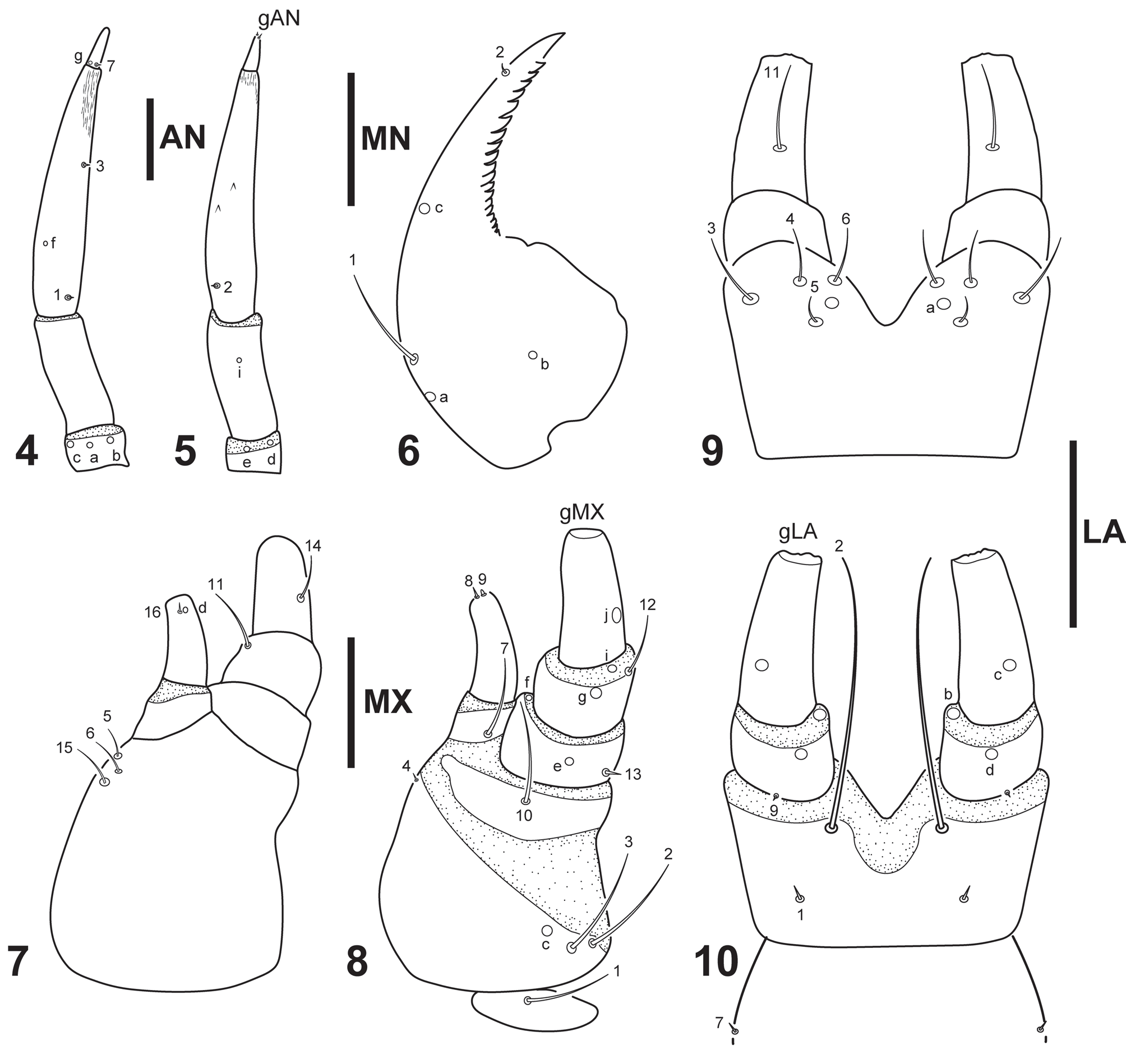
Head: Cephalic capsule ( Figs 2–3 View FIGURES 1–3 ) subprognathous, broader than long; maximum width at level of stemmata, not constricted posteriorly, with large occipital foramen; coronal suture absent, ecdysial sutures arising from occipital foramen; occipital suture absent; posterior tentorial pits visible ventrally, oblique, well separated from each other, contiguous to occipital foramen ( Fig. 3 View FIGURES 1–3 ); six stemmata at each side, four dorsal and two ventral. Frontoclypeus subrectangular, with one spine-like egg burster near each posterolateral angle ( Fig. 2 View FIGURES 1–3 ); anterior margin emarginate medially, lateral lobes well produced beyond central portion; frontal margin with well developed spinulose epipharingeal band. Antenna ( Figs 4–5 View FIGURES 4–10 ) short, robust, shorter than HW, composed of four antennomeres; A3 the longest, much longer than A2, narrowing to apex, with rugged area on distal portion; A1 and A4 the shortest, subequal in length (approximately 10 times shorter than A3). Mandibles ( Fig. 6 View FIGURES 4–10 ) symmetrical; short, basal half robust, distal half curved inwards, relatively slender, narrowing to pointed apex, inner dorsal margin serrate. Maxilla ( Figs 7–8 View FIGURES 4–10 ): cardo small, subovate; stipes well developed, subtrapezoidal, bearing a galea on distal inner margin and a palpus on distal outer margin; galea well developed, second galeomere longer and more slender than first galeomere; palpifer not clearly differentiated from stipes ( Fig. 8 View FIGURES 4–10 ); palpus short, robust, composed of three palpomeres, MP1 shortest, MP3 longest. Labium ( Figs 9–10 View FIGURES 4–10 ): prementum well developed, subrectangular, broader than long, anterior margin deeply indented medially; palpus short, robust, composed of two palpomeres, LP2 longer than LP1.
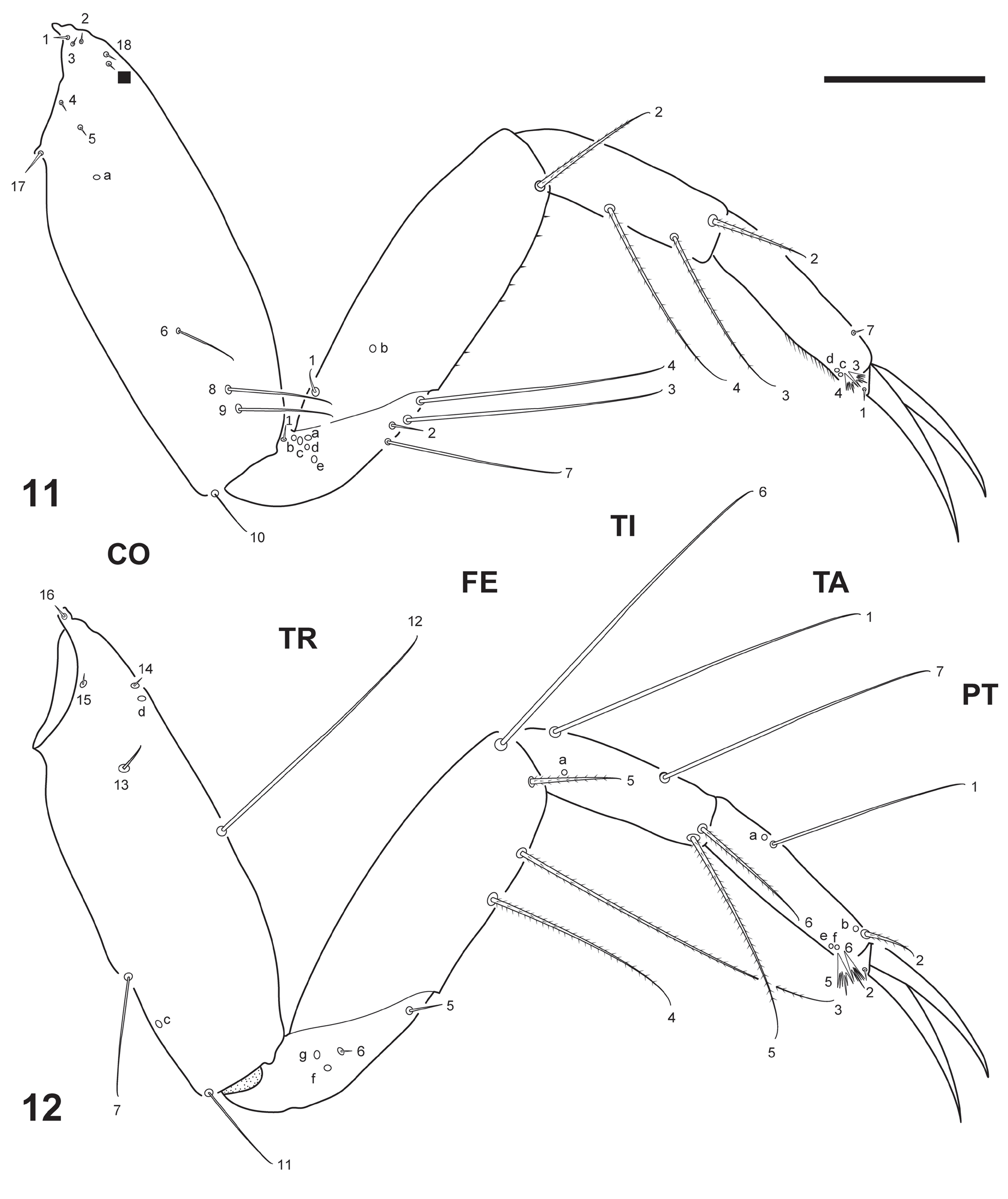
Thorax ( Fig. 1 View FIGURES 1–3 ): Terga fully sclerotized, convex; pronotum about as long as meso- and metanotum combined, meso- and metanotum subequal in length, wider than pronotum; protergite subrectangular, margins rounded, more developed than meso- and metatergite; meso- and metatergite with anterotransverse carina; sterna with a ventral plate between coxae, plates of meso- and metasterna notched anteromedially; meso- and metathorax with minute non functional spiracles. Legs ( Figs 11–12 View FIGURES 11–12 ) short, composed of six articles, L1 shortest, and L3 longest; coxa elongate, trochanter lacking annulus, femur, tibia and tarsus slender, subcylindrical, pretarsus with two long, slender, slightly curved claws, posterior claw shorter than anterior claw on L1 and L2, posterior claw longer than anterior claw on L3.
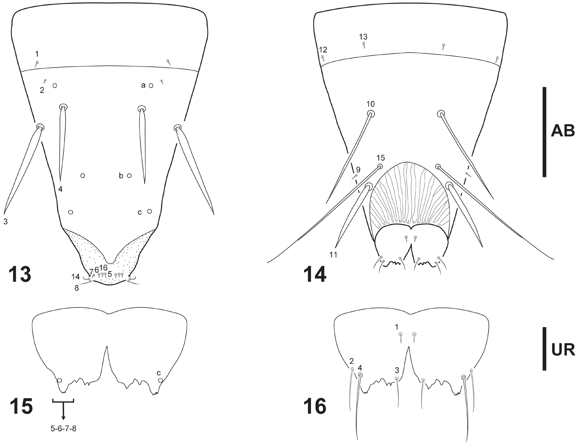
Abdomen ( Fig. 1 View FIGURES 1–3 ): Eight-segmented; segments I–VII completely sclerotized, ring-like (segment I with a narrow membranous sagittal band ventrally), with minute non functional spiracles lateroventrally on anterior half; sclerites I-VII with anterotransverse carina; segment VIII ( Figs 13–14 View FIGURES 13–16 ) the longest and narrowest, completely sclerotized except for a U-shaped wavy membranous area ventrally contiguous to urogomphi; sclerite VIII with anterotransverse carina; siphon very short, not protruding. Urogomphus ( Figs 15–16 View FIGURES 13–16 ) very short, flat, not visible in dorsal view; urogomphi fused to each other proximally along inner margin.
Chaetotaxy: Head: Frontoclypeus ( Fig. 2 View FIGURES 1–3 ): Central portion of anterior margin with three very short spine-like setae (FR9, FR10, FR11), two hair-like setae (FR7, FR8), and one pore (FRf); lateral lobe of anterior margin with one very short spine-like seta (FR6); lateral margin with one hair-like seta (FR1) and one pore (FRa); central portion with three hair-like setae (FR2, FR4, FR5) and one pore (FRc) on distal third and one hair-like seta (FR3) and one pore (FRb) at mid length. Parietal ( Figs 2–3 View FIGURES 1–3 ): Dorsal surface with three hair-like setae (PA8, PA13, PA14) on lateral margin, one hair-like seta (PA10) on anterior portion, three hair-like setae (PA6, PA7, PA9) and three pores (PAc, PAd, PAe) on medial portion, and three short spine-like setae (PA1, PA2, PA3) and three pores (PAa, PAb, PAp) on basal region. Ventral surface with three hair-like setae (PA11, PA12, PA17) and one pore (PAf) on anterolateral angle, three pores (PAh, PAi, PAo) on anterior margin, three hair-like setae (PA16, PA18, PA19) and one pore (PAm) on central portion of anterior half, one hair-like seta (PA15) and two pores (PAj, PAk) at mid length of external margin, and one pore (PAl) on basal region. Antenna ( Figs 4–5 View FIGURES 4–10 ): A1 with three pores (ANa, ANb, ANc) on dorsal surface and two pores (ANd, ANe) on ventral surface; A2 with on minute pore (ANi) on ventromedial region; A3 with two short hair-like setae (AN1, AN3) and one pore (ANf) on dorsal surface and one short hair-like seta (AN2) on ventral surface; A4 with one short hair-like seta (AN7) and one pore (ANg) on dorsobasal portion, and three apical setae (gAN). Mandible ( Fig. 6 View FIGURES 4–10 ): Dorsal surface with one hair-like seta (MN1) and two pores (MNa, MNb) on basal third, one pore (MNc) at mid length, and one short hair-like seta (MN2) near tip. Maxilla ( Figs 7–8 View FIGURES 4–10 ): Cardo with one hair-like seta ( MX 1); stipes with two hair-like setae ( MX 2, MX 3) and one pore (MXc) on ventroexternal margin, and one very short seta ( MX 4), and three hair-like setae ( MX 5, MX 6, MX 15) on internal margin, near base of galea; first galeomere with one hair-like seta ( MX 7) on ventral surface; second galeomere with one very short seta ( MX 16) and one pore (MXd) dorsally near apex and two short spine-like setae ( MX 8, MX 9) at apex; PPF with one hair-like seta ( MX 10) on ventral surface; MP1 with one minute setae ( MX 13) and one pore (MXe) on ventroproximal portion, and one pore (MXf) on ventrodistal portion; MP2 with one hair-like seta ( MX 11) on dorsodistal portion, and one hair-like seta ( MX 12) and two pores (MXg; MXi) on ventrodistal portion; MP3 with one hair-like seta ( MX 14) on dorsoexternal margin, one pore (MXj) on ventroexternal margin, and several minute sensilla at apex (gMX). Labium ( Figs 9–10 View FIGURES 4–10 ): Postmentum with one seta (LA7) on lateral margin; prementum with four hair-like setae (LA3, LA4, LA5, LA6) and one pore (LAa) on dorsal surface, and one short seta (LA1), and one hair-like seta (LA2) on ventral surface; LP1 with one short seta (LA9) and two pores (LAb, LAd) on ventral portion; LP2 with one hair-like seta (LA11) on dorsomedial portion, one pore (LAc) on ventromedial portion, and several minute sensilla at apex (gLA).
Legs ( Figs 11–12 View FIGURES 11–12 ): Anterior surface of CO with eight short setae (CO1, CO2, CO3, CO4, CO5, CO17, CO18 and one additional seta) and one pore (COa) on proximal portion, and four setae (CO6, CO8, CO9, CO10) on distal portion; posterior surface of CO with four short setae (CO13, CO14, CO15, CO16) and one pore (COd) on proximal portion, one seta (CO12) at mid length, and two hair-like setae (CO7, CO11) and one pore (COc) on distal portion; anterior surface of TR with one short seta (TR1) on dorsal margin, four setae (TR2, TR3, TR4, TR7) on ventrodistal margin, and five pores (TRa, TRb, TRc, TRd, TRe) on central portion; posterior surface of TR with one seta (TR6) and two pores (TRf, TRg) on central portion and one seta (TR5) on distal margin; anterior surface of FE with one seta (FE1) and one pore (FEb) on proximal portion and one spinose seta (FE2) on distal portion; posterior surface of FE with two spinose setae (FE3, FE4) on ventral margin, and one spinose seta (FE5) and one seta (FE6) on distal margin; anterior surface of TI with two spinose setae (TI3, TI4) on ventral margin and one spinose seta (TI2) on distal margin; posterior surface of TI with two setae (TI1, TI7) on dorsal margin, two spinose setae (TI5, TI6) on distal margin, and one pore (TIa) on proximal portion; anterior surface of TA with two multifid setae (TA3, TA4), one seta (TA7) and two pores (TAc, TAd) on distal portion; posterior surface of TA with one seta (TA1) and one pore (TAa) on proximal portion, and one spinose seta (TA2), two multifid setae (TA5, TA6) and three pores (TAb, TAe, TAf) on distal portion; anterior surface of PT with one seta (PT1) on basoventral portion; posterior surface of PT with one seta (PT2) on basoventral portion.
Abdomen: Dorsal surface of segment VIII ( Figs 13–14 View FIGURES 13–16 ) with two short setae (AB1, AB2) and one pore (ABa) on basal region, two setae (AB3, AB4) and two pores (ABb, ABc) on central portion, and six setae (AB5, AB6, AB7 AB8, AB14, AB16) on distal region; ventral surface of segment VIII with two short setae (AB12, AB13) on basal region, and four setae (AB9, AB10, AB11, AB15) on medial region. Urogomphus ( Figs 15–16 View FIGURES 13–16 ): With one pore (URc) on dorsolateral margin, four setae (UR1, UR2, UR3, UR4) on ventral surface, and four minute setae (UR5, UR6, UR7, UR8) at apex.
Instar II ( Tables 1–2). As for instar I except for following features:
Color: Darker in general, predominantly light brown, abdominal segments I-VI and VIII slightly darker.
Body: Measurements and ratios that characterize the body shape are shown in Table 1.
Head: Ecdysial suture not arising from occipital foramen, but coronal suture not clearly distinguishable; egg bursters absent.
Thorax: Protergite with anterotransverse carina.
Chaetotaxy: Frontoclypeus with 2–3 minute secondary sensilla on anterior portion; dorsal surface of parietal with one minute secondary sensillum near seta PA6; ventral surface of parietal with one hair-like secondary seta near seta PA18; coxa with one minute secondary pore on anterior surface, near seta CO8; secondary leg setation detailed in Table 2; abdominal segments I–VII with several secondary setae; abdominal segment VIII with two spine-like secondary setae and several minute setae on dorsal surface and three spine-like secondary setae on ventral surface.
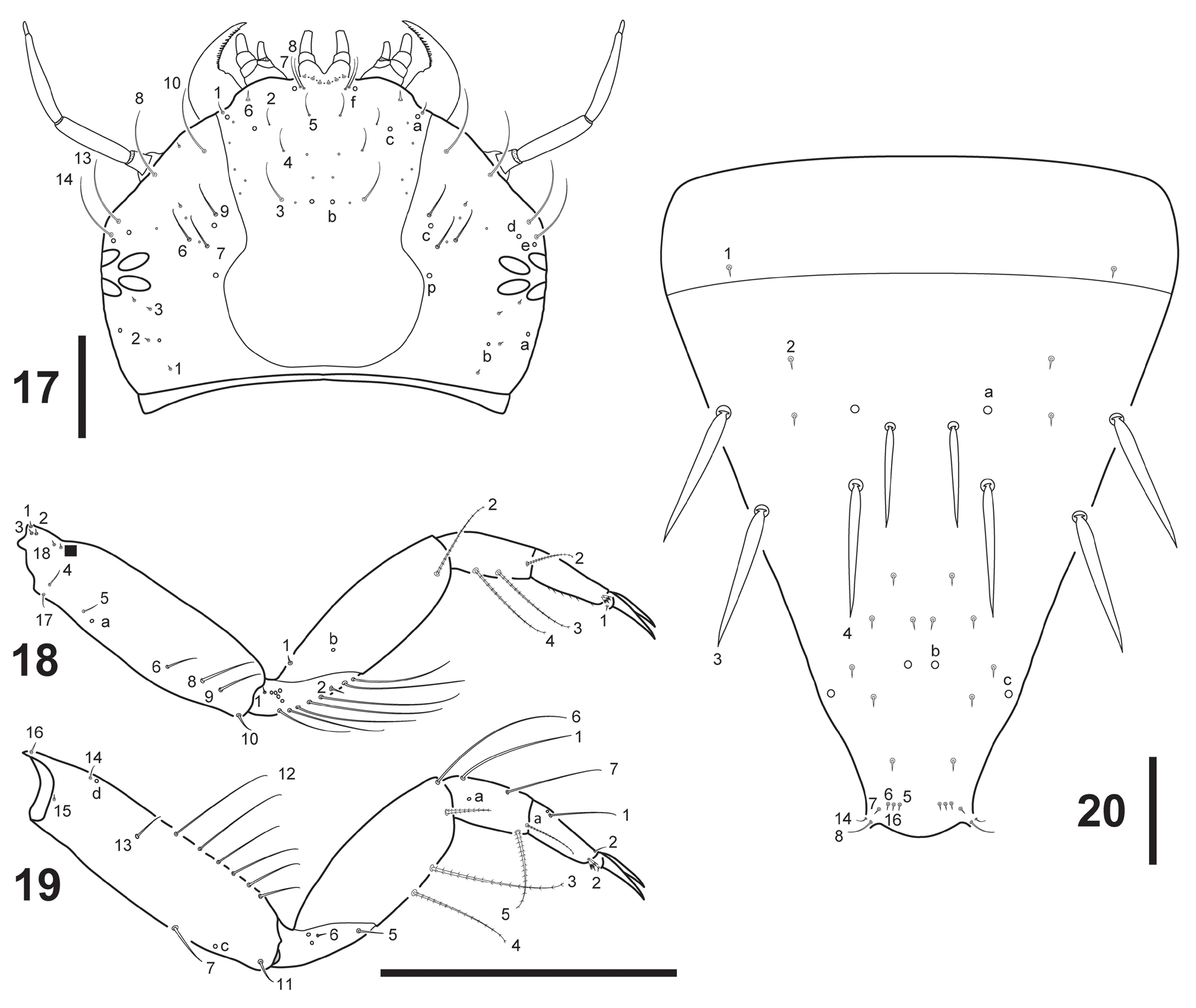
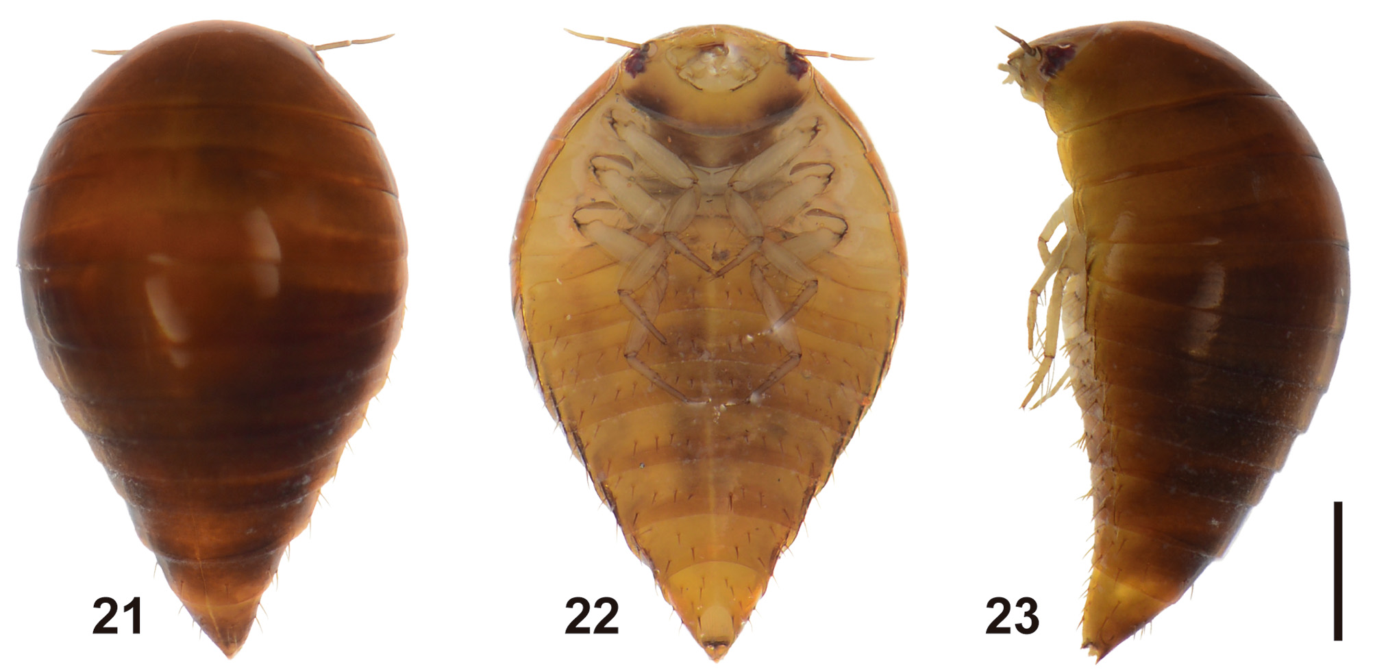
Instar III ( Figs 17–23 View FIGURES 17–20 View FIGURES 21–23 ; Tables 1–2). As for instar II except for following features:
Color: Darker in general, predominantly brown, abdominal segments I-VI and VIII slightly darker.
Body ( Figs 21–23 View FIGURES 21–23 ): Measurements and ratios that characterize the body shape are shown in Table 1.
Head ( Fig. 17 View FIGURES 17–20 ): A3 slightly longer than A2.
Abdomen: Segment VIII ( Fig. 20 View FIGURES 17–20 ).
Chaetotaxy: Frontoclypeus with 4–6 minute secondary sensilla on anterior portion; dorsal surface of parietal with two minute secondary sensilla near seta PA6 and one minute secondary seta near seta PA3; secondary leg setation detailed in Table 2 and Figs 18–19 View FIGURES 17–20 .
Comparative notes. Out of S. cimicoides described in this paper, the mature larva of S. inflatus represents the only other species of the genus known to date ( Spangler & Folkerts 1973). Except for the relative width of the head capsule (0.77–0.82 mm in S. cimicoides compared to 0.68 mm in S. inflatus ) no reliable morphological differences could be found to separate the mature larvae of these two species. Chaetotaxic characters could have proven to be useful in providing additional diagnostic characters. The previous description of S. inflatus , however, did not emphasize this character system.
It is worth stressing the presence of egg bursters in S. cimicoides ( Fig. 2 View FIGURES 1–3 ), which contradicts a former observation that egg bursters are lacking in the Noteridae ( Ruhnau 1985) .

No known copyright restrictions apply. See Agosti, D., Egloff, W., 2009. Taxonomic information exchange and copyright: the Plazi approach. BMC Research Notes 2009, 2:53 for further explanation.
|
Kingdom |
|
|
Phylum |
|
|
Class |
|
|
Order |
|
|
Family |
|
|
Genus |