
Arthropeina Lindner, 1949
|
publication ID |
https://doi.org/ 10.11646/zootaxa.3827.2.6 |
|
publication LSID |
lsid:zoobank.org:pub:D6C800AA-8127-41D1-ACEA-52254F7CE89D |
|
DOI |
https://doi.org/10.5281/zenodo.6124270 |
|
persistent identifier |
https://treatment.plazi.org/id/5E0C3871-7558-FFD6-B0D0-6CF71760E31C |
|
treatment provided by |
Plazi |
|
scientific name |
Arthropeina Lindner, 1949 |
| status |
|
Arthropeina Lindner, 1949 View in CoL
( Figs. 2–89 View FIGURES 1 – 7 View FIGURE 8 View FIGURES 9 – 14 View FIGURES 15 – 20 View FIGURES 21 – 26 View FIGURES 27 – 31 View FIGURES 32 – 37 View FIGURES 38 – 42 View FIGURES 43 – 49 View FIGURES 50 – 55 View FIGURES 56 – 59 View FIGURES 60 – 65 View FIGURES 66 – 77 View FIGURES 78 – 83 View FIGURES 84 – 89 )
Arthropeina Lindner, 1949: 789 View in CoL . Type-species: Arthropeina fulva Lindner, 1949 View in CoL (mon.).
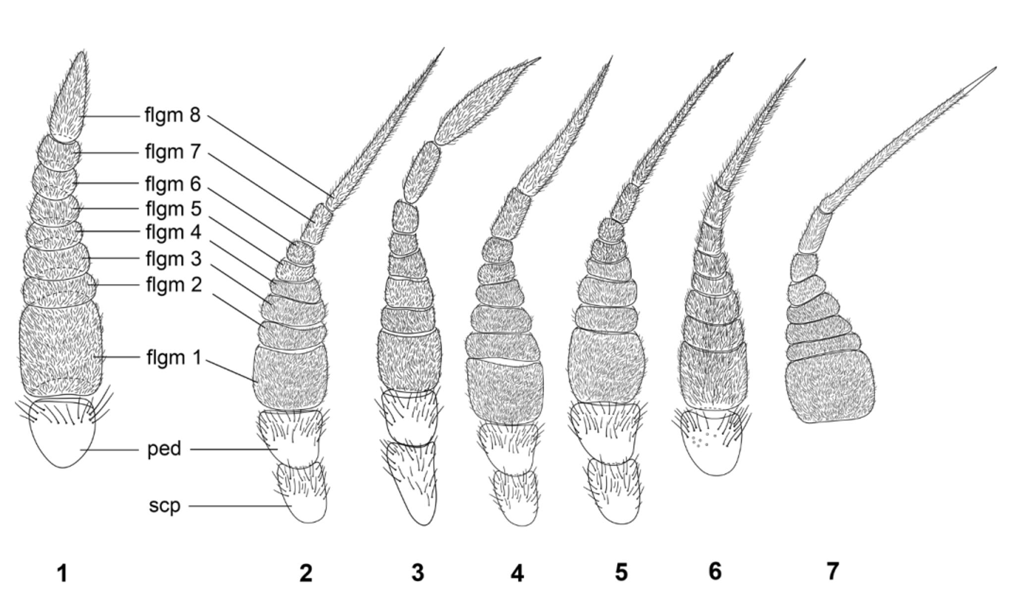
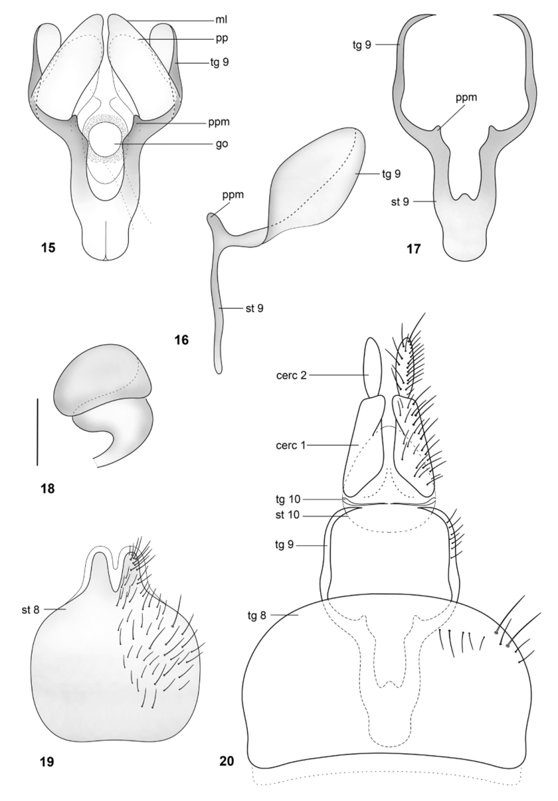
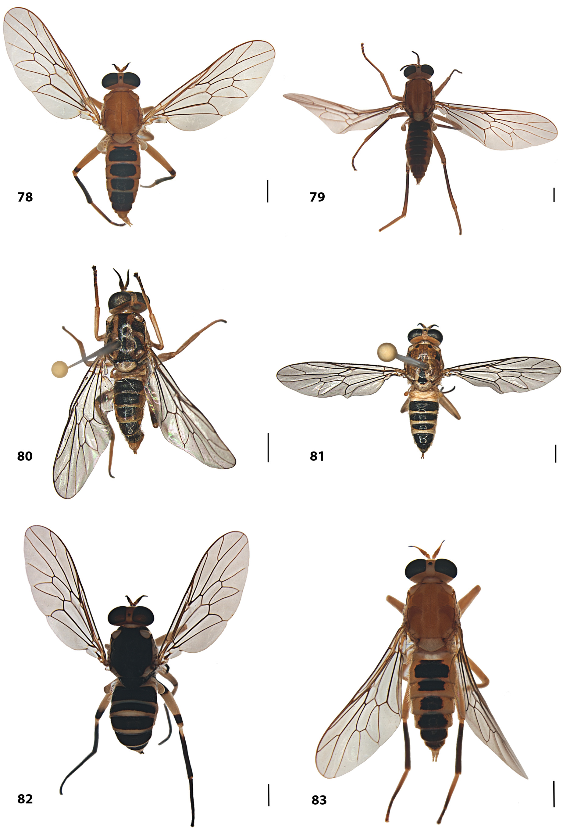
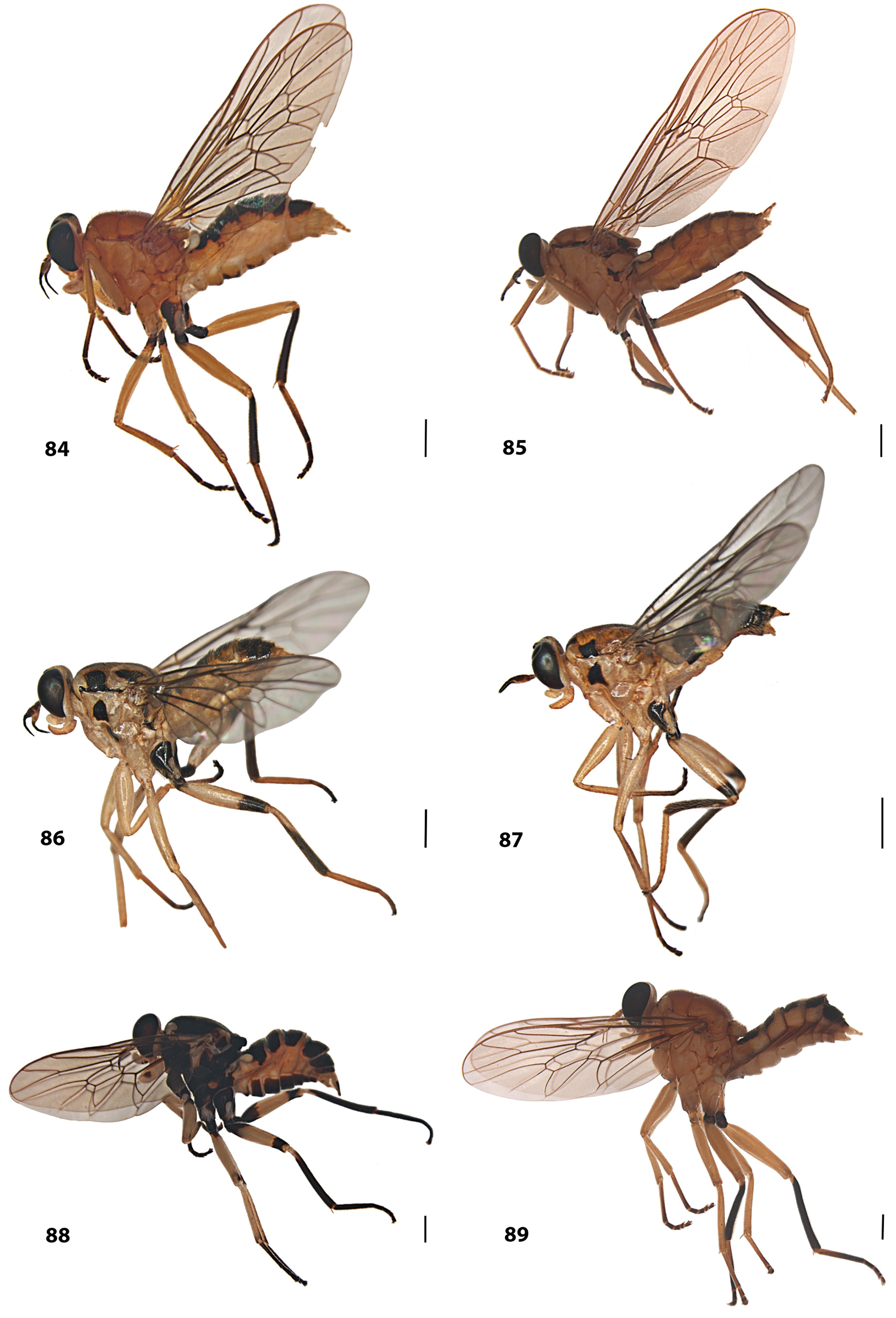
Diagnosis. Body color mostly yellow to reddish yellow. Antennae conically tapered towards apex; apical two flagellomeres (7–8) forming a stylus (e.g., Fig. 2 View FIGURES 1 – 7 ), as long as or longer than the remaining of the flagellomeres. Hind femur without row of tubercles, frequently slender, not differing from the other femora. Membranous lobes present in the female genital fork (e.g., Fig. 15 View FIGURES 15 – 20 ).
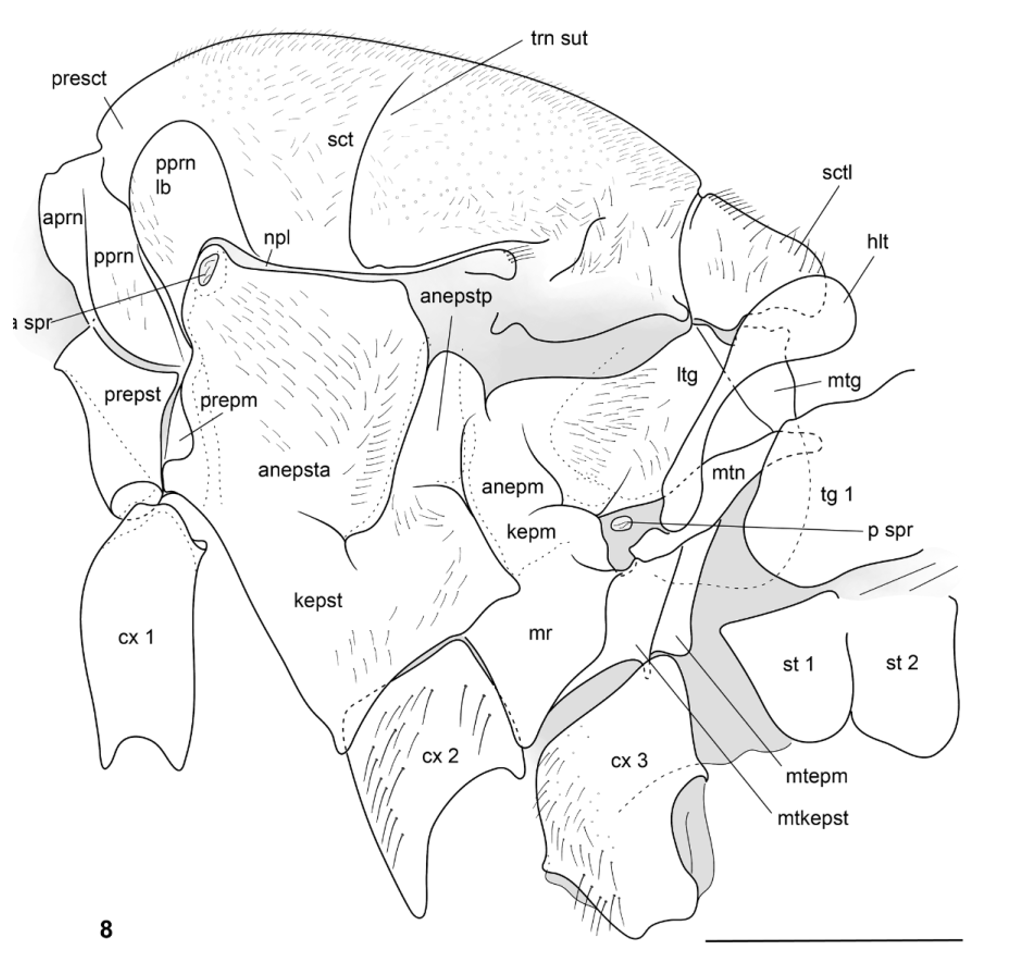
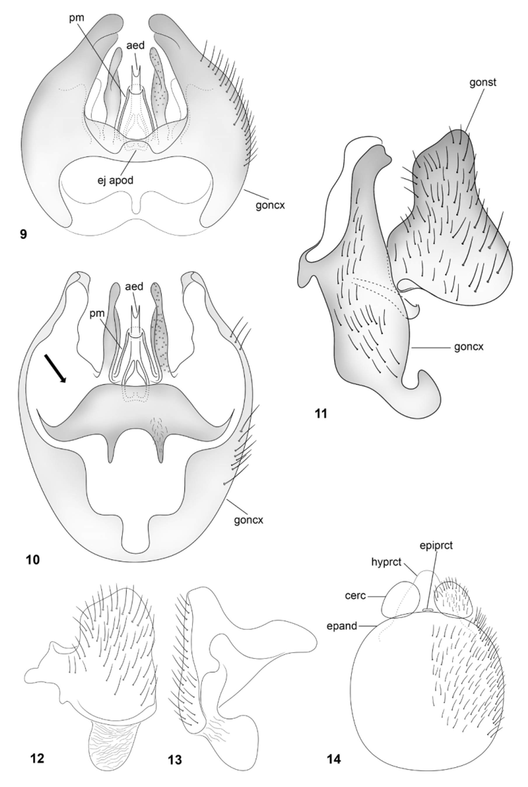
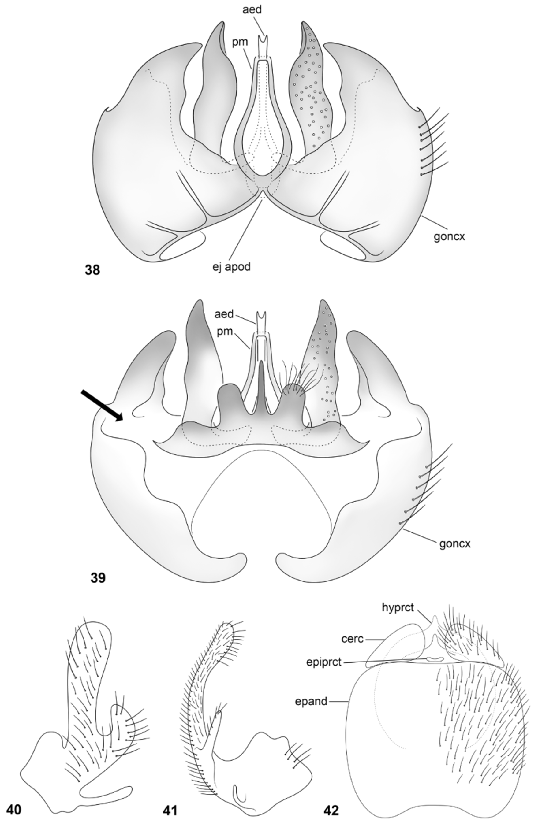
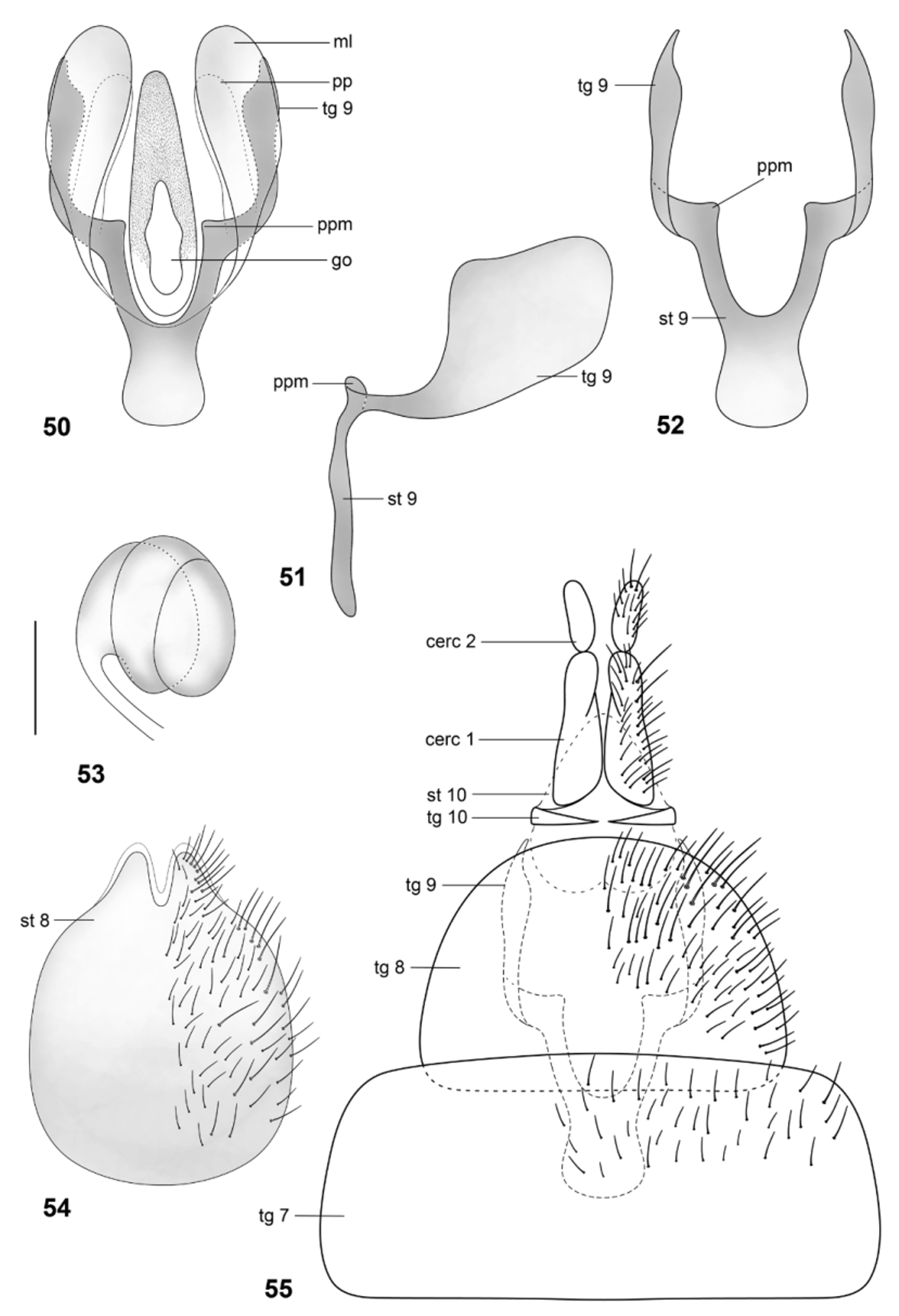
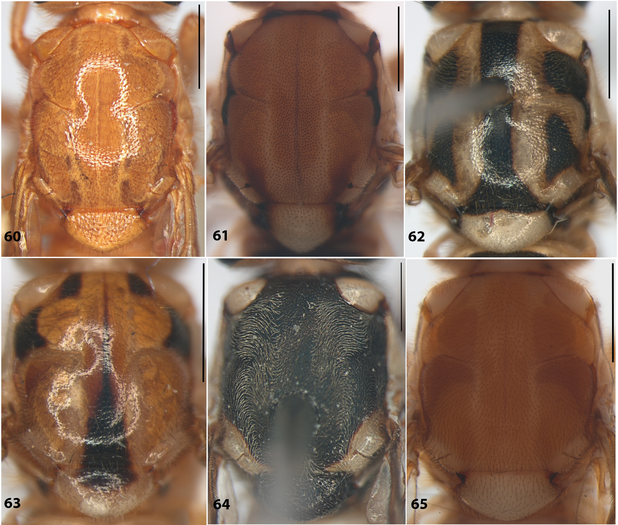
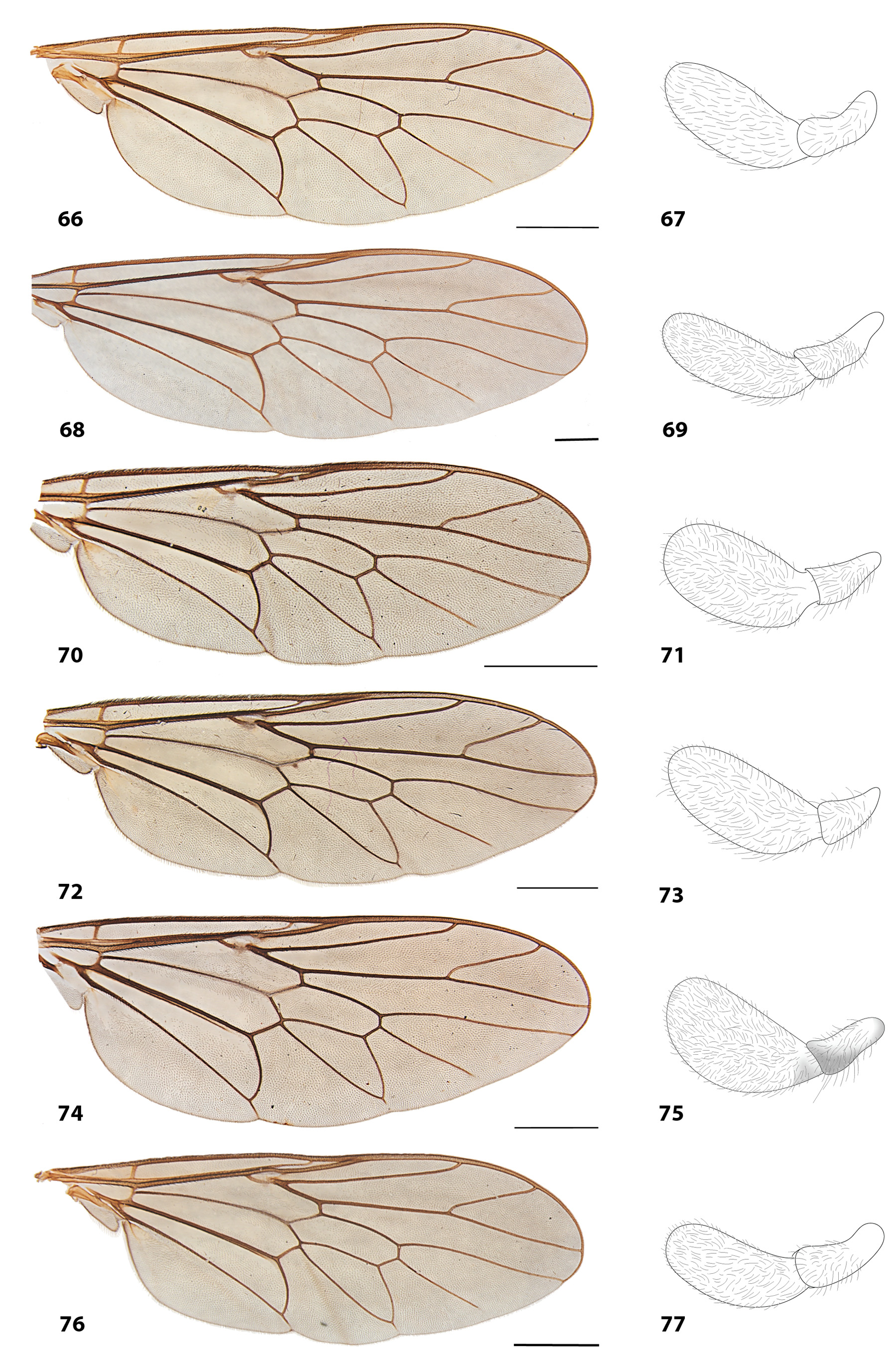
Redescription. Male. Length: body, 6.5–7.0 mm; wing, 6.5–7.0 mm. Head. Ocellar tubercle black, only slightly prominent, short setae at vertex. Eyes black, bare. Upper frons concave, gradually widening ventrally. Upper part of face silver tomentose. Pilosity short on frons, erect on genal region, and on posterior part of head. Antenna narrowing towards apex, conical (e.g., Fig 2 View FIGURES 1 – 7 ); scape slightly shorter than pedicel, reddish yellow with yellow setae; first flagellomere longer than subsequent flagellomeres except last; eighth flagelomere considerably longer than seventh, length ratio ranging from 2:1 to 5:1; flagellomeres 7–8 forming a stylus as long as or longer than rest of flagellomeres combined. Palpus elongate, basal (first) segment cylindrical, longer than wide; apical (second) segment oval, at least two times length of basal segment; pale hairs on both segments. Proboscis yellowish with pale hairs. Thorax ( Fig. 8 View FIGURE 8 ). Postpronotal lobes and notopleural strip frequently contrasting with color of scutum. Scutum and scutellum unicolorous or with dark bands or spots; pleura frequently with dark spots, pilosity short. Legs. Frequently yellow with some dark marks, or almost completely dark; pilosity on legs short, dense, yellowish, conforming to color of surface, except tarsomeres, mostly with brownish hairs. Wing. Venati on relatively uniform among the species (e.g., Fig. 66 View FIGURES 66 – 77 ). Veins brown to dark brown; C extending to M1. M2 and cell m3 not reaching wing margin. Cells bm, br and cup well defined. Cell cup not reaching cubital fork. Alula narrow, gradually widening towards apex. Abdomen. Pale to reddish yellow, with dark transverse bands on tergites; sternites weakly sclerotized; six to eight segments visible; pilosity conforming to color of surface, frequently yellowish. Male genitalia. Gonocoxite rounded laterally, distal margin strongly projected; inner plate with acuminate projections ventrally to phallus (aedeagus + parameres) (e.g., Fig 10 View FIGURES 9 – 14 ); gonocoxal apodeme absent. Gonostylus long and wide, varying in shape, but frequently with inner projections. Aedeagus elongate, completely enclosed by the parameres (e.g., Fig 10 View FIGURES 9 – 14 ); ejaculatory apodeme short, not exceeding basal margin of gonocoxite; a pair of long, acuminate projections lateral to phallus (e.g., Fig 10 View FIGURES 9 – 14 ). Epandrium quadrangular, wide, without lateral projections (surstylus) (e.g., Fig. 14 View FIGURES 9 – 14 ). Hypoproct (S10) strongly developed, epiproct (T10) limited to small sclerotized plate dorsally; cercus well developed, wider on distal half.
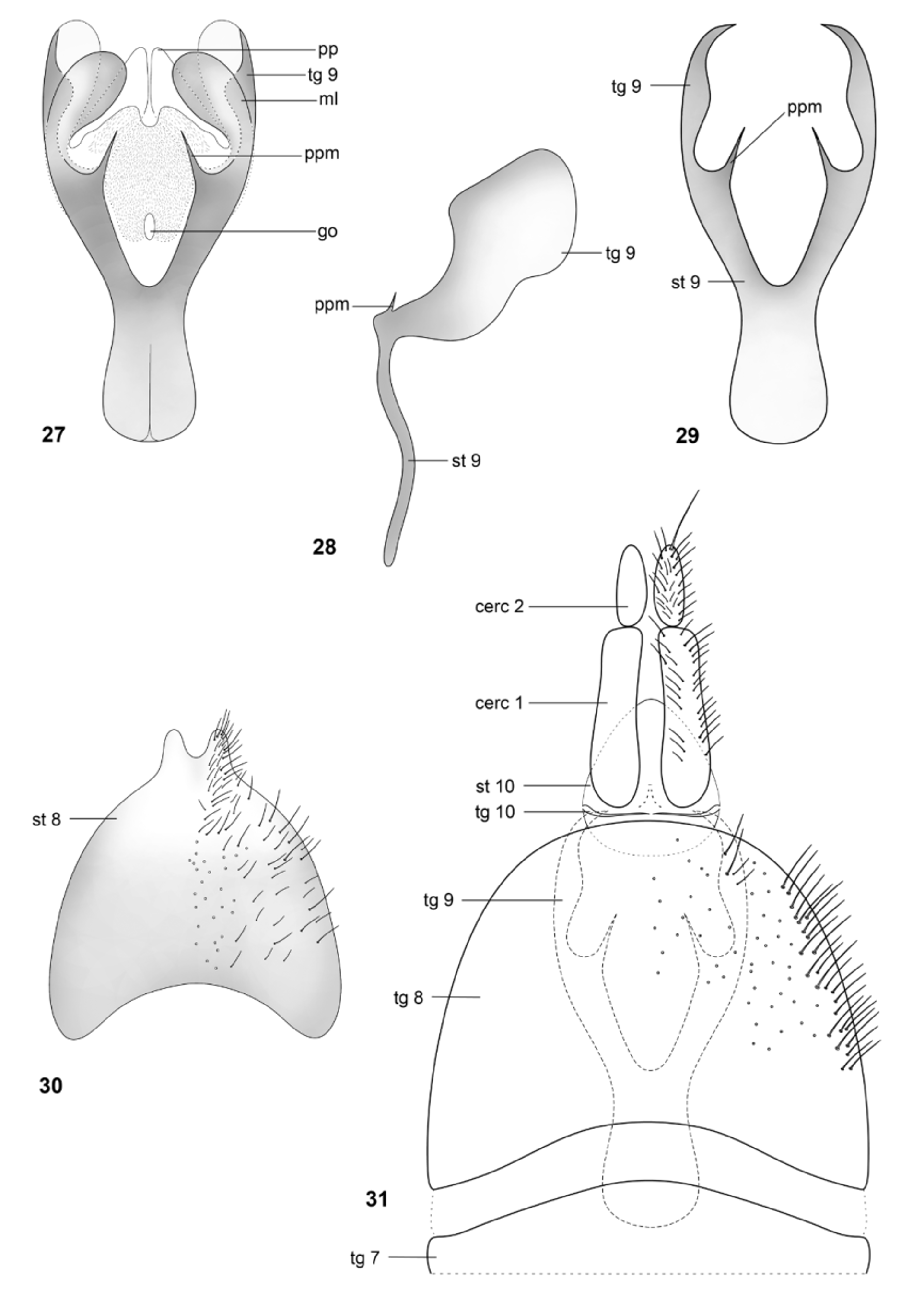
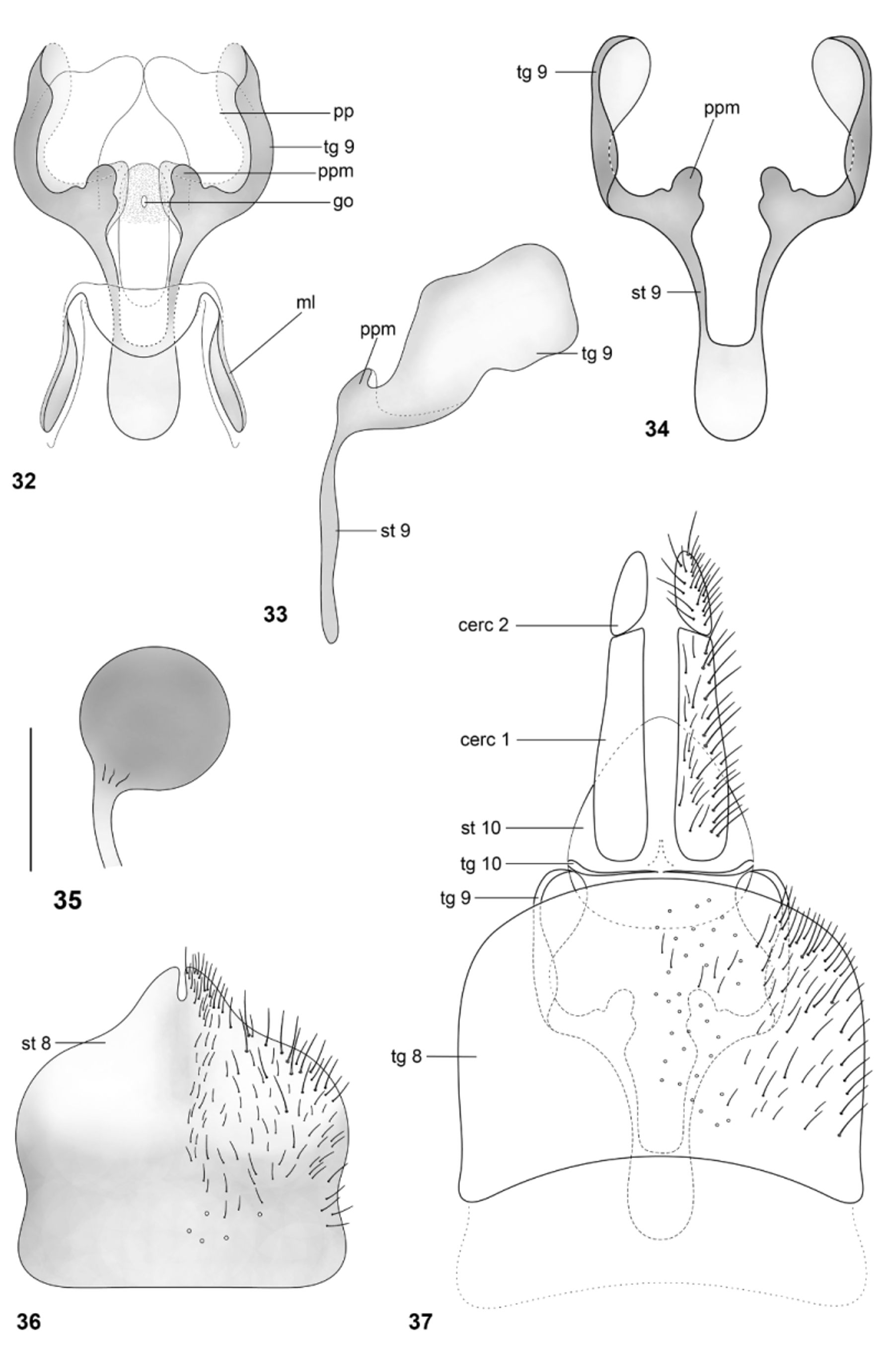
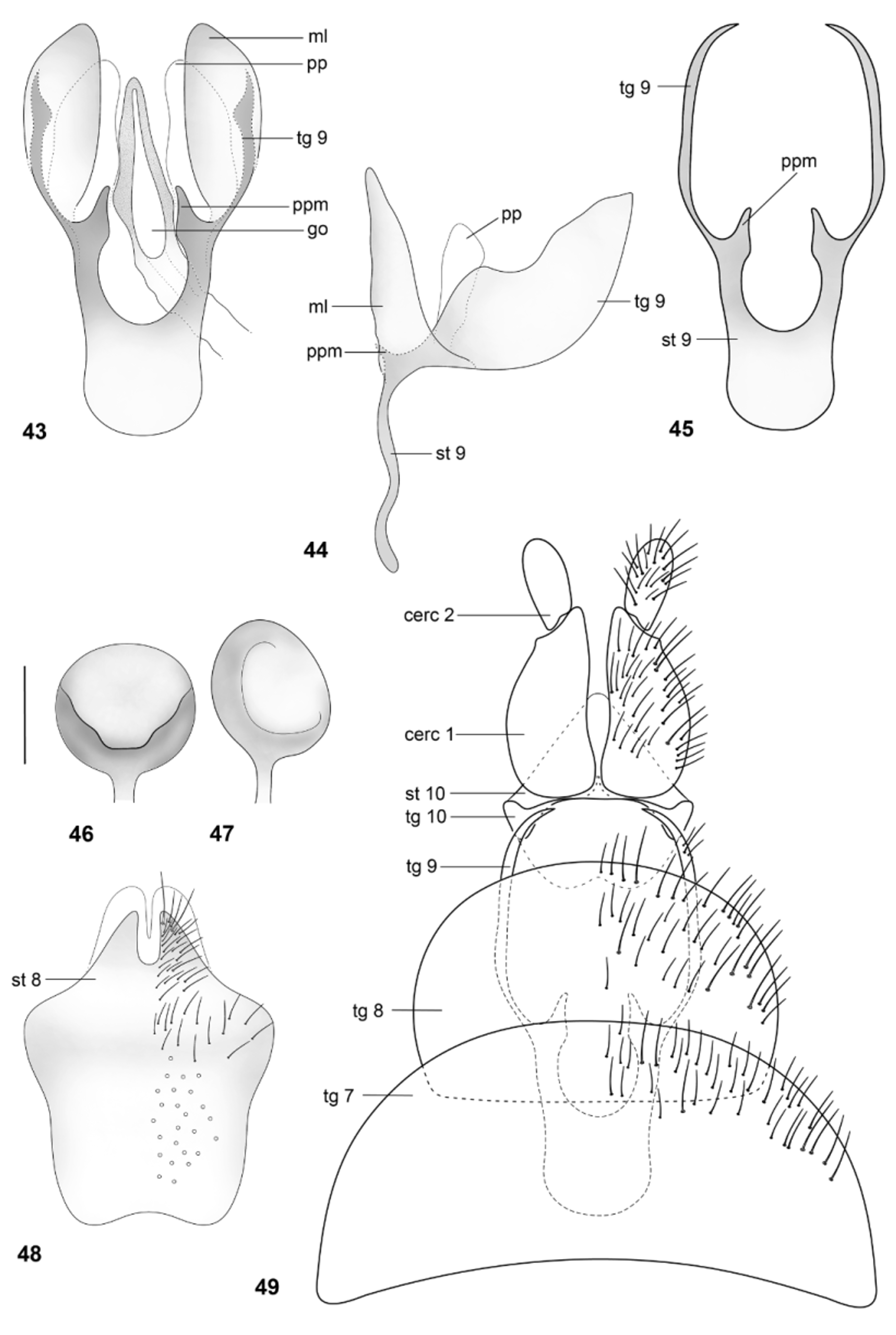
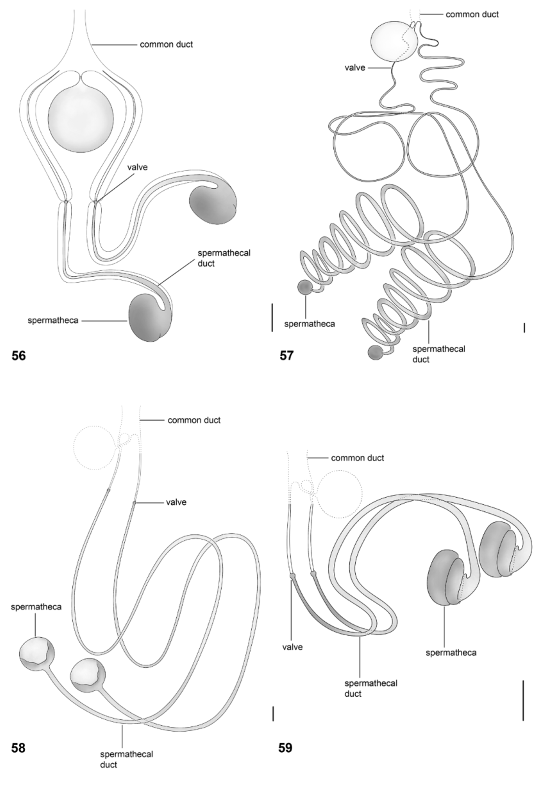
Female. Differs from male as follow. Length: body, 6.0– 7.5 mm; wing, 6.0–8.0 mm. Female genitalia. Tergite 8 wide, rounded anteriorly. Sternite 8 more or less quadrate, weakly sclerotized distally; gonapophyses unsclerotized, with short lobes. Tergite 9 laterally more sclerotized, completely fused to sternite 9 (genital fork) anterolaterally (e.g., Figs. 15–17 View FIGURES 15 – 20 ). Genital fork strongly sclerotized, anterior arm rounded basally; posterolateral processes clearly membranous, covering the genital opening distally; membranous lobes covering the genital fork ventrally (e.g., Fig. 15 View FIGURES 15 – 20 ), with some variation in shape; projection of posterior margin frequently prominent; margin between the posterior process with a medial incision; genital opening varying from small to quite large, placed between projections of posterior margin of genital fork, with short microtrichia on bursa. Three spermathecae present ( Figs. 56–59 View FIGURES 56 – 59 ), lateral ones with moderately long ducts, median one large, with a short duct; common duct membranous, unpigmented; posterior part of lateral ducts more sclerotized, with a valve medially; spermathecal capsules circular, more or less ovoid, strongly sclerotized. Tergite 10 not well sclerotized. Sternite 10 well developed, more or less triangular, distal margin rounded. First segment of cercus longer than second segment. Geographic distribution. The genus Arthropeina is known only from the Neotropical region. Up to now, the genus was known only from the type-locality, Nova Teutônia, Santa Catarina, in southern Brazil. The geographic distribution of this genus is now greatly expanded, with species known from northern Brazil and north and northwest South America, reaching Guyana, Colombia, Ecuador, Peru and Bolivia ( Fig. 92 View FIGURE 92 ). These are the first records of the genus for these last five listed countries.
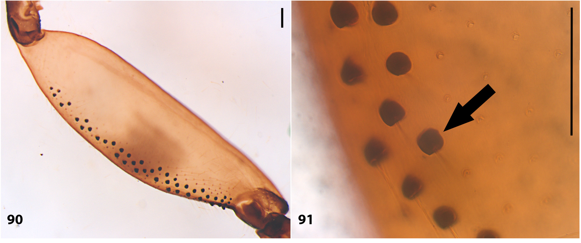
Comments. The genus can be easily separated from the remaining xylomyid genera by having the antenna conically tapered to the apex, with the apical two flagellomeres (7–8) forming a stylus (e.g., Fig. 2 View FIGURES 1 – 7 ), which is as long as or longer than the basal six flagellomeres—an autapomorphy of the genus. This condition is clearly distinct from that of Solva species ( Fig. 1 View FIGURES 1 – 7 ). Additionally, Arthropeina species do not present the row of tubercles ventrally on hind femora found in Solva ( Figs. 90–91 View FIGURES 90 – 91 ).
The male genitalia of Arthropeina , illustrated for the first time in this paper, show great resemblances to the genitalia of Solva and Xylomya ( Webb, 1984) . The gonocoxites are well developed, distally projected, the gonostylus is wide and frequently elongate, and the aedeagus is entirely enclosed by the parameres, forming a triangular structure that narrows towards the apex. Some inner gonocoxite projections in Solva and Xylomya ,—usually named as lateral (“interbasis” – Nagatomi & Tanaka, 1971), median and ventral parameres ( Webb, 1984),—have uncertain homology in the genitalia of Arthropeina . Apparently, the “lateral paramere” may be the lateral projections of the aedeagus (e.g., Figs. 10 View FIGURES 9 – 14 , 39 View FIGURES 38 – 42 ), while the “ventral paramere” may be homologous to the inner plate of the gonocoxite (e.g., Figs. 10 View FIGURES 9 – 14 , 39 View FIGURES 38 – 42 ) that in Arthropeina has distal projections.
A more rigorous understanding of homology in xylomyid male genitalia is still necessary to properly understand their correspondence to structures illustrated in detail by Webb (1984) and Nagatomi & Tanaka (1971). There is apparently some confusion on structures in Xylomyidae , especially on the inner projections of the gonocoxite, resulting in some difficulty to establish homology and character polarity in male genitalia of Arthropeina .


No known copyright restrictions apply. See Agosti, D., Egloff, W., 2009. Taxonomic information exchange and copyright: the Plazi approach. BMC Research Notes 2009, 2:53 for further explanation.
|
Kingdom |
|
|
Phylum |
|
|
Class |
|
|
Order |
|
|
Family |
Arthropeina Lindner, 1949
| Fachin, Diego Aguilar & Amorim, Dalton De Souza 2014 |
Arthropeina
| Lindner 1949: 789 |