
Neelus klisurensis, Kováč, Ľubomír & Papáč, Vladimír, 2010
|
publication ID |
https://doi.org/ 10.5281/zenodo.276298 |
|
DOI |
https://doi.org/10.5281/zenodo.6212258 |
|
persistent identifier |
https://treatment.plazi.org/id/5E348799-1A58-A32C-FF0D-596E614BEC12 |
|
treatment provided by |
Plazi |
|
scientific name |
Neelus klisurensis |
| status |
sp. nov. |
Neelus klisurensis sp. nov.
Figs 17–30 View FIGURES 17 – 20 View FIGURES 21 – 23 View FIGURES 24 – 26 View FIGURES 27 – 30
Diagnosis. Posterior part of head behind antennae with mesosetae, dorsal side of hind abdomen covered with meso- and macrosetae. Prelabral/labral setae formula (p-, m-, a-row): 4/5, 5, 4. Anterior labral setae R1 and R2 thick, curved, R1 medially with 2 strong teeth progressing ventrally, R2 with external edge finely serrate in its distal half. Ventral side of head with posterior macrosetae of postmedian area smooth, straight, not thickened. Sensory field on thorax furnished with numerous surrounding spines and setae. Ant. I segment with 2 setae, external side of Ant. IV with 12 thin and curved macrosensilla finely blunt at the tip. Unguis extremely elongated furnished with 2 long lateral teeth and 1 inner tooth in distal 1/3, weak incision in basal 1/3 absent. Manubrium with 4+4 dorsal setae, dens with dorsal spines E1 and J1 modified. Mucro with both dorsal lamellae serrated, apparently split at the tip.
Type material. Holotype: female on slide (No. VP–04–07), Serbia, western Kosovo, Prokletje Mts., Velika Klisura Cave, central part of the cave, aphotic zone, 400 m from the cave entrance, surface of the water pools, hand collecting, 4.ii.2007, leg. V. Papáč. Paratypes: 1 female and 2 juveniles on slides (No. VP–04–07), the same data as holotype. Type material (holotype and 1 paratype) saved in collection of the MNHN in Paris; 2 paratypes (juveniles) kept in the Department of Zoology, Institute of Biology and Ecology, Faculty of Science, P. J. Šafárik University, Košice.
Description. Body length up to 0.7–0.9 mm, habitus usual for the genus. Eyes absent, body colour white in ethylalcohol, without pigmentation. Cuticle finely granulated, integumentary channels on head and thorax not seen.
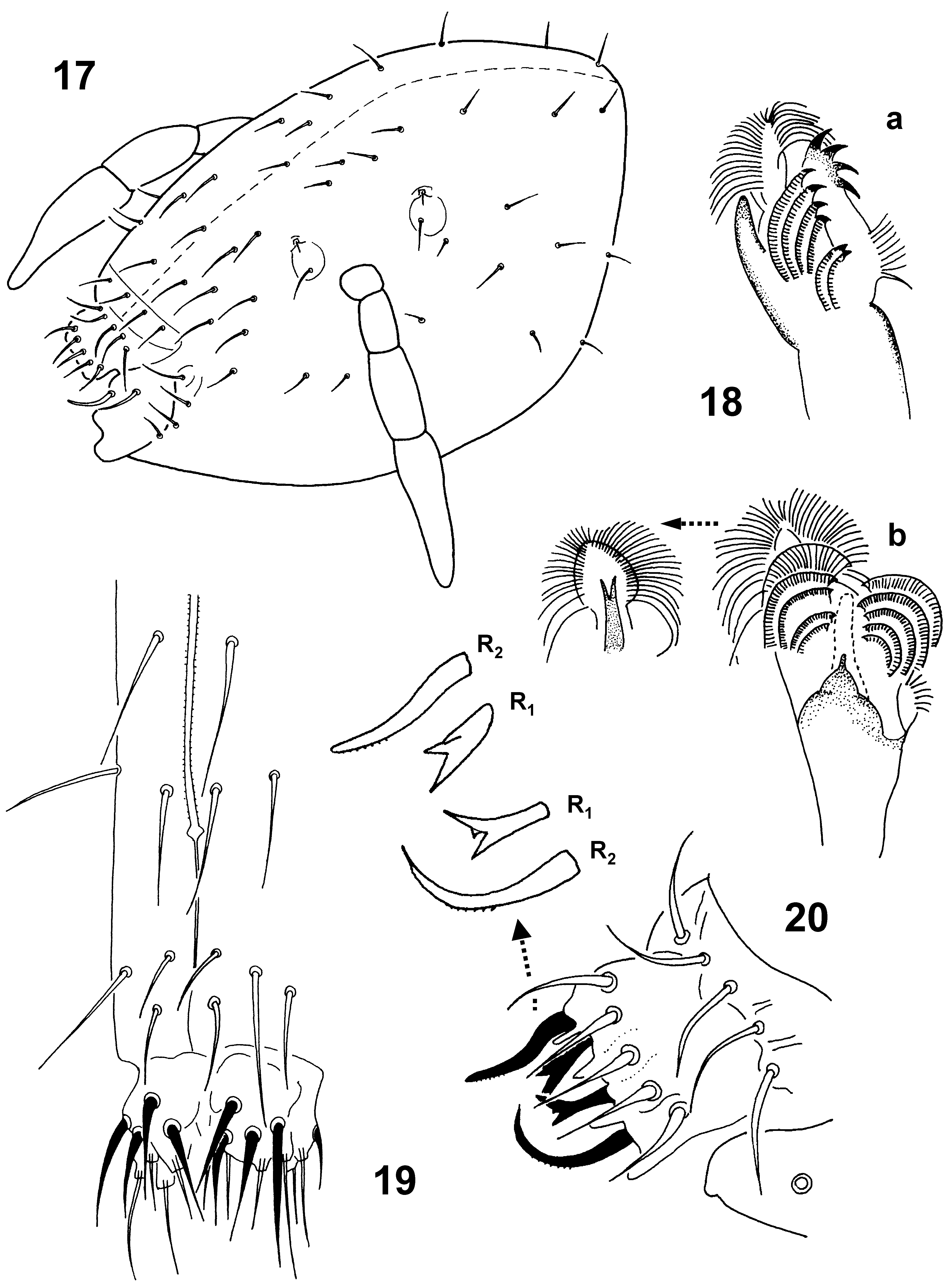
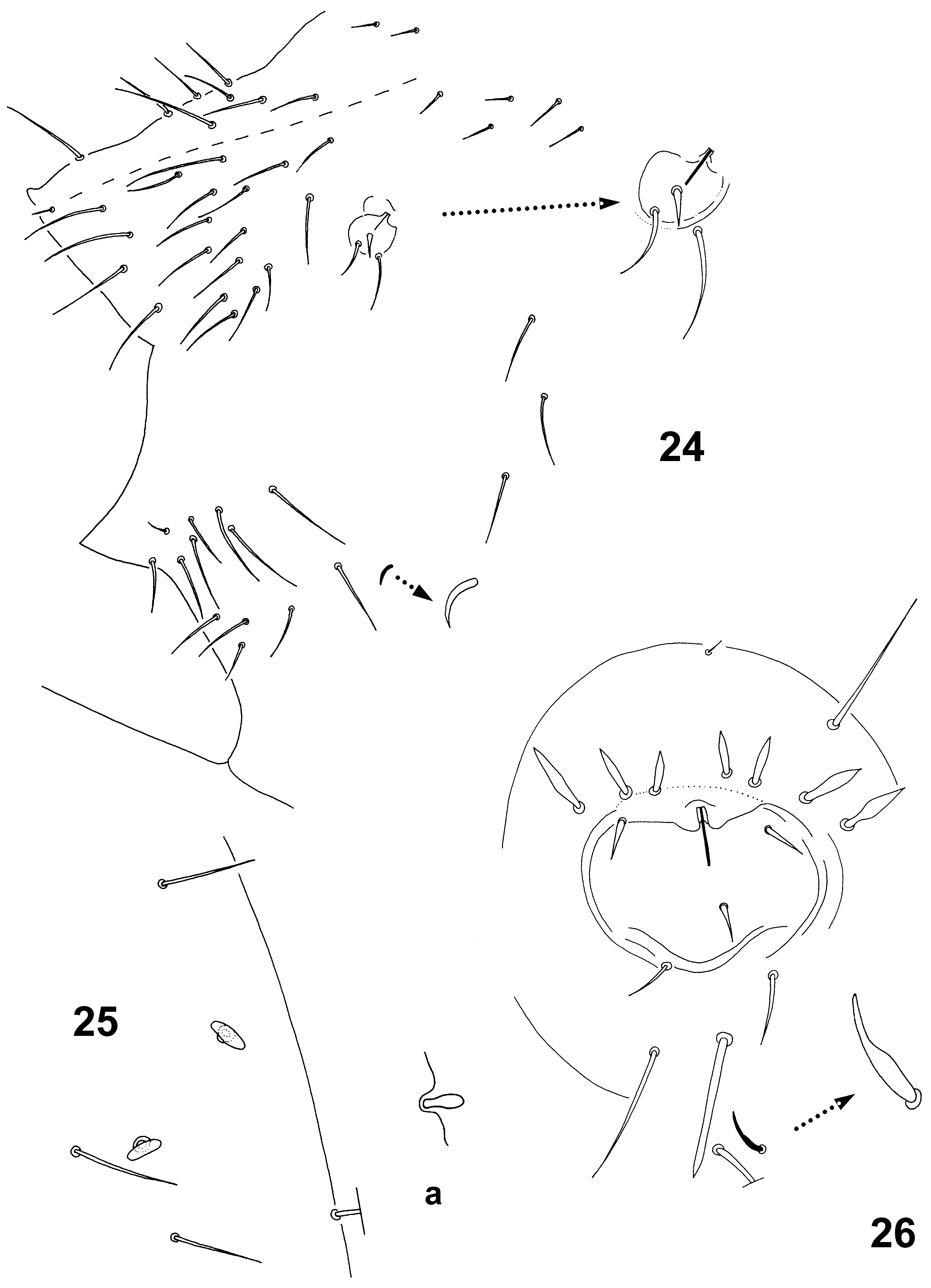
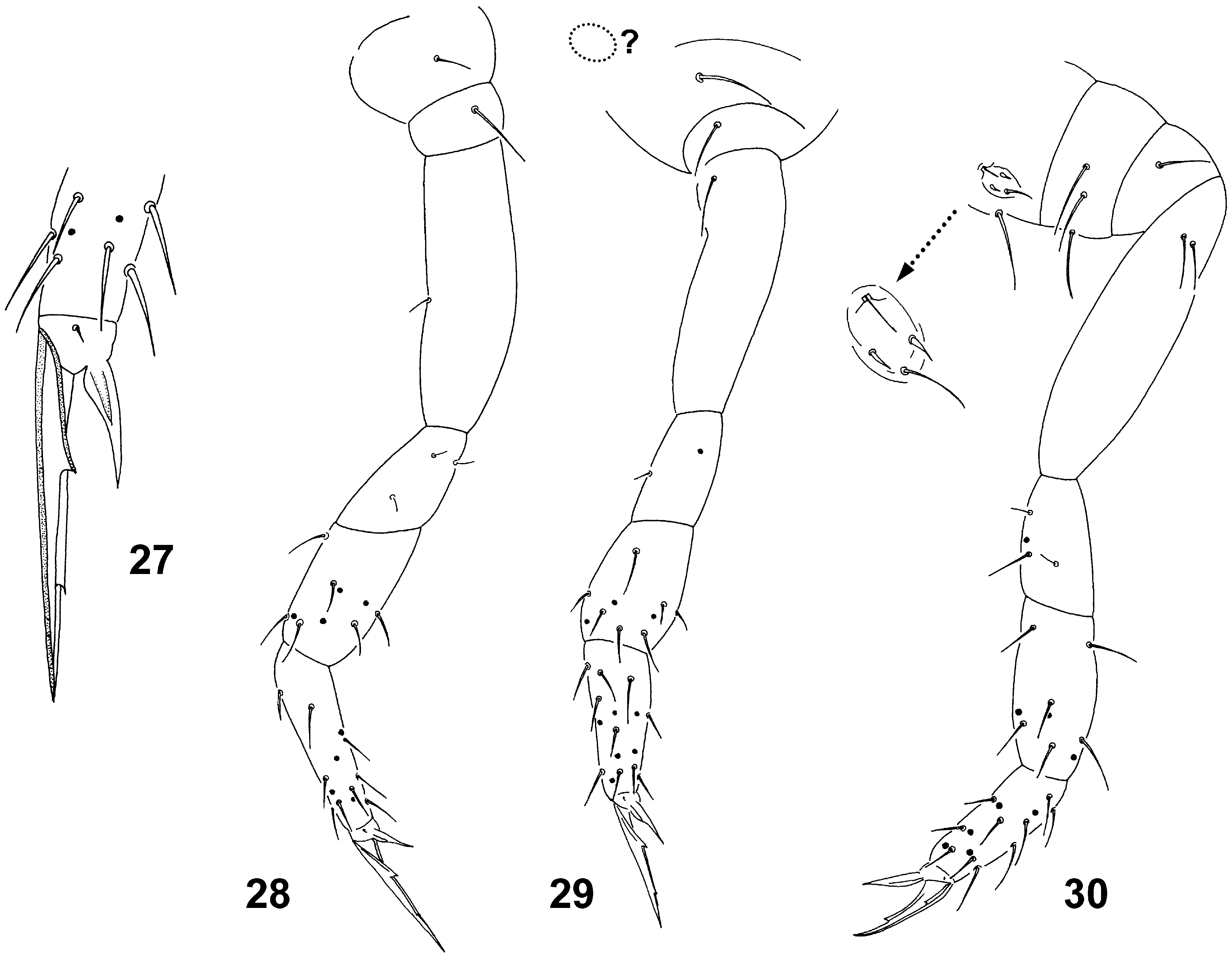
Sensory fields. Sensory fields placed in small depressions each with secretory rod (8 µm), i.e. blunt, straight seta with basal part inserted in cuticle and placed in upper margin of field. Fields’ arrangement: (a) on head—anterior and posterior fields (20 x 12 µm; Fig. 17 View FIGURES 17 – 20 ) each with secretory rod (12 µm) and 1 internal seta (18 µm); (b) large thoracal field (45 x 60 µm; Fig. 26 View FIGURES 24 – 26 ) with secretory rod (16 µm), 3 curved spines (10 µm) arranged in triangle and 2 marginal external setae (20 µm each); above the field 1 macroseta (36 µm) and 7 arrowhead-like (rhomboid) spines (13–18 µm) situated in one row; below the field 1 strong pointed rod-like macroseta (36 µm), 2 macrosetae (35 and 45 µm, respectively), 1 central microseta (5 µm) and 1 curved sensillum (11µm); (c) abdominal field (20 x 20 µm; Fig. 24 View FIGURES 24 – 26 ) with secretory rod (10 µm), 1 curved spine (11 µm), 2 marginal setae (internal—20 µm, external—28 µm); (d) field at base of leg III (30 x 15 µm; Fig. 30 View FIGURES 27 – 30 ) with secretory rod (10 µm), 2 curved spines (4 µm each) and 1 marginal external seta (15 µm); field at base of leg II not seen. Small sensory field on lateral part of abdomen above base of leg III with small sensory field furnished with short secretory rod (5 µm), stronger rod in cup-like depression above it absent.
Head. Head 270 µm long (width not seen in adult specimens). Eyes absent. Dorsal side with smooth and pointed setae ( Fig. 17 View FIGURES 17 – 20 ), frontal ones longer (20–25 µm) than those posterior placed behind antennae (8–18 µm); in the frontal part 2 unpaired axial setae present. Labrum with 5,5,4 setae, 4 prelabrals ( Fig. 20 View FIGURES 17 – 20 ). Pattern of labral setae: a-row 2R1 + 2R2, m-row m + 2r1 + 2r2 and p-row with 5 ordinary setae. Anterior labral setae R1 and R2 thick, curved; R2 longer than R1 (18 and 12 µm, respectively). Seta R1 medially with 2 strong teeth progressing ventrally in about 120o angle, R2 with external edge finely serrate in its distal half. Medial setae (m-row) equal (18 µm), smooth, spine-like. Posterior setae (p-row) smooth, lateral ones thicker and longer than those axial (20 and 18 µm, respectively). Ventral side of head with 3+3 smooth postmedian macrosetae ( Fig. 19 View FIGURES 17 – 20 ): 2+2 anterior equally long (26 µm), 1+1 posterior slightly longer (28 µm) than anterior ones. Basomedian field of labium with 4+4 setae ( Fig. 19 View FIGURES 17 – 20 ), medial setae longer (26 µm) and thicker than others (15 µm); basolateral field with 2+2 setae (1+1 axial shorter). Maxillary palp simple, with 1 enlarged sublobal seta. Mandible normal, strong, with 5 apical teeth. Maxilla as in Fig. 18 View FIGURES 17 – 20 a, b.
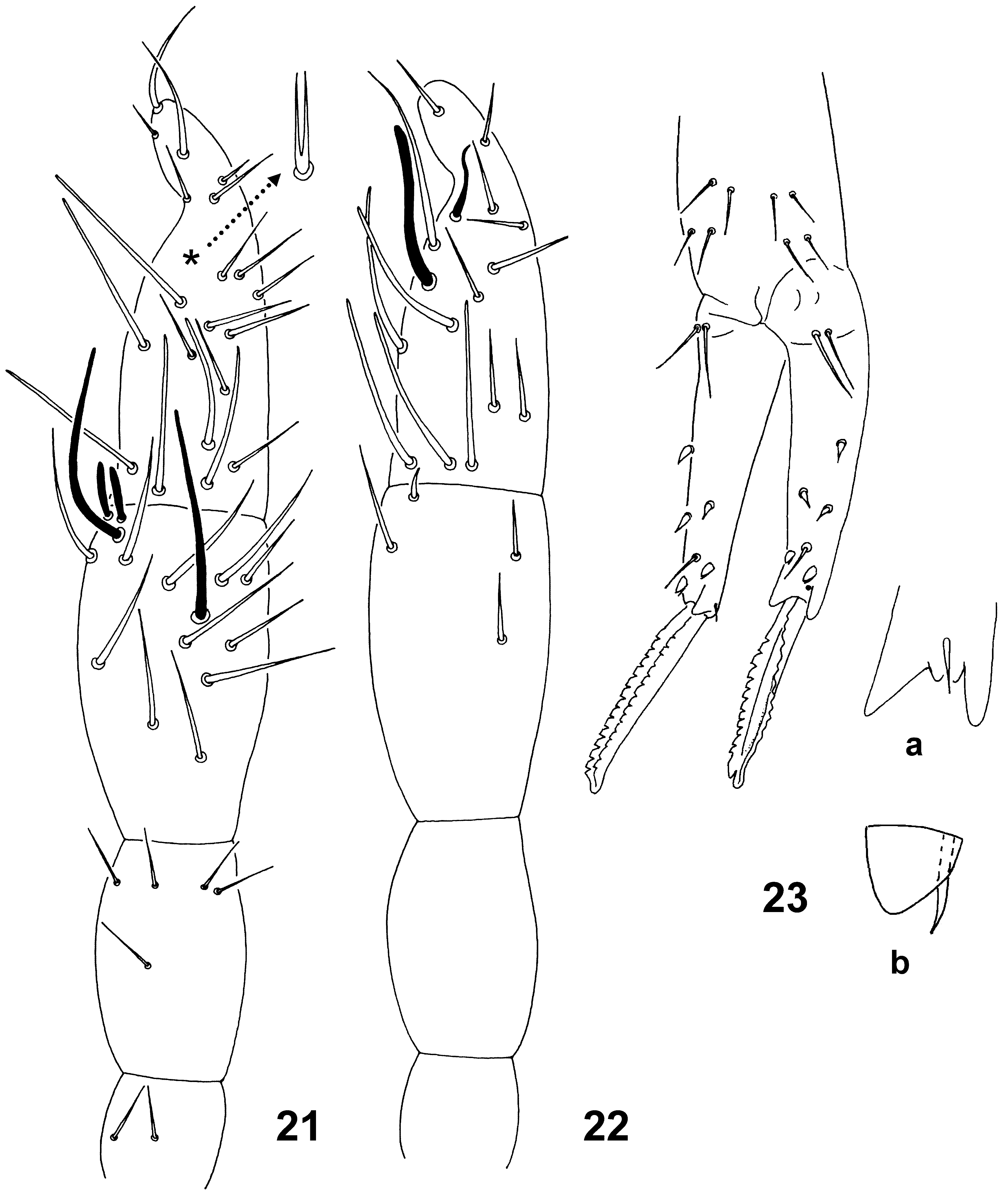
Antennae. Ant. III–IV distinctly separated ( Figs 21and 22 View FIGURES 21 – 23 ). Length of antennae 190 µm, ratio antenna/ head = 0.7. Length of Ant. I, II, III and IV as 17, 38, 60 and 75 µm. Ant. I furnished with 2 short setae (13 µm). Ant. II with 1 medial seta and 4 apical setae arranged in a whorl. Ant. III organ consists of 2 rather long sensory rods (25 µm), 2 long guard sensilla (32 µm) and short ventral spine-like seta (5 µm). Ant. IV with ordinary setae mostly on the internal side, external side with 12 thin and curved macrosensilla finely blunt at the tip (up to 38 µm); ventrally with 1 long and thick medial sensillum (27 µm) and 1 apical, basally thick shorter sensillum (14 µm) with abruptly narrowed and curved tip; dorsally with forked subapical seta (12 µm); subapically with 5 short setae (10 µm).
Thorax and abdomen. Dorsal side of thorax and abdomen sparsely covered with ordinary setae (20 μm); hind part of abdomen with longer setae (30 μm), axially with macrosetae (30–40 μm; Fig. 24 View FIGURES 24 – 26 ). Mid-abdomen dorsally with at least 2 pairs of swollen T-shaped microsensilla (6 µm; Fig. 25 View FIGURES 24 – 26 ). Upper edge of prefurcal area with 1+1 short, sharply pointed and curved neosminthuroid setae (9 µm; Fig. 24 View FIGURES 24 – 26 ). Abdominal segments V and VI cryptic. Genital plate not clearly seen in both adult females. Anal opening transversal; upper anal valve with 4+4 macrosetae (43 µm) and 1 unpaired posterior microseta (10 µm); lower anal valve only partly seen ( Fig. 24 View FIGURES 24 – 26 ).
Appendages. Setae of leg I–III ( Figs 28–30 View FIGURES 27 – 30 ; longer setae in parenthesis): subcoxae I 1, 1(1), 3(3); subcoxae II 1 (1), 1, 1; coxae 1, 1, 2; trochantera 3, 2, 4; femora 10, 10, 9 and tibiotarsi 13, 16, 15. Some of them as thin meso- or microsetae: leg I—coxa with 1, trochanter with 3, femur with 2; leg II—trochanter with 2; leg III—trochanter with 2. Tibiotarsal tenent seta pointed. Unguis narrow and extremely elongated, both unguis and unguiculus unequally long in leg I, II and III: unguis 74, 68 and 54 µm, respectively, unguiculus 22, 24 and 30 µm, respectively. Length ratio unguis I (inner margin) / Ti. I width (65/15 µm) = 4.3. Unguis furnished with 2 long lateral teeth and 1 inner tooth in distal 1/3, weak incision in basal 1/3 absent ( Fig. 27 View FIGURES 27 – 30 ). Unguiculus untoothed without apical filament. Tubus ventralis with 2+2 distal setae and posterior lobe. Retinaculum with 3+3 teeth, seta on corpus absent. Furca well developed ( Fig. 23 View FIGURES 21 – 23 ), length of manubrium, dens and mucro: 105, 140 and 87 µm, respectively. Manubrium dorsally with 4+4 setae, lateral ones (22 µm) slightly shorter than those axial (25 µm). Dens in basal part with 2+2 dorsal setae, lateral ones (28 µm) slightly shorter than those axial (32 µm); apically with 1+1 broad, blunt lateral spines and 1 medial sharp spine (10 µm; Fig. 23 View FIGURES 21 – 23 a) on ventral side; distal part dorsally with 3 external (E1–E3) and 2 internal (J1–J2) spines (9 µm each), and 1 medial, subapical seta (20 µm). In modified spines E1 and J1 basal circle absent, both constisting of sharp spine behind the protective leaf-shaped blunt structure ( Fig. 23 View FIGURES 21 – 23 b). Mucro with both dorsal lamellae serrated, apparently split at the tip.
Only females known.
Etymology. The new species is named after the type locality, the Velika Klisura Cave situated in western Kosovo ( Serbia).
Biology. Gut with four broadened compartments (diverticula) was filled mainly with clay and organic particles, fungal hyphae were recognized in smaller extent.
Distribution. Up till present Neelus klisurensis sp. nov. is known only from the Velika Klisura Cave situated in the Rugova Canyon near Peje town at foothill of the Lumbardska Planina. Its broader range covering another caves of the Lumbardska Planina or surrounding part of the Prokletje Mts. is highly probable. The species represents an obligate subterranean form (troglobiont), the obvious troglomorphic characters compared to other species of the genus show strong adaptations to cave life similar as in many other invertebrate inhabitants of caves of the Balkan Peninsula.
Discussion. N. klisurensis sp. nov. is similar to N. koseli sp. nov. in the number of prelabral and labral setae and setal pattern of dorsal manubrial setae. N. koseli sp. nov. differs from other species of the genus by the presence of spine-like microsetae dorsally on hind part of abdomen and by number of setae in apical whorl on Ant. II segment (five and four setae, respectively). N. klisurensis sp. nov. differs from others of the genus by the presence of large sensory field on thorax with complex of modified surrounding setae. Both N. klisurensis sp. nov. and N. koseli sp. nov. exhibit clear morphological adaptation to life in subterranean environment, i.e. elongated ungua and unguicula on legs, the length ratio of unguis I/Ti. I width being 4.3 and 2.7, respectively. Moreover, N. klisurensis sp. nov. has longer thick sensillum on Ant. IV (27 and 23 µm, respectively) and more numerous macrosensilla on the same segment (12 and 8, respectively). For other diagnostic characters see Table 1.
| MNHN |
Museum National d'Histoire Naturelle |
No known copyright restrictions apply. See Agosti, D., Egloff, W., 2009. Taxonomic information exchange and copyright: the Plazi approach. BMC Research Notes 2009, 2:53 for further explanation.
|
Kingdom |
|
|
Phylum |
|
|
Class |
|
|
Order |
|
|
Family |
|
|
Genus |