
Cephalaeschna yanagisawai, Sasamoto & Lien, 2018
|
publication ID |
https://doi.org/ 10.11646/zootaxa.4471.2.5 |
|
publication LSID |
lsid:zoobank.org:pub:44EF0661-851A-4B9B-89DA-98AAE9197C93 |
|
DOI |
https://doi.org/10.5281/zenodo.5980017 |
|
persistent identifier |
https://treatment.plazi.org/id/5E38F063-FF8A-DB1A-A1A5-3004FBE3F925 |
|
treatment provided by |
Plazi |
|
scientific name |
Cephalaeschna yanagisawai |
| status |
sp. nov. |
Cephalaeschna yanagisawai View in CoL sp. nov.
( Figs. 1–3 View FIGURE 1 View FIGURE 2 View FIGURE 3 )
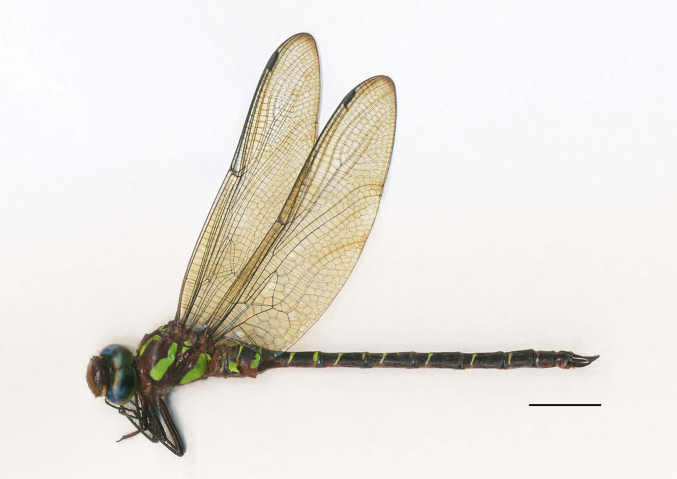
Holotype. ♂ (NSWT-I-Od-32335) ( Fig. 1 View FIGURE 1 ), Hoang Lien National Park , Lai Chau Province, N . Vietnam (103°45’E, 22°20’N, 1900 ~ 2000 m a.l.s.), 29-VI-2017; Takashi Yanagisawa leg. The holotype will be deposited in the National Museum of Nature and Science, Tokyo GoogleMaps .
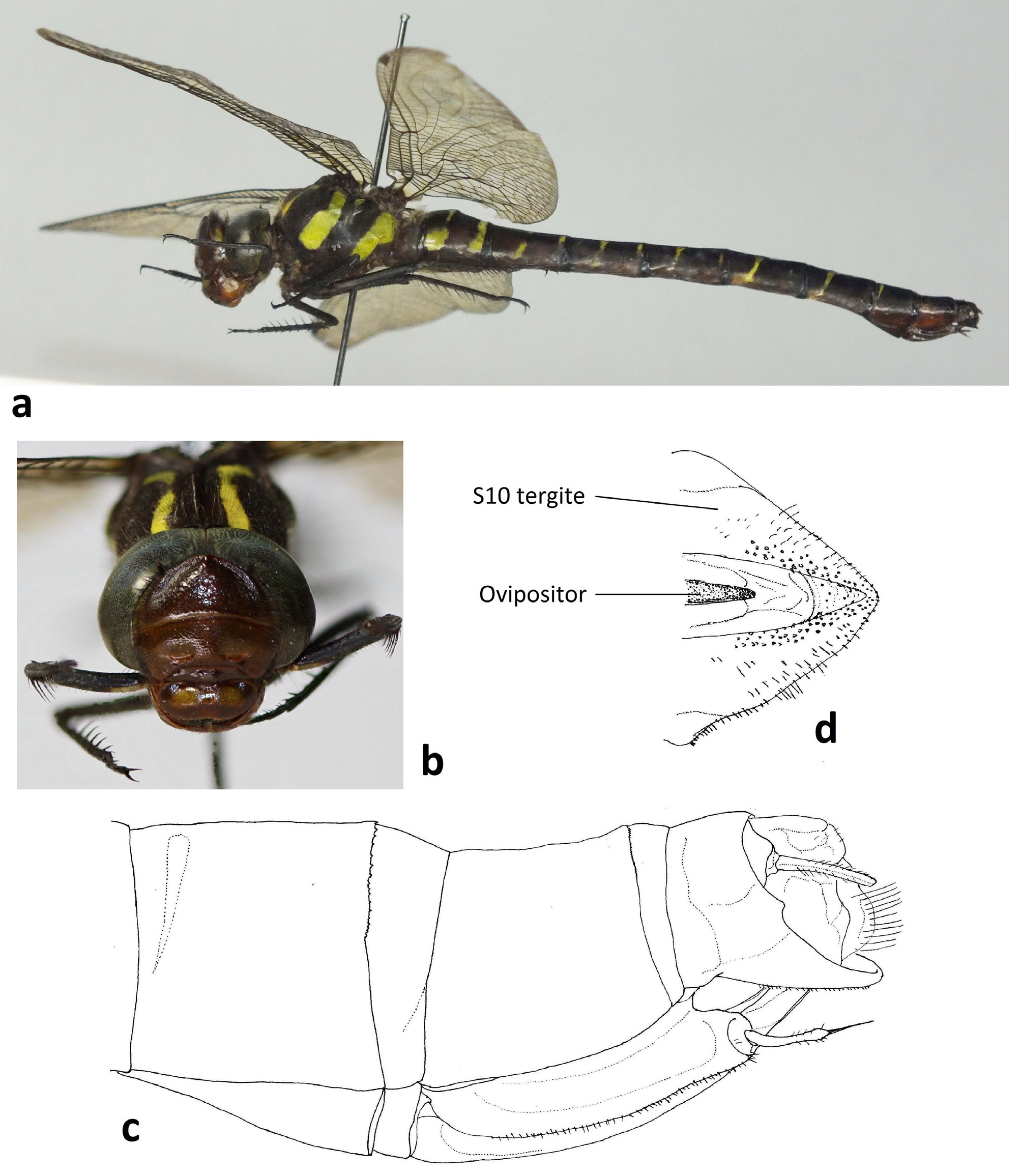
Paratypes. 1♂, same place as the holotype, 16-VIII-2017, Kenji Yashiro leg.; 1♀ ( Fig. 3a View FIGURE 3 ), same place as the holotype, 29-X-2017, Kenji Yashiro leg. The paratypes are preserved in Mr. Kozo Yashiro’s collection.
Etymology. The species name, a noun in the genitive case, is dedicated to Mr. Takashi Yanagisawa, collector of the holotype.
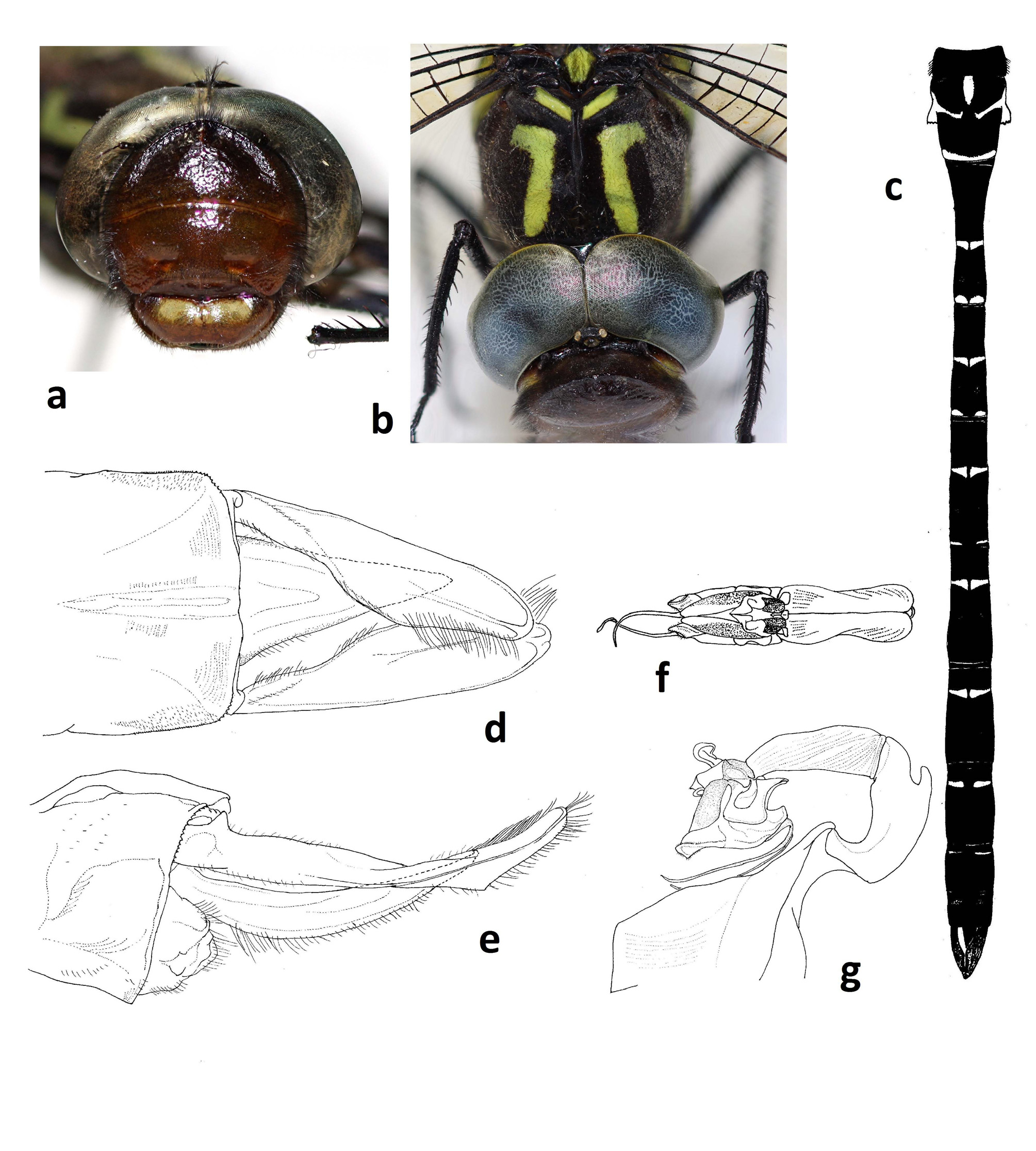
Description of the holotype. Head ( Fig. 2a View FIGURE 2 ). Labium brown with partly blackish shades; labrum brown with a large transverse oblong greenish yellow spot, a little split at the center by vertical brown line; genae brown with largely greenish yellow on upper part; ante- and postclypeus brown, with dark brown at the lower border of anteclypeus, and vaguely yellowish in the pits of lower side of postclypeus and on the upper part of the same; antefrons brown, much darker on upper frontal and dorsal sides; dorso-frontal apex of antefrons is obtusely but distinctly angled; vertex and antennae black, partly dark brown. The width of anteforns is about 0.65 times of the distance between the ends of compound eyes. The compound eye in life is of emerald blue, margined with green color.
Thorax. Prothorax brown, bordered with black. Synthorax dark brown with greenish yellow markings as follows; mesepisternum (ref. Fig. 2b View FIGURE 2 ) with antehumeral stripe, turning gently inwards and upwards, and its upper portion abruptly curving outwards; antealar sinus with greenish yellow above the ridge; mesepimeron with broad stripe, slightly constricted at a little above the middle, then narrower in upper portion and changing direction a little anteriorly; metaepisternum with a small inverted triangular spot at the dorsal border and with tiny vague vertical spot at the middle; interalar sclerite greenish; metaepimeron broadly greenish, except for the ventral and anterior margin; mesoinfraepisternum and metainfraepisternum each with a tiny spot at posterior corner; ventral side pale brown. Legs black, coxa, trochanter and basal femur tinted with dark brown; fore-leg with small greenish spots at inner side of trochanter and the basal femur.
Wings. Hyaline, faintly tinted with pale brown. Nodal index: antenodal veins (An) 21 (right) and 22 (left) in forewing (Fw), 16 (both) in hind wing (Hw); postnodal veins (Pn) 15 (both) in Fw, 18 (right) and 19 (left) in Hw. Pterostigma (Pt) reddish brown, covering 2.5 to 3 cell-length in Fw and 2 to 2.5 in Hw, with an embraced vein situated at or slightly interior to the proximal vein of Pt in both wings. Median space with 4 cross-veins in both wings; triangle 5 cells; anal loop contains 9 cells; anal triangle 4 cells.
Abdomen ( Fig. 2c View FIGURE 2 ). Slender cylindrical, most swelling at 2nd segment (S); colored dark brown or black, slightly pale ventrally, with greenish yellow maculation as follows. S1 hairy dorsally, with vertical thin green spot near postero-ventral border; S2 with arrowhead-like spot on the anterior half of dorsum, with a pair of thin median dorsal (MD) markings obliquely continuous to the antero-ventral marking, covering auricle, and with annular postero-dorsal (PD) marking, which is expanded ventrally; MD oblique markings present on S2 to S9, gradually inconspicuous and situating anteriad in segment towards the back; at antero-ventral part of each tergite of S3 to S10, faintly yellow vague tints, connecting with MD marking; PD markings present on S2 to S6, those on S3 and S4 semi-circular, that on S5 almost linear, and that on S6 rudimentary; A pair of inconspicuous yellow spots on distal fourth of dorsa of S3 to S7; S10 without markings, except for dark yellowish at antero-ventral corner.
Penis ( Fig. 2f–g View FIGURE 2 ). dark brown with whitish membranous parts; vesicle robust, trapezoidal shape in lateral view; 2nd and 3rd segments are almost similar length, with a dorsal protuberance on antero-dorsum of 2nd; distal segment triangular in lateral view with a pair of long flagelliform projections originating from ventral corner and with ear-like lobules on dorsum.
Anal appendages. Dark brown. Cercus (superior appendages), in dorsal view ( Fig. 2d View FIGURE 2 ), basally narrow, from basal 2/5 expanded inwards, then from its half gradually tapering distad; the apex round; the dorsal longitudinal ridge running from middle to the apex. Cercus, in lateral view ( Fig. 2e View FIGURE 2 ), slender and gently warped downwards, with an obtusely shallow triangular ventral protrusion at apical forth. Epiproct (inferior appendage), about 0.72 times as the length of cercus, narrow triangle shape in dorsal view, and bow shape, warped downwards, in lateral view.
Measurements (mm): Hw 44.0, abdomen plus anal appendages 53.8.
Variation in a paratype male. There are only small variations in a paratype male as follows. Wing nodal index: An 24 in Fw, 17 and 18 in Hw; Pn 15 and 16 in Fw, 17 and 18 in Hw; anal loops 10 or 11 cells, anal triangle 3 or 4 cells. Abdominal maculation is similar to the holotype, but PD markings on S6 invisible. Measurements (mm): Hw 41.9, abdomen plus anal appendages 50.6.
Female. The body shape is generally thicker than in male. The background color is brownish and maculation is more yellowish than in male, but its pattern is similar to that of male ( Fig. 3a View FIGURE 3 ). Head: the yellow markings of labium are thinner and more ambiguous ( Fig. 3b View FIGURE 3 ); postcylepus with a pair of two dark yellow spots around the pits; the width of antefrons is about 0.67 times as wide as the distance between the ends of compound eyes. Wing generally tinted with brown and basally yellow; nodal index: An 23 and 25 in Fw, 18 and 19 in Hw; Pn 15 and 17 in Fw, 16 and 19 in Hw; irregular halfway subcostal extension is found in only left Fw; triangle and median space are crossed same as in male; anal loops contain 11 or 13 cells. Abdomen ( Fig. 3a View FIGURE 3 ) thick cylindrical, and less constricted in S3 than male; ground color dark brown and maculation patterns are almost same as males, but S9 without any markings: the postero-ventral part of tergite of S10 projecting posteriorly, each tip of which on both side is fused ( Figs. 3c–d View FIGURE 3 ); semi-membranous structure is formed between each projections. The tip of ovipositor valves slightly beyond the level of dorsum of S10. Measurements (mm): Hw 45.0, abdomen plus cerci 47.0.
Diagnosis. The external morphology of most Cephalaeschna species more or less resemble each other, but can be identified by combinations of the following characters in male ( Asahina 1983; Fraser 1936; Joshi & Kunte 2017): (a) pointedness and width of antefrons, (b) marking on labrum, (c) maculation patterns on synthorax and abdomen, (d) the number of cells of anal triangle (3 or 4 vs ± 5), and (e) morphology of anal appendages, which can roughly be classified into two types, i.e. the apex of cercus ending rounded (or obtuse) vs acutely angled.
The degree of acuteness in dorsal antefrons is characteristic in each species. The antefrons of C. yanagisawai is obtusely angled ( Fig. 2a View FIGURE 2 & 3b View FIGURE 3 ), but neither pointed upwards nor smoothly rounded. This state a little resembles those of C. viridifrons (Fraser, 1922) ( Myanmar to Himalaya), C. klapperichi Schmidt, 1961 ( Afghanistan to W. Himalaya), and C. patrorum Needham, 1930 (Beijing to Sichuan), as far as we know. However C. yanagisawai can easily be discriminated from the latters by maculation and other features.
The following species have somewhat similar maculation to C. yanagisawai , sharing with it an obtuse apex of cercus, but can be differentiated as described below: C. klotsi Asahina, 1982 (Zhejiang to Guangxi, Hong Kong), which has narrow antefrons, i.e. the width of which is less than the half of the head width, exceptional in this genus ( Asahina 1983 Wilson & Xu 2008), whereas the width of antefrons of C. yanagisawai is much broader ( Fig. 2a View FIGURE 2 & 3b View FIGURE 3 ). Cephalaeschna masoni (Martin, 1909) (NE India & Nepal) has a “roundly inflated antefrons” ( Asahina 1981a), not dorsally angled like C. yanagisawai . Cephalaeschna obversa Needham, 1930 (Hubei, Sichuan & Guizhou) and C. solitaria Zhang, Cai & Liao, 2013 (Hubei) have PD markings on S2–S8 (Zhang et al. 2013), but C. yanagisawai has those only on S2–S6 ( Fig. 2c View FIGURE 2 ). Cephalaeschna patrorum has the cercus, the inner side of distal half of which is almost linear and PD markings on S2 to S8 ( Needham 1930; Asahina 1981b; Zhang et al. 2013), whereas that of C. yanagisawai is gently concaved inwards ( Fig. 2d View FIGURE 2 ), and lacking PD markings on S7 and S8 ( Fig. 2c View FIGURE 2 ).
The female of C. yanagisawai is peculiar in that postero-ventral part of S10 tergite projects posteriorly ( Fig. 3c View FIGURE 3 ). Although there are not many species in the genus for which female morphology is described, the females with such structure are exceptional, only known in C. acutifrons (Martin, 1909) (Himalaya) ( Asahina 1983), C. cornifrons Zhang & Cai, 2013 (Yunnan) (Zhang & Cai 2013) and C. patrorum (Zhang et al. 2013) .
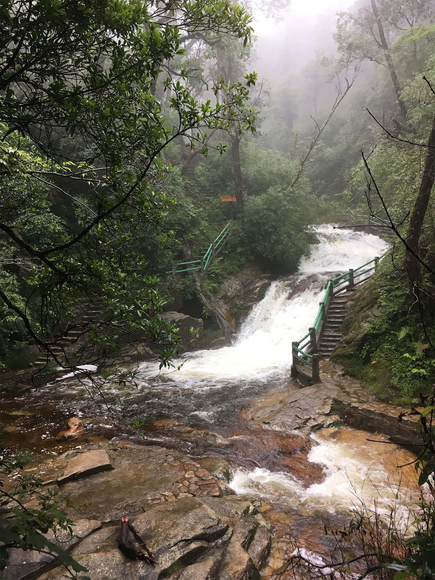
Notes on habitat. According to Mr Yanagisawa (pers. comm.), he collected the holotype male at around 4 p. m. following rainy conditions from morning. The specimen was flying over a wet grassy place near the stream ( Fig. 4 View FIGURE 4 ). In the same place, Cephalaeschna aritai Karube, 2003 was observed the day before.
No known copyright restrictions apply. See Agosti, D., Egloff, W., 2009. Taxonomic information exchange and copyright: the Plazi approach. BMC Research Notes 2009, 2:53 for further explanation.
|
Kingdom |
|
|
Phylum |
|
|
Class |
|
|
Order |
|
|
Family |
|
|
Genus |