
Shivaphis pteroceltis, Jiang, Liyun, An, Guangchi, Li, Weiwei & Qiao, Gexia, 2014
|
publication ID |
https://doi.org/ 10.11646/zootaxa.3753.4.6 |
|
publication LSID |
lsid:zoobank.org:pub:0C9A3954-6BA0-4B3F-A6E2-28829DDB25C6 |
|
DOI |
https://doi.org/10.5281/zenodo.6133651 |
|
persistent identifier |
https://treatment.plazi.org/id/5E3B87D0-C631-FFF5-FF69-4E9C2BCAFD5D |
|
treatment provided by |
Plazi |
|
scientific name |
Shivaphis pteroceltis |
| status |
sp. nov. |
Shivaphis pteroceltis sp. n.
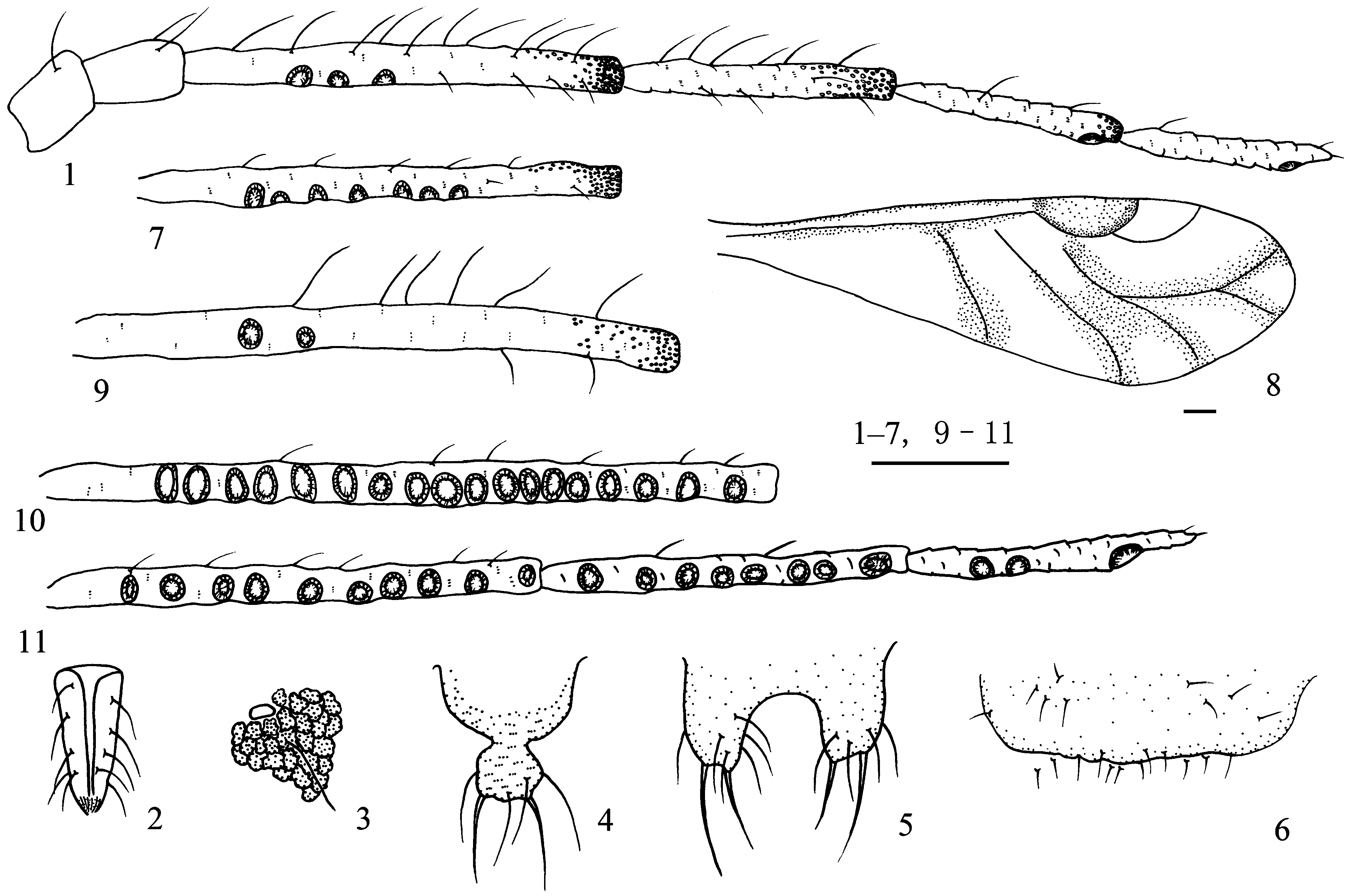
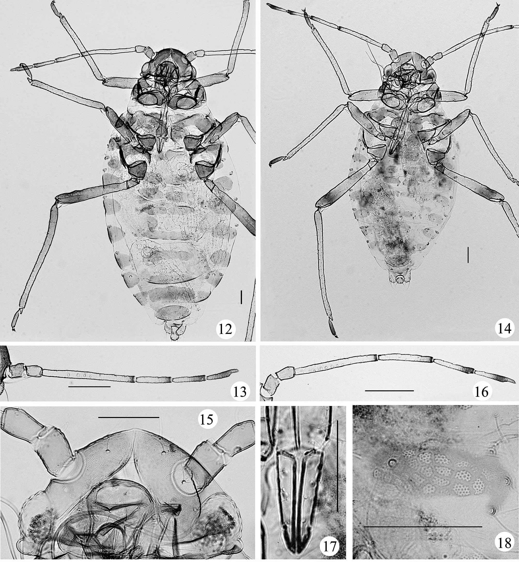
Apterous viviparous females ( Figs 1–6 View FIGURES 1 – 11 , 14–18 View FIGURES 12 – 18 , 34 & Table 1). Body oval ( Fig. 14 View FIGURES 12 – 18 ), red brown in life (Fig. 34), covered with thick wax powder or filaments. Head, thorax and abdominal segments each with 1 pair of dorsal dark brown scleroites on both spinal and marginal regions.
Mounted specimens: Body pale, with distal ends of antennal segments III–VI, apex of ultimate rostral segment, distal 1/3 of hind femora, second tarsal segments and claws brown; dorsal head, pronotum, antennal segments I and II, remaining parts of antennal segments III–VI, remaining parts of rostrum, remaining parts of legs, wax plates, spiracular plates, cauda, anal plate and genital plate pale brown. Dorsum of body smooth, with wax glands on each segment, distal parts of antennal segments III–VI, coxae, trochanters, femora and tibiae. Wax glands composed of visible wax pores, forming wax facets on antenna and legs, and wax plates on dorsum ( Figs 3 View FIGURES 1 – 11 , 18 View FIGURES 12 – 18 ); head with 1 pair of large oval wax plates ( Fig. 15 View FIGURES 12 – 18 ); thoracic nota and abdominal tergites I–VII each with 1 pair of spinal and 1 pair of marginal wax plates; tergite VIII with one spinal wax plate forming a band across the segment. Dorsal setae of body long and fine ( Fig. 3 View FIGURES 1 – 11 ). Head with 2 pairs of frontal and 3 pairs of dorsal setae; abdominal tergites I–V each with 3–6 pairs of spino-pleural and 2 or 3 pairs of marginal setae; tergite VI with 2 or 3 pairs of spino-pleural and marginal setae; tergite VII with 1 pair of spinal, pleural and marginal setae; tergite VIII with 1 pair of spinal and marginal setae. Frontal setae, marginal setae on abdominal tergite I, setae on tergite VIII 3.00–4.25, 3.00–5.00 and 3.50–5.25 times as long as basal diameter of antennal segment III, respectively. Spiracles oval and closed, on kidney shaped spiracular plates.
Head. Frons round, antennal tubercles undeveloped. Eyes normal, with numerous facets. Antennae 6- segmented ( Figs 1 View FIGURES 1 – 11 , 16 View FIGURES 12 – 18 ), 0.58–0.69 times as long as body, with sparse spinules on segments III–VI. Processus terminalis 0.19–0.29 times as long as base of the segment VI. Segments I–VI each with 3 or 4, 2–6, 16–23, 10–13, 2–7, 2–5+0 or 1 setae, respectively. Processus terminalis with 2–4 apical setae. Outer setae much longer than inner setae on segment III–V, longest setae on segment III 3.00–4.00 times as long as basal diameter of the segment. Primary rhinaria small and round, ciliated; segment III with 2–7 secondary rhinaria. Rostrum reaching hind coxae. Ultimate rostral segment wedge-shaped ( Figs 2 View FIGURES 1 – 11 , 17 View FIGURES 12 – 18 ), 1.92–2.40 times as long as its basal width, 0.82–0.96 times as long as second hind tarsal segment; with 6 primary setae and 10–18 secondary setae.
Thorax. Legs normal but with fore coxae distinctly enlarged. Hind femur 1.34–1.54 times as long as antennal segment III, hind tibia 0.43–0.51 times as long as body; setae on hind tibia 1.88–2.86 times as long as its middiameter. First tarsal chaetotaxy: 7, 7, 7, including 2 dorsal and 5 ventral setae.
Abdomen. Siphunculi ring-like ( Fig. 3 View FIGURES 1 – 11 ), on abdominal tergite VI, widest diameter 0.75–1.00 times as long as basal diameter of antennal segment III. Cauda, anal plate and genital plate with spare spinules. Cauda knobbed ( Fig. 4 View FIGURES 1 – 11 ), constricted in distal 1/3, 0.89–1.04 times as long as its basal width, with 6–10 setae, including 2 long and thick setae. Anal plate bilobed ( Fig. 5 View FIGURES 1 – 11 ), each lobe with 9–14 setae, including 2–4 long and thick setae. Genital plate broad band-shaped ( Fig. 6 View FIGURES 1 – 11 ), with 20–26 setae, including 8–10 anterior setae and 12–16 posterior marginal setae. Gonapophyses fused as one, with 7–11 short setae.
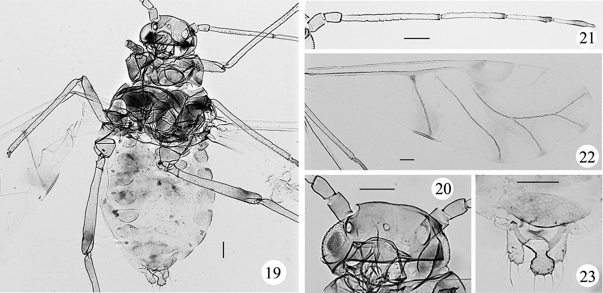
Alate viviparous females ( Figs 7, 8 View FIGURES 1 – 11 , 19–23 View FIGURES 19 – 23 , 35 & Table 1). Body oval ( Fig. 19 View FIGURES 19 – 23 ), red brown in life (Fig. 35), covered with thick wax powder or filaments.
Mounted specimens: Dorsal of head, thorax, distal ends of antennal segments III–VI, apex of ultimate rostral segment, distal 1/3 of hind femora, second tarsal segments and claws brown; antennal segments I and II, most of antennal segments III–VI, remaining parts of legs, wax plates, spiracular plates, cauda and anal plate pale brown; the other parts of body pale. Dorsum of body smooth, with wax glands on each segment of body, distal parts of antennal segments III–VI, coxae, trochanters, femora and tibiae. Wax glands composed of visible wax pores, with separate facets on antenna and legs, or forming wax plates on dorsum; dorsal head and thorax covered with large wax plates; abdominal tergites I–III each with 1 spinal band and 1 pair of marginal wax plates; tergites IV–VII each with 1 pair of marginal wax plates, and tergite VIII with 1 wax plate as a broad band across the segment ( Fig. 23 View FIGURES 19 – 23 ). Dorsal setae of body long and fine. Head with 2 pairs of frontal and 3 pairs of dorsal setae; abdominal tergites I–V each with 3–6 pairs of spino-pleural and 2–5 pairs of marginal setae; tergite VI with 2 or 3 pairs of spino-pleural and 2 pairs of marginal setae; tergite VII each with 1 pair of spinal, pleural and marginal setae; tergite VIII each with 1 pair of spinal and marginal setae. Frontal setae, marginal setae on abdominal tergite I, setae on tergite VIII 2.00–2.75, 2.50–3.50 and 2.75–4.00 times as long as basal diameter of antennal segment III, respectively.
Head. Frons rounded, antennal tubercles undeveloped ( Fig. 20 View FIGURES 19 – 23 ). Antenna 6-segmented ( Fig. 21 View FIGURES 19 – 23 ), 0.66–0.79 times as long as body, with sparse spinules on segments III–VI. Processus terminalis 0.23–0.30 times as long as base of the segment VI. Segments I–VI each with 3 or 4, 3–5, 15–23, 10–15, 3–7, 1–4+0 or 1 setae, respectively. Processus terminalis with 2 or 3 apical setae. Segment III with 7–12 secondary rhinaria ( Fig. 7 View FIGURES 1 – 11 ). Longest setae on segment III 1.00–1.50 times as long as basal diameter of the segment. Rostrum reaching mid-coxae. Ultimate rostral segment 1.85–2.60 times as long as its basal width, 0.85–0.96 times as long as second hind tarsal segment; with 6 primary setae and 6–16 secondary setae.
Thorax. Legs normal, but with fore coxae distinctly enlarged. Hind trochanter 1.27–1.36 times as long as antennal segment III, hind tibia 0.48–0.56 times as long as body; setae on hind tibia 1.50–2.71 times as long as its mid-diameter. Fore wings ( Figs 8 View FIGURES 1 – 11 , 22 View FIGURES 19 – 23 ) with media twice branched, radial sector normal or absent; veins very thickly fuscous-bordered, except for radial sector and basal of Cu1; with confluence of pigment between media and Cu1; pterostigma short and round. Hind wings with two obliques.
Abdomen. Siphunculi ring-like, widest diameter 0.88–1.00 times as long as basal diameter of antennal segment III. Cauda, anal plate and genital plate with sparse spinules. Cauda knobbed, constricted in distal 1/3, 0.89–1.04 times as long as its basal width, with 7–10 setae, including 2 or 3 long and thick setae. Anal plate bilobed, each lobe with 8–12 setae, including 2–4 long and thick setae. Genital plate broadly band-shaped, with 15–24 setae. Gonapophyses fused as one, with 8–12 short setae.
Fundatrix ( Figs 12, 13 View FIGURES 12 – 18 , 33 & Table 1). Body oval ( Fig. 12 View FIGURES 12 – 18 ), yellow brown in life (Fig. 33), covered with thick wax powder. Head, thorax and abdominal segments each with 1 pair of dark brown scleroites on both spinal and marginal regions.
Mounted specimens: Dorsum of body smooth, with wax glands developed on each segment of body, distal parts of antennal segments III–VI, coxae, trochanters, femora and tibiae. Head with 1 pair of large wax plates; each thoracic notum and abdominal tergites I–III with 1 pair of spinal and marginal wax plates; tergites IV–VII each with 1 fused spinal and 1 pair of marginal wax plates; tergite VIII with 1 wax plate forming band across the segment.
Head. Antenna 6-segmented ( Fig. 13 View FIGURES 12 – 18 ), with sparse spinules on segments III–VI. Antenna 0.51–0.56 times as long as body. Processus terminalis 0.30–0.33 times as long as base of the segment VI. Segments I–VI each with 3, 4, 11–15, 3 or 4, 3 or 4, 1 or 2+0 setae, respectively. Processus terminalis with 2 or 3 apical setae. Segment III with 1–4 secondary rhinaria. Rostrum reaching mid-hind coxae. Ultimate rostral segment 2.08–2.36 times as long as its basal width, 0.83–0.87 times as long as second hind tarsal segment; with 6 primary setae and 12–14 secondary setae.
Thorax. Legs normal, but with fore coxae distinctly expanded.
Abdomen. Cauda knobbed, constricted in distal 1/3, with 6–9 setae. Anal plate bilobed, each lobe with 7–11 setae.
The other characteristics similar to apterous viviparous females.
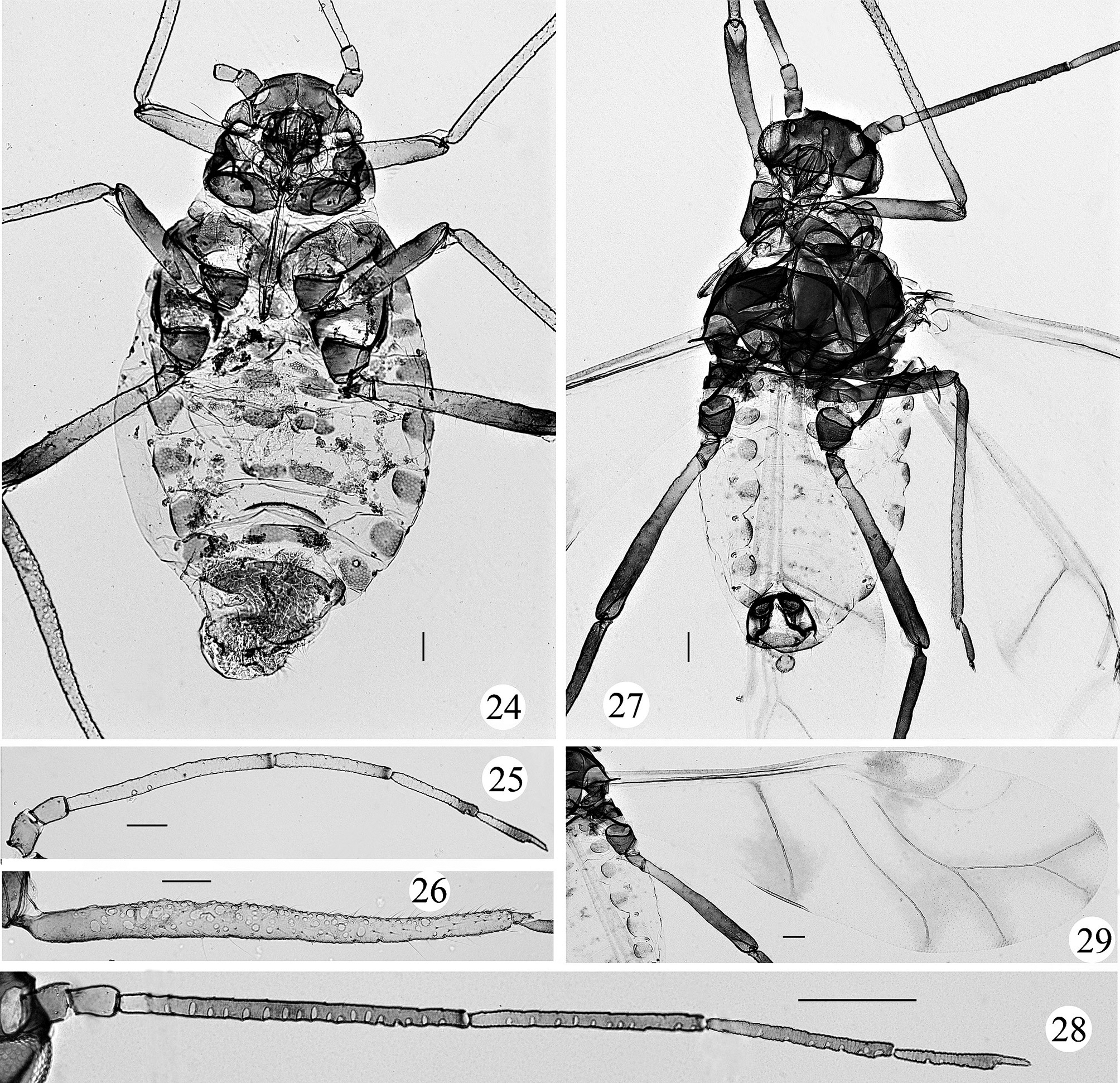
Apterous oviparous females ( Figs 9 View FIGURES 1 – 11 , 24–26 View FIGURES 24 – 29 , 36, 37 & Table 1). Body oval ( Fig. 24 View FIGURES 24 – 29 ), terminal part of abdomen elongated, dark yellow in life, covered with wax powder (Figs 36, 37).
Mounted specimens: Dorsum of body smooth, with wax glands developed on all segments of body, distal parts of antennal segments III–VI, coxae, trochanters, femora and tibiae. Head with 1 pair of large oval wax plates; pronotum with 2 pairs of spinal and 1 pair of marginal wax plates; meso-, metanotum and abdominal tergites I–VII each with 1 pair of spinal and 1 pair of marginal wax plates, and tergite VIII with 1 wax plate forming a band across the segment.
Head. Antenna 6-segmented ( Fig. 25 View FIGURES 24 – 29 ), 0.65–0.67 times as long as body, with spare spinules on segments III–VI. Processus terminalis 0.30–0.33 times as long as base of the segment VI. Segments I–VI each with 3–5, 4 or 5, 17– 24, 9–16, 5–8, 1+0 or 1 setae, respectively. Processus terminalis with 3 or 4 apical setae. Segment III with 2 or 3 secondary rhinaria ( Fig. 9 View FIGURES 1 – 11 ). Rostrum reaching mid-coxae. Ultimate rostral segment 2.17–2.60 times as long as its basal width, 0.79–0.87 times as long as second hind tarsal segment; with 6 primary setae and 10–14 secondary setae.
Thorax. Hind tibia swollen, with numerous rhinaria ( Fig. 26 View FIGURES 24 – 29 ).
Abdomen. Cauda knobbed, constricted in distal 1/3, with 10–13 setae. Anal plate round, with 52–69 setae.
The other characteristics similar to apterous viviparous females.
Alate males ( Figs 10, 11 View FIGURES 1 – 11 , 27–29 View FIGURES 24 – 29 , 37 & Table 1). Body oval ( Fig. 27 View FIGURES 24 – 29 ), dark greenish yellow in life, covered with wax powder (Fig. 37).
Mounted specimens: Head. Frons rounded. Antenna 6-segmented ( Fig. 28 View FIGURES 24 – 29 ), 0.84–1.03 times as long as body. Processus terminalis 0.19–0.26 times as long as base of the segment VI. Segments I–VI each with 3–5, 5, 19–21, 11–14, 7 or 8, 1 or 2+0 setae, respectively. Processus terminalis with 3 or 4 apical setae. Segments III–VI each with 18 or 19, 6–10, 6 or 7, 2 or 3 secondary rhinaria, respectively ( Figs 10, 11 View FIGURES 1 – 11 ). Ultimate rostral segment 2.27–2.50 times as long as its basal width, 0.81–0.87 times as long as second hind tarsal segment.
Thorax. Fore wings ( Fig. 29 View FIGURES 24 – 29 ) as in alate viviparous females.
Abdomen. Cauda knobbed, constricted in distal 1/3, with 11 or 12 setae. Anal plate bilobed, each lobe with 13–18 setae.
The other characteristics similar to alate viviparous females.
Egg (Figs 32, 36). Eggs are orange when first laid, then turning dark brown or black.
Holotype. Apterous viviparous female, CHINA, Shandong Province, Zaozhuang City, Xiwangzhuang, Xicun Village, 34.8°N, 117.6°E, altitude 60m, on Pteroceltis tatarinowii, 6.ix.2011 (An GC) [No. Y 9152–1–3]. Paratypes. 11 apterous and 13 alate viviparous females (slides), also 1 apterous viviparous female ( COI: KF772792 View Materials ), [No. Y 9152], with same data as holotype; Shandong Province, Zaozhuang City, same host and locality data as holotype, 1 apterous and 1 alate viviparous females, [No. Y 9348], 20.iv.2013 (Li WW); 1 apterous and 1 alate viviparous females (slides), and 1 apterous viviparous female ( COI: KF772794 View Materials ), [No. Y 9325], 21.viii.2013 (Li WW); 2 alate viviparous females, [No. Y 9346], 13.x.2013, (Li WW); 4 apterous oviparous females, [No. Y 9374], 20.x.2012 (Li WW); 4 alate males (slides), and 1 male ( COI: KF772793 View Materials ), [No. Y 9373], 20.x.2012 (Li WW); 3 fundatrix, [No. Y 9347], 18.iii.2013, (Li WW); Anhui Province, Chuzhou City, same host as holotype, 1 apterous, Quanjiao County, Longshan Temple, 32.02°N, 117.58°E, altitude 243m, [No. Y 9349] and 1 alate viviparous females, Mt. Langyashan, 32.18°N, 118.18°E, altitude 118m, [No. Y 9350], 30.ix.2013 (Li WW).
The holotype and most paratypes are deposited in the National Zoological Museum of China, Institute of Zoology, Chinese Academy of Sciences, Beijing, except for 2 apterous and 2 alate viviparous females [No. Y 9152], 1 apterous oviparous female [No. Y 9374] and 2 alate males [No. Y 9373] which are deposited in Forestry Bureau of Central District, Zaozhuang City, Shandong Province.
FIGURES 30–37. Shivaphis pteroceltis sp. n. in life. (30) a colony covered with wax powder, visited by ants; (31) a colony covered with wax filaments, not visited by ants; (32) eggs and 1st instar of fundatrix on the branch; (33) fundatrix and nymphs, with ants and parasitic mites; (34) apterous viviparous female on the upper side of leaf; (35) alate viviparous female feeding on the leaf vein; (36) apterous oviparous female, laying eggs; (37) apterous oviparous female, mating with alate male.
Distribution. China: Shandong Province (Zaozhuang City), Anhui Province (Chuzhou City).
Biology. The species forms large colonies on Pteroceltis tatarinowii. Colonies that are visited by ants bear thick wax powder (Figs 30, 34), but colonies not visited by ants bear thick wax filaments (Figs 31, 35). The aphids damage the leaves along the veins, young branches and fruits of the host. Fundatrices hatch in March (Fig. 32), and usually have parasitic mites (Fig 33). In November, after mating, oviparae lay eggs in cracks of the bark or around the overwintering leaf-buds (Fig. 36).
Remarks. This species belongs to subgenus Sinishivaphis by having confluent pigmented zones on media and vein Cu 1 in fore wing ( Figs 8 View FIGURES 1 – 11 , 22 View FIGURES 19 – 23 , 29 View FIGURES 24 – 29 ). It differs from the others in the genus by bearing numerous long dorsal setae and feeding on Pteroceltis tatarinowii (the other species bear spare short dorsal setae and feed on Celtis , Tilia or Ulmus ). Abdominal tergites I–V each have 3–6 pairs of spino-pleural setae and 2–5 pairs of marginal setae (in the other species, abdominal tergites I–VII each have 1 pair of spinal and 1 pair of marginal setae). Body dorsal setae length is 3.00–5.25 times longer than the basal diameter of antennal segment III in apterous viviparous females, and 2.00–4.00 times longer in alate viviparous females (in the other species 1.10–1.30 times in apterous viviparous females, and 1.40–2.10 times in alate viviparous females) ( Quednau, 2003, Qiao et al., 2005).
| COI |
University of Coimbra Botany Department |
No known copyright restrictions apply. See Agosti, D., Egloff, W., 2009. Taxonomic information exchange and copyright: the Plazi approach. BMC Research Notes 2009, 2:53 for further explanation.
|
Kingdom |
|
|
Phylum |
|
|
Class |
|
|
Order |
|
|
Family |
|
|
Genus |