
Tubulanus cf. lutescens Cantell, 2001
|
publication ID |
https://doi.org/10.5852/ejt.2022.845.1959 |
|
publication LSID |
lsid:zoobank.org:pub:214BF6A7-233B-47F6-8819-A1284093B0DB |
|
DOI |
https://doi.org/10.5281/zenodo.7268374 |
|
persistent identifier |
https://treatment.plazi.org/id/5F0F3852-FFA8-FFF1-FDC3-FC8EFD2A2912 |
|
treatment provided by |
Felipe |
|
scientific name |
Tubulanus cf. lutescens Cantell, 2001 |
| status |
|
Tubulanus cf. lutescens Cantell, 2001 View in CoL
Figs 3A–B View Fig , 4 View Fig
Diagnosis
Tubulanus cf. lutescens can be clearly identified as a member of the genus Tubulanus based on the amount of body wall, proboscis, and rhynchocoel muscle layers, the position of the nervous system, the composition of the cerebral organs, and several other internal characteristics ( Gibson 1982; Sundberg & Hylbom 1994; Gibson & Sundberg 1999). As in other members of the genus, the body wall is composed of three muscle layers (outer circular, median longitudinal, and inner circular musculature), whereas the proboscis is composed of two layers (outer longitudinal, inner circular) and the rhynchocoel wall of only one circular muscle layer. The nervous system is located between the epidermal basement membrane and the outer circular body wall musculature and the cerebral organs consist of ciliated canals in the epidermis. Moreover, a mid-dorsal blood vessel and eyes are absent. The collected specimens are of bright red coloration and lacks a conspicuous colour pattern. A lower dorsal nerve is absent.
Material examined
COSTA RICA • 1 spec. (fixed in paraformaldehyde and prepared as 67 slides with transverse sections); methane seep Mound 12; 8.930° N, 84.313° W; depth 990–999 m; 22 May 2017; Lisa Levin and Charlotte Seid leg.; collected by HOV Alvin, Dive 4907; SIO-BIC N277 (ex SIO-BIC N233) (image series: https://doi.org/10.5281/zenodo.6368041) GoogleMaps • 1 spec. (anterior fixed in paraformaldehyde, posterior fixed in ethanol); same collection data as for preceding; GenBank: ON186263 View Materials , ON036061 View Materials , ON021853 View Materials , ON182046 View Materials ; SIO-BIC N233 GoogleMaps • 1 spec. (fixed in paraformaldehyde); same collection data as for preceding; MZUCR Nemertea Collection MZUCR-103-01 (ex SIO-BIC N233) GoogleMaps • 1 spec. (piece in ethanol); methane seep Mound 11; 8.920° N, 84.306° W; depth 1019–1045 m; 26 Feb. 2009; Greg Rouse and Mike Skowronski leg.; collected by HOV Alvin, Dive 4505; GenBank: ON186261 View Materials , ON036062 View Materials , ON021852 View Materials , ON182043 View Materials ; SIO-BIC N107 GoogleMaps • 1 spec. (piece in ethanol); methane seep Mound 12; 8.931° N, 84.313° W; depth 982–1000 m; 7 Jan. 2010; Lisa Levin and Graham Nash leg.; collected by HOV Alvin, Dive 4586; GenBank: ON186262 View Materials , ON036063 View Materials , ON021854 View Materials , ON182044 View Materials ; SIO-BIC N120 GoogleMaps • 1 spec. (ethanol); methane seep Mound 12; 8.93037° N, 84.31319° W; depth 996 m; 20 Oct. 2018; Lisa Levin and Kyle Metcalfe leg.; collected by HOV Alvin, Dive 4974; GenBank: ON186264 View Materials , ON036060 View Materials , ON021851 View Materials , ON182045 View Materials ; SIO-BIC N257 GoogleMaps .
Description
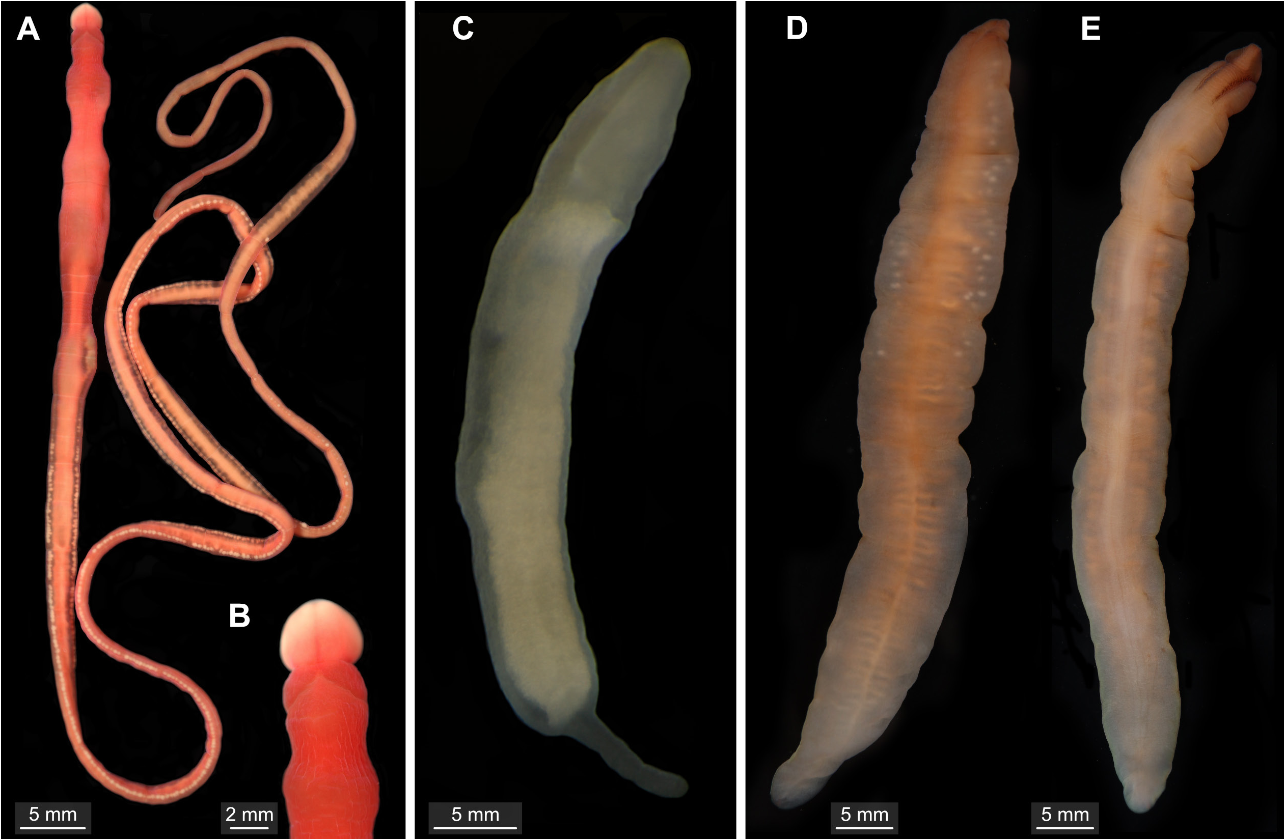
Specimens up to 20 cm in length, up to 4 mm in diameter. Body more or less cylindrical. Tapered head slightly wider than rest of body ( Fig. 3A View Fig ). One pair of dorsal, V-shaped cephalic furrows in front of brain lobes ( Fig. 3B View Fig ). Body bright red, trunk slightly translucent. Anterior tip brighter than rest of head ( Fig. 3A–B View Fig ). Epidermal constrictions along whole body length. Gut and gonads visible through body wall ( Fig 3A View Fig ).
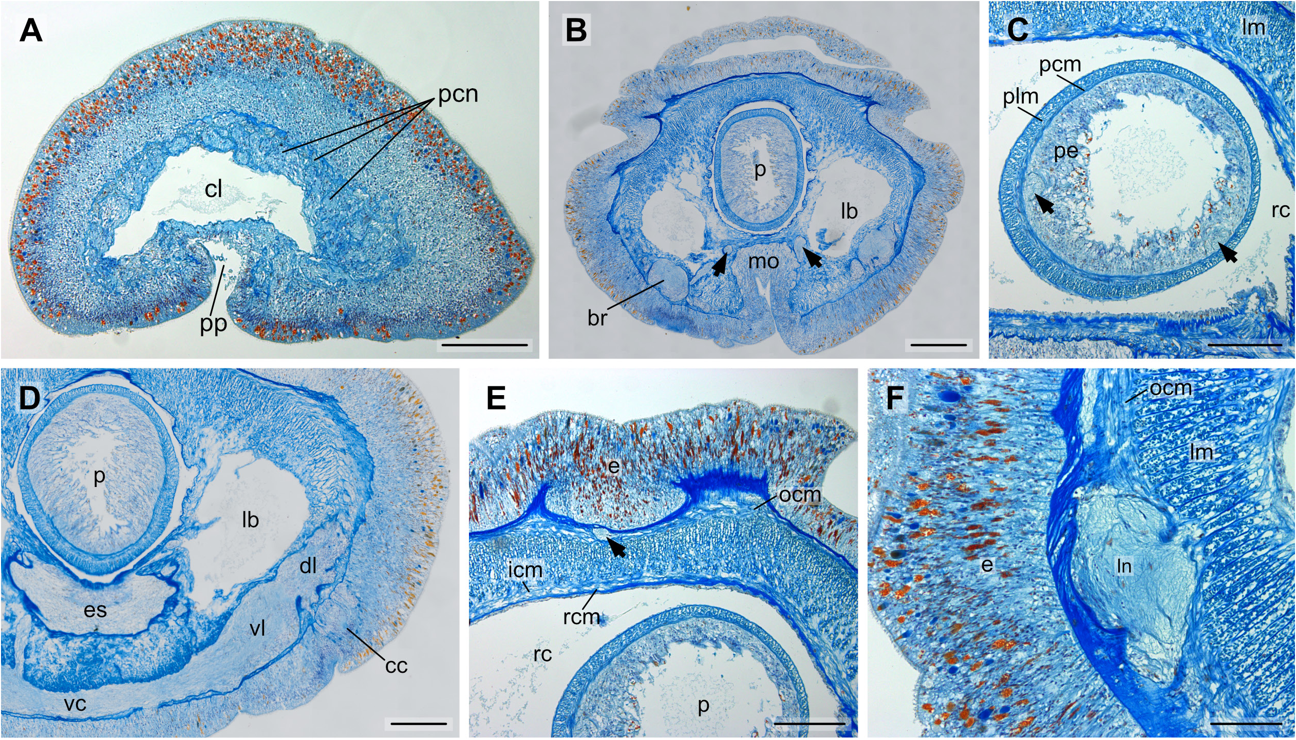
Epidermis in foregut region 150–200 µm thick. Body wall composed of outer circular, middle longitudinal and inner circular musculature ( Fig. 4E View Fig ). Rhynchocoel wall only with circular muscle layer ( Fig. 4E View Fig ). Proboscis with two layers of musculature (outer longitudinal, inner circular), with two nerves, no proboscis armature ( Fig. 4C View Fig ). Proboscis pore subterminal, ventral ( Fig. 4A View Fig ); mouth opening separate, behind cerebral ganglia ( Fig. 4B View Fig ). Cerebral ganglia with only outer neurilemma, neurochord cells absent. Dorsal and ventral ganglia fused, located in ventral part of head ( Fig. 4D View Fig ). Lateral nerve cords without accessory nerves, myofibrillae absent ( Fig. 4F View Fig ). Precerebral nerves numerous, not grouped ( Fig. 4A View Fig ); buccal nerves paired. Upper dorsal nerve present ( Fig. 4E View Fig ). Nervous system between epidermal basement membrane and outer circular body wall musculature ( Fig. 4D, F View Fig ). Eyes lacking. Cerebral organs as simple ciliated canals in epidermis, posterior to ventral ganglia ( Fig. 4D View Fig ). Apical organ as depression frontal of rhynchopore, cephalic glands in anterior half of head, scattered between muscle fibres of head. Blood vascular system consisting of cephalic loop giving rise to two paired postcerebral, lateral vessels ( Fig. 4A–B View Fig ). Mid-dorsal blood vessel absent, smaller vessels splitting from lateral blood vessels in foregut region.
Remarks
The Costa Rican specimens are attributed to the genus Tubulanus , based on the phylogenetic analyses and the similarity of external and internal characteristics. In the phylogenetic analysis, this species belongs to a highly supported clade containing Tubulanus eozensis Yamaoka, 1940 , Tubulanus polymorphus , and an undescribed tubulanid from the Sea of Okhotsk ( Fig. 3 View Fig ). A clade of undescribed abyssal and hadal tubulanids is sister to that clade. The pairwise distances of the COI gene between the four deepsea specimens are below 1%. Interestingly, pairwise distances between Tubulanus cf. lutescens and two sequences of Tubulanus lutescens , a shallow water species collected in Sweden, are only 1–1.3%. In a TCS haplotype network analysis of the mitochondrial COI gene Tubulanus lutescens is only separated from our specimens by six nucleotide substitutions ( Fig. 3 View Fig ). However, the two species differ by body coloration as Tubulanus cf. lutescens is bright red, whereas Tubulanus lutescens is yellow. Moreover, Tubulanus cf. lutescens lacks a lower dorsal nerve which is present in Tubulanus lutescens . The markedly dissimilar habitats of both species ( T. lutescens intertidal from Northern Europe vs T. cf. lutescens from ca 1000 m depth in Eastern Pacific) might indicate that they represent different species. Future analyses of nuclear sequences of T. lutescens are warranted to determine whether Tubulanus cf. lutescens and T. lutescens are indeed two different species.
No known copyright restrictions apply. See Agosti, D., Egloff, W., 2009. Taxonomic information exchange and copyright: the Plazi approach. BMC Research Notes 2009, 2:53 for further explanation.
|
Kingdom |
|
|
Phylum |
|
|
Class |
|
|
Order |
|
|
Family |
|
|
Genus |