
Rhinolekos schaeferi Martins & Langeani, 2011
|
publication ID |
https://doi.org/ 10.1590/S1679-62252011000100005 |
|
persistent identifier |
https://treatment.plazi.org/id/5F4887C1-FFCD-CB41-1773-524AFC7C30A4 |
|
treatment provided by |
Carolina |
|
scientific name |
Rhinolekos schaeferi Martins & Langeani |
| status |
sp. nov. |
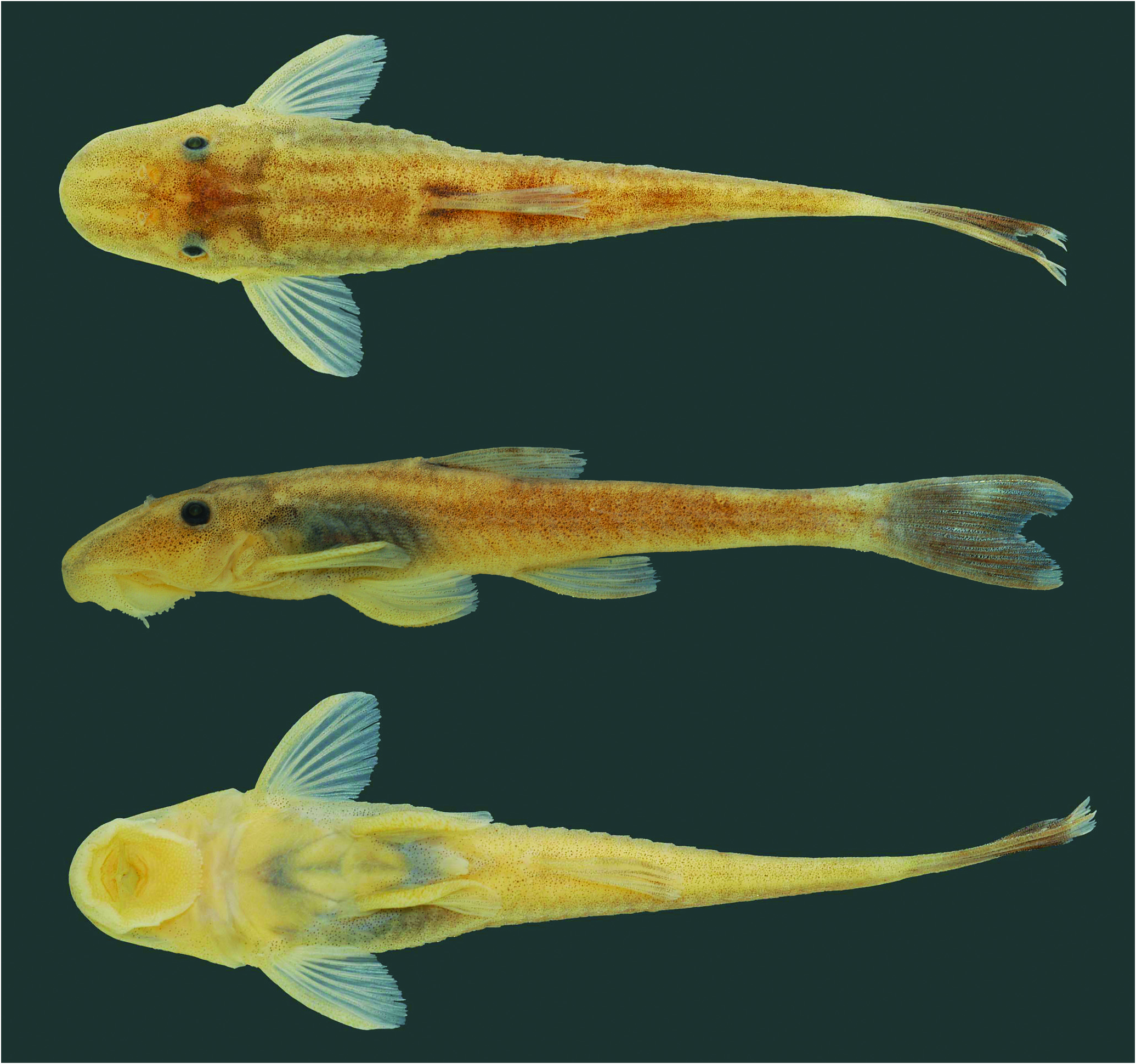
Rhinolekos schaeferi Martins & Langeani View in CoL , new species Fig. 6 View Fig
Holotype. MCP 26939, 35.4 mm SL, male, Brazil, Goiás State, Alexânia, rio Paranaíba drainage, córrego Fazenda at Chácara Fernanda , ca. 16º07’S 48º31’W, 31 Dec 2000, R. E. Reis. GoogleMaps
Paratypes. All collected with holotype. DZSJRP 12192 , 3 , 1 c&s, 36.5-38.4 mm SL (2, 37.0- 38.4 mm SL). MCP 44056, 12 View Materials , 2 View Materials c&s, 27.2-37.3 mm SL (10, 27.2-37.3 mm SL) GoogleMaps .
Diagnosis. Rhinolekos schaeferi differs from its congeners by the following characters: premaxillary and dentary accessory teeth present (vs. absent); shallower caudal peduncle (7.7-8.7% in SL vs. 9.0-10.8% in R. britskii and 10.2- 13.0% in R. garavelloi ); 18-20 mid-dorsal plates (vs. 21-24 in R. britskii and 23-27 in R. garavelloi ). Furthermore, it differs from R. britskii by having transverse dark bands in pectoral-, pelvic- and anal-fin rays absent, (vs. present) 32 vertebrae (vs. 31), and anterior portion of the compound supraneuralfirst dorsal-fin proximal radial contacting the neural spine of the 10 th vertebra (vs. 9 th). It is further distinguished from R. garavelloi by having 26-28 dorsal plates (vs. 30-35); 20-22 mid-ventral plates (vs. 24-28); larger postanal length (35.7- 41.3% in SL vs. 29.0-34.6%); shorter thoracic length (13.7- 17.6% in SL vs. 18.1-20.8%); and slender head, width 20.5- 22.9% in SL (vs. 24.6-28.1%).
Description. Morphometric and meristic data are given in Tables 5 and 6. Dorsal body profile convex from tip of snout to dorsal-fin origin; concave at dorsal-fin base; almost straight to caudal-fin origin. Small elevation at supraoccipital immediately posterior to eyes. Ventral head profile slightly concave. Ventral body profile almost straight from pectoral-fin origin to anal-fin origin, ascending at anal-fin base. Greatest body depth at dorsalfin origin. Greatest body width at opercular region, gradually tapering toward snout and caudal fin. Head without longitudinal crests, anterior margin rounded in dorsal view. Snout without rostral plate, bearing numerous small plates with thin odontodes; most anterior portion of head naked. Odontodes equal in size and uniformly distributed, not forming rows, on head and body. Eye small; dorsolaterally placed, not visible in ventral view. Iris operculum present. Infraorbital canal entering infraorbital series via compound pterotic. Compound pterotic roughly quadrangular in shape, without elongate posterior extension, its posteroventral margin with irregular and median to large perforations. Supraoccipital not contributing to the dorsal portion of the swimbladder capsule.
Body entirely covered with bony plates, except on ventral part of head, region overlying opening of swim bladder capsule, and around anus and pelvic-fin origin. Abdomen covered with small plates irregularly arranged.
Lips roundish, papillose; lower lip larger than upper lip, with papillae gradually smaller towards edges. Maxillary barbel free from lower lip and reduced. Teeth slender, bifid; median cusp larger and rounded, lateral cusp smaller and pointed. Premaxillary teeth 27-30. Dentary teeth 24-30. Number of teeth increasing with size of specimen. Premaxillary and dentary accessory teeth present.
Dorsal-fin rays ii,6-7; originating approximately at vertical through urogenital opening; its length surpassing vertical through middle of anal fin; spinelet small, approximately rectangular, with posterior margin slightly convex; locking mechanism non-functional. Anterior portion of compound supraneural-first dorsal-fin proximal radial contacting neural spine of 10 th vertebra ( Fig. 1c View Fig ). Pectoral-fin rays i,6; originating immediately posterior to opercular opening; surpassing pelvicfin origin. Cleithrum and coracoid exposed and supporting odontodes only laterally, near pectoral-fin insertion. Arrector fossae partially enclosed by ventral lamina of coracoid; opening relatively large, extending laterally halfway towards pectoralfin base. Pectoral-fin axillary slit present only in juvenile specimens. Pelvic-fin rays i,5, not reaching anal-fin origin when depressed. Pectoral- and pelvic-fin unbranched rays smaller than branched rays; enlarged odontodes at tip of pectoral-fin unbranched ray and at mesial margin of pelvic-fin unbranched ray. Anal-fin rays i,5. Caudal-fin rays i,14,i; lobes equal in size; three or four dorsal and three or four ventral procurrent rays. Adipose fin and azygous plates absent. Median lateral plates 26-29. Median-plate series complete, from compound pterotic to caudal-fin base. Vertebrae 32.
Color in alcohol. Ground color of dorsal surface light brown. Trunk with four transverse inconspicuous bars: first at dorsalfin origin; second, ventral to dorsal-fin rays; third at vertical through end of anal fin; last near caudal-fin insertion. Clear area between tip of snout and nares. Opercle region unpigmented. Lateral portion of body light brown with inconspicuous longitudinal stripe, from compound pterotic to caudal-fin origin. Ventral surface of body mostly unpigmented. Dorsal-, anal-, pectoral- and pelvic-fins membranes hyaline; dorsal- and anal-fin rays with dense concentration of chromatophores over rays. Caudal fin uniformly dark; tip of lobes unpigmented; some specimens, mainly juveniles, with a circular unpigmented area on each lobe. Procurrent rays sometimes with yellowish white coloration, extending laterally to caudal peduncle.
Sexual dimorphism. Males with conspicuous urogenital papillae immediately posterior to anus, and with a dorsal expanded flap of skin in all pelvic-fin rays.
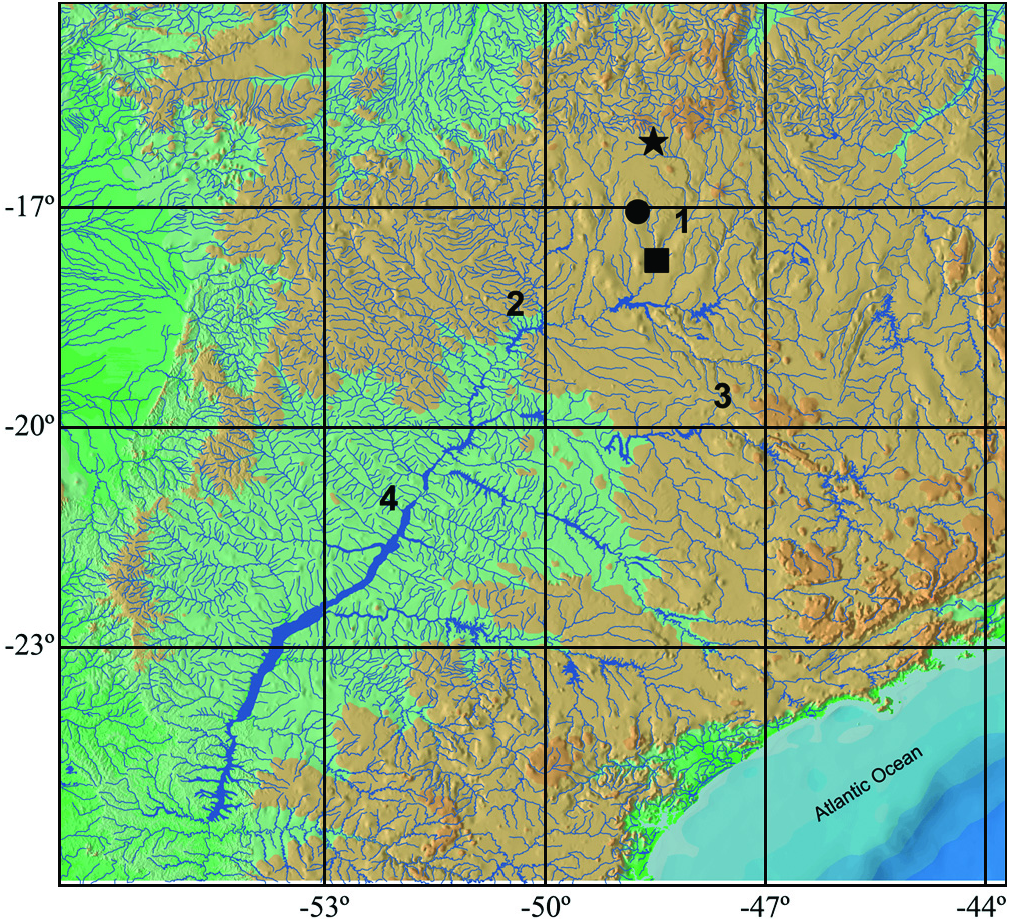
Distribution. Known from type locality, a stream tributary to rio Paranaíba drainage, upper rio Paraná system, Alexânia, Goiás State, Brazil ( Fig. 3 View Fig ).
Etymology. Named after ScottA. Schaefer, American Museum of Natural History, for his remarkable contributions for the Hypoptopomatinae systematics.
Remarks. All examined specimens of R. schaeferi present iris operculum extremely reduced, almost inconspicuous. Since the iris operculum may vary according to level of illumination ( Douglas et al., 2002), this reduction is probably a result of the light intensity at the moment these specimens were collected.

| MCP |
Pontificia Universidade Catolica do Rio Grande do Sul |
| R |
Departamento de Geologia, Universidad de Chile |
No known copyright restrictions apply. See Agosti, D., Egloff, W., 2009. Taxonomic information exchange and copyright: the Plazi approach. BMC Research Notes 2009, 2:53 for further explanation.