
Semigothograptus meganassa ( Rickards & Palmer, 2002 )
|
publication ID |
https://doi.org/ 10.11646/zootaxa.4208.6.2 |
|
publication LSID |
lsid:zoobank.org:pub:32BF47BA-AFE5-4C6F-816F-1EAECB3DA3BB |
|
DOI |
https://doi.org/10.5281/zenodo.5670208 |
|
persistent identifier |
https://treatment.plazi.org/id/5F6B647F-CB46-FFC4-1ACD-DCFD3F1237E2 |
|
treatment provided by |
Plazi |
|
scientific name |
Semigothograptus meganassa ( Rickards & Palmer, 2002 ) |
| status |
|
Semigothograptus meganassa ( Rickards & Palmer, 2002)
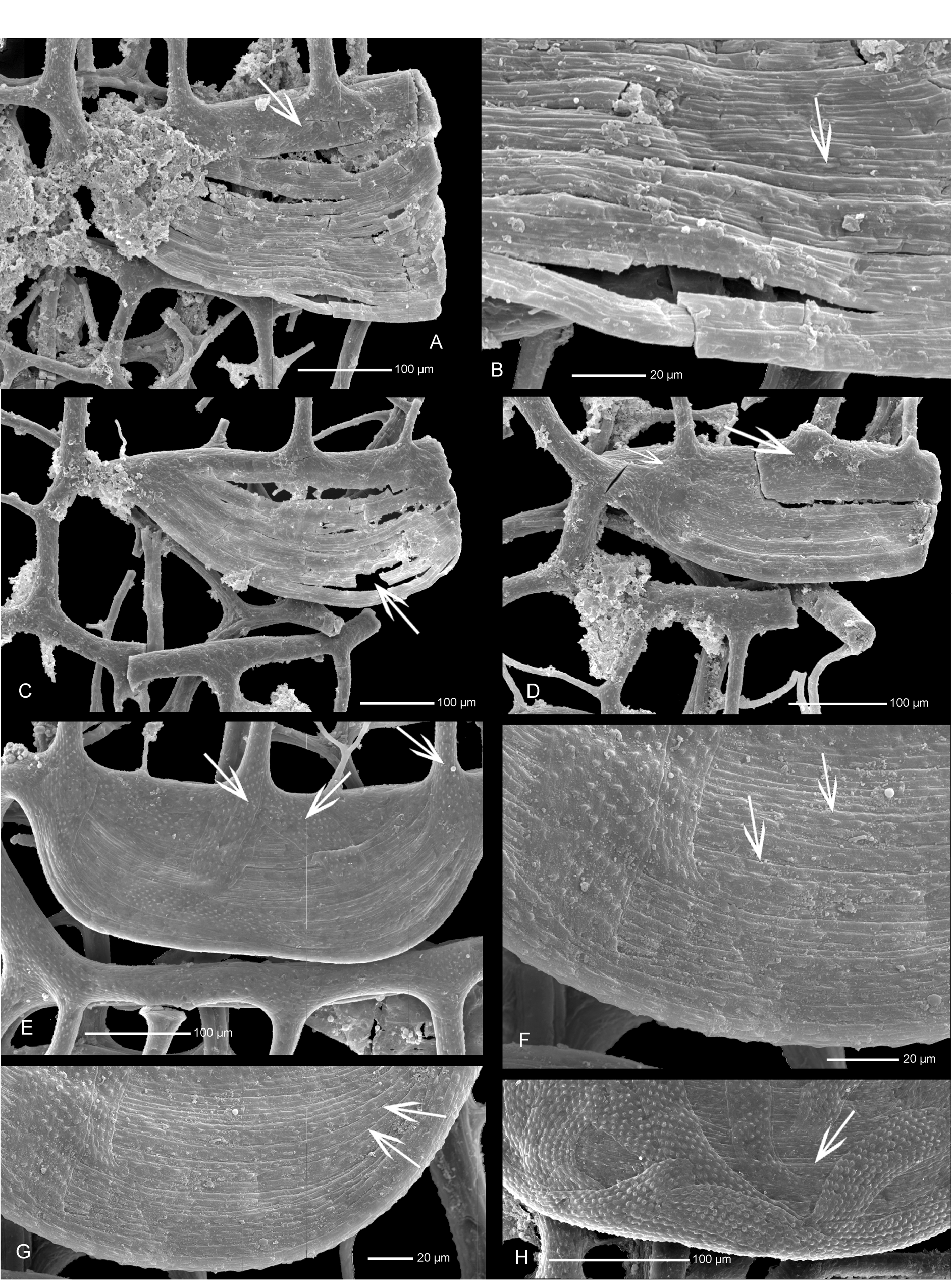
( Figs 1 View FIGURE 1 D, 2B, 3, 4B, 7A–D)
2001 Gothograptus nassa Holm, 1890 (wide form)—Kozłowska-Dawidziuk, Lenz & Štorch, pp. 151–152, fig. 4: 11–12. 2002 Gothograptus ? meganassa Rickards & Palmer , pp. 228–231, text-fig. 4.
Diagnosis. Genicular hoods of nassa type, reticulum dense in mature colonies.
Material. Four fragments of isolated tubaria with up to six pairs of thecae, from laminated marls, Bartoszyce IG-1 drill core, depth 1649.2 m and 1649.0 m, geographical coordinates 54˚14’ N, 20˚57’S, Poland, eastern part of Baltica, East European Platform. Based on flattened graptolites Tomczyk (1974) assigned the interval 1636.5– 1650.0 m to the dubius / nassa Biozone , upper Homerian, Wenlock. The material comes from the uppermost part of the dubius / nassa Biozone. The studied material was isolated from the rock though slow dissolution in 10–30% hydrochloric acid (HCl), for cleaning specimens with sediment attached 10% hydrofluoric acid (HF) was used. Specimens are stored in glycerine in plastic containers, and on SEM stubs in the Institute of Paleobiology, Polish Academy of Sciences in Warsaw.
Description. Length of immature tubarium with proximal end, ZPAL G.54/1, consisting of four pairs of thecae is 5.4 mm ( Fig. 1 View FIGURE 1 D). The distance from the ancora umbrella rim to the lip of th1 1 is 1 mm. Length of lateral apertural rods increases gradually from 0.94 mm in the first theca up to 1.1 mm in the fourth. Width of tubarium measured between thecal lip and lateral apertural rod also increases distally. Width of tubarium on the th1 1 level is 0.7 mm, on th2 1 is 0.8 mm, on th3 1 is 1.1 mm, and th4 1 level is 1.2 mm.
Specimen ZPAL G.54/2, representing possibly a more distal part of tubarium, is parallel-sided, about 1 mm wide.
The proximal ventral orifices are of triangular shape ( Fig. 1 View FIGURE 1 D, 4B). The proximal lateral orifices are located centrally, between looping meshes of the ancora umbrella. The looping meshes are developed on both sides of tubarium forming a wavy shape of the ancora umbrella rim ( Figs 2 View FIGURE 2 , 4 View FIGURE 4 B). They are smaller on the th1 1 side of tubarium ( Fig. 2 View FIGURE 2 B). Within the looping meshes are some thin additional lists ( Fig. 2 View FIGURE 2 B).
Inside specimen ZPAL G. 54/2 there is a free list, which may represent a fragment of the free virgella or nema ( Fig. 3 View FIGURE 3 B). Lateral apertural rods (pleural lists of Rickards & Palmer 2002) are long, and distinctly convex towards the longitudinal axis of the lateral walls. Clathrial lists of the lateral walls of tubarium are relatively thin, oblique, going from lateral apertural rod to lateral apertural rod of tubarium ( Fig. 4 View FIGURE 4 B).
Thecal apertures are almost rectangular. Genicular hoods are of nassa type. On the immature specimen the hoods are present on the first thecae, becoming larger in the medial part of tubarium and absent in the two distal growing thecae ( Figs 1 View FIGURE 1 D, 4B). In some thecae there are some narrow open spaces between bandages, possibly due to taphonomic changes ( Fig. 7 View FIGURE 7 A–D). Some irregular bandages are secreted on the outer surface of the upper part of the hoods ( Fig. 7 View FIGURE 7 A, D).
The largest specimen, of six pairs of thecae, is preserved in two fragments with no proximal end ( Fig. 3 View FIGURE 3 ). The specimen possibly represents the medial part of the tubarium.
The reticulum of the tubarium is composed of thin lists, dense in the proximal and medial part of the growing colony, becoming weaker distally, and not developed in the most distal immature part of the tubarium.


No known copyright restrictions apply. See Agosti, D., Egloff, W., 2009. Taxonomic information exchange and copyright: the Plazi approach. BMC Research Notes 2009, 2:53 for further explanation.
|
Kingdom |
|
|
Phylum |
|
|
Class |
|
|
Order |
|
|
Family |
|
|
Genus |
Semigothograptus meganassa ( Rickards & Palmer, 2002 )
| Kozłowska, Anna 2016 |
Gothograptus nassa
| Holm 1890 |